Abstract
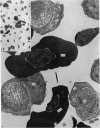
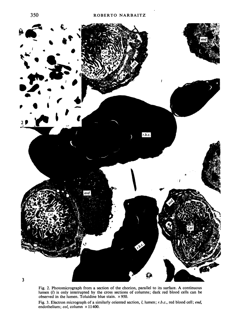
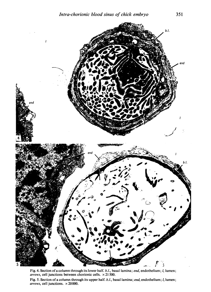
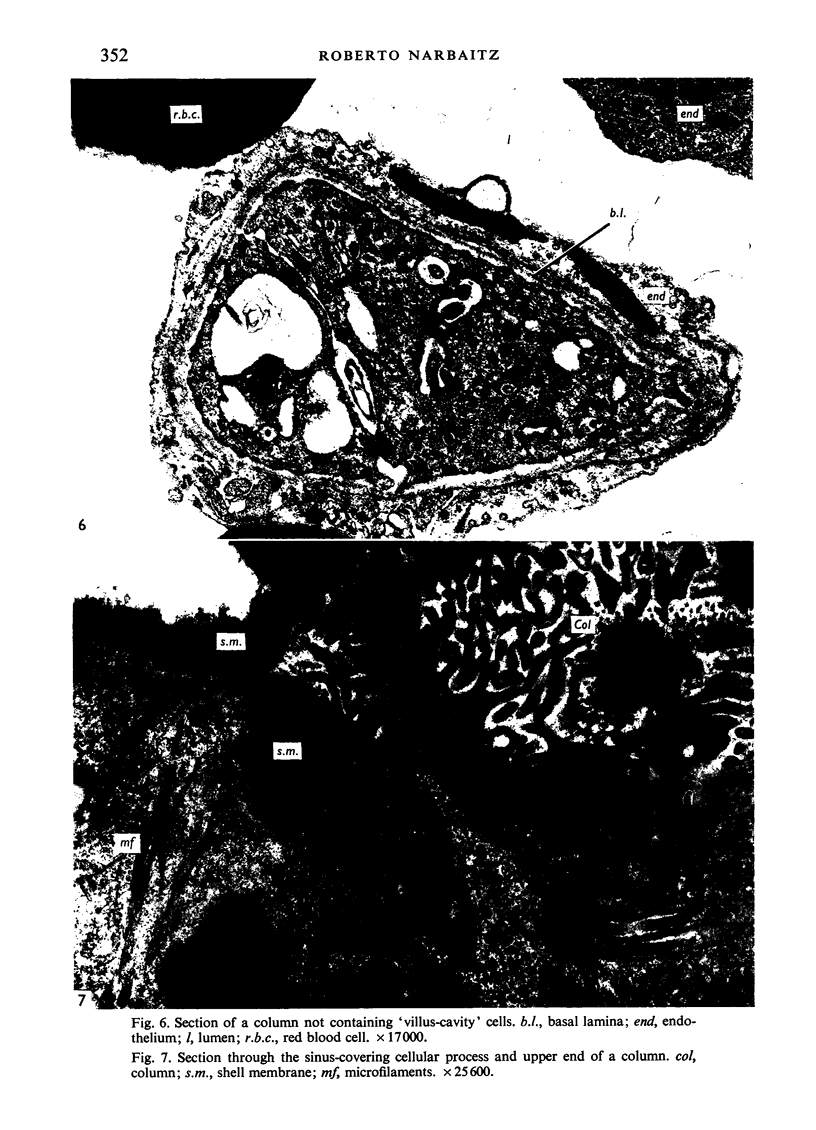
Portions of the chorio-allantoic membranes from 15 day old chick embryos were processed for electron microscopical examination. The analysis of both 1 micrometer thick sections stained with toluidine blue, and of thin sections stained with uranyl acetate and lead citrate, showed that the lumen of the intraepithelial vascular spaces in the chorion constitutes a single cavity extending over the whole membrane. The vascular arrangement can thus best be described as a single blood sinus, and not as a network of capillaries or sinusoids. The large lumen of the sinus is interrupted by cylindrical columns connecting its floor with its roof. Each column is enveloped in a layer of endothelium, a basal lamina intervening. The core of the column is formed by cytoplasm from two to five different cells ('villuscavity' cells, 'capillary-covering' cells or various combinations of both).
Full text
PDF
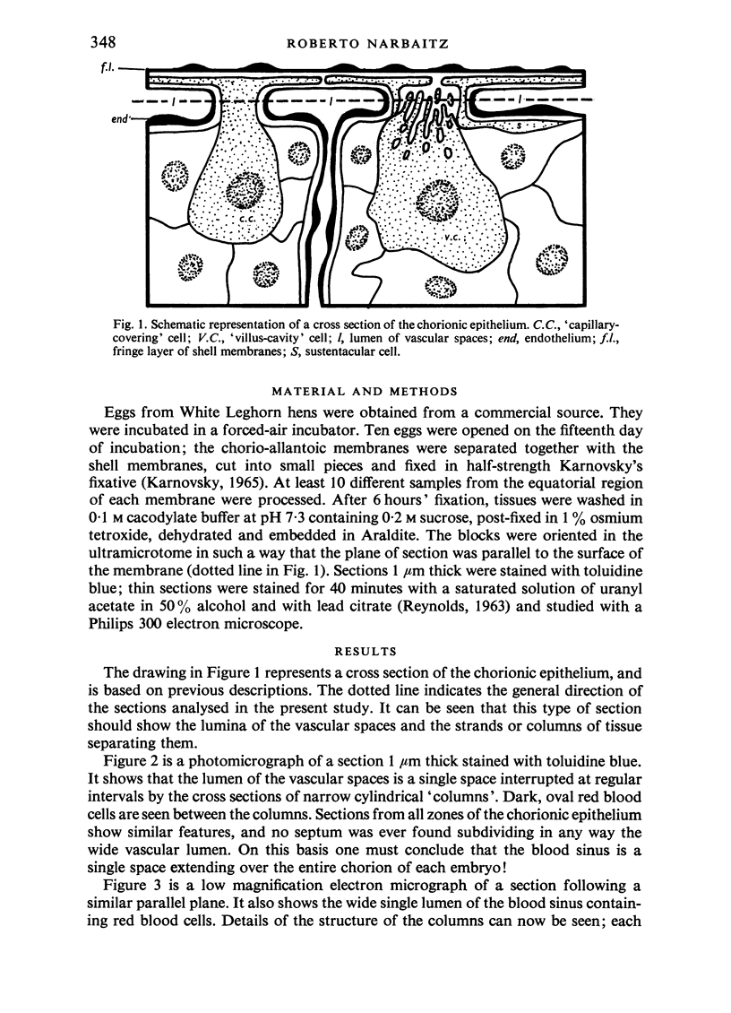
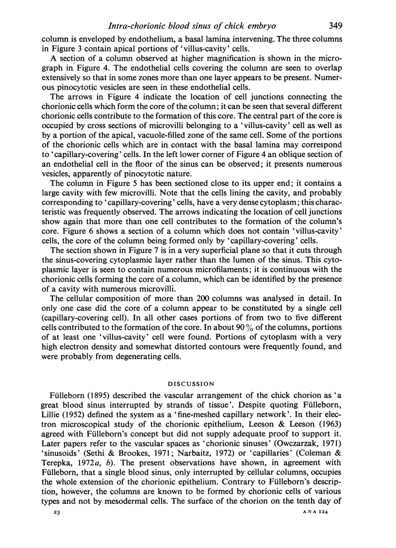
Images in this article
Selected References
These references are in PubMed. This may not be the complete list of references from this article.
- Coleman J. R., Terepka A. R. Electron probe analysis of the calcium distribution in cells of the embryonic chick chorioallantoic membrane. II. Demonstration of intracellular location during active transcellular transport. J Histochem Cytochem. 1972 Jun;20(6):414–424. doi: 10.1177/20.6.414. [DOI] [PubMed] [Google Scholar]
- LEESON T. S., LEESON C. R. THE CHORIO-ALLANTOIS OF THE CHICK. LIGHT AND ELECTRON MICROSCOPIC OBSERVATIONS AT VARIOUS TIMES OF INCUBATION. J Anat. 1963 Oct;97:585–595. [PMC free article] [PubMed] [Google Scholar]
- Owczarzak A. Calcium-absorbing cell of the chick chorioallantoic membrane. I. Morphology, distribution and cellular interactions. Exp Cell Res. 1971 Sep;68(1):113–129. doi: 10.1016/0014-4827(71)90593-3. [DOI] [PubMed] [Google Scholar]
- REYNOLDS E. S. The use of lead citrate at high pH as an electron-opaque stain in electron microscopy. J Cell Biol. 1963 Apr;17:208–212. doi: 10.1083/jcb.17.1.208. [DOI] [PMC free article] [PubMed] [Google Scholar]
- SKALINSKY E. I., KONDALENKO V. F. ELECTRON MICROSCOPIC STUDIES OF THE CHICK CHORIO-ALLANTOIS DURING EMBRYOGENESIS. Acta Morphol Acad Sci Hung. 1964;12:247–259. [PubMed] [Google Scholar]
- Sethi N., Brookes M. Ultrastructure of the blood vessels in the chick allantois and chorioallantois. J Anat. 1971 May;109(Pt 1):1–15. [PMC free article] [PubMed] [Google Scholar]


