Abstract
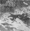
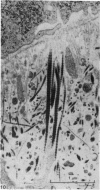
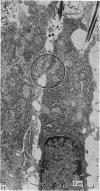
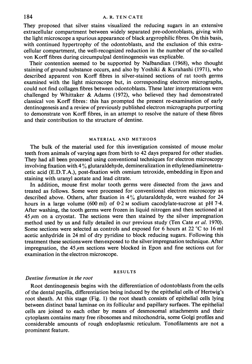
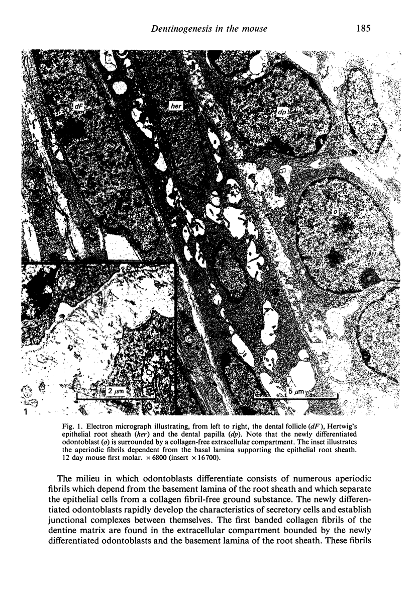
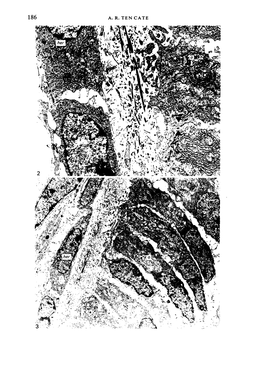
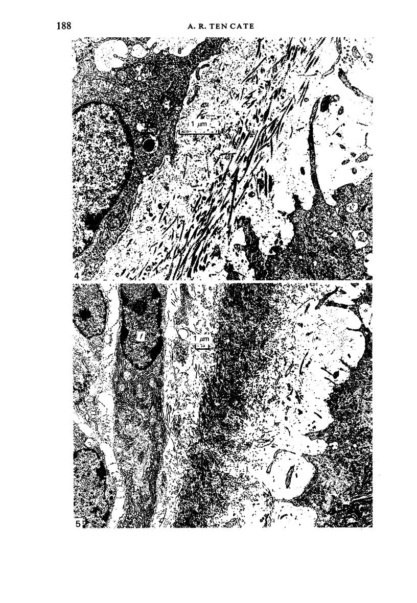
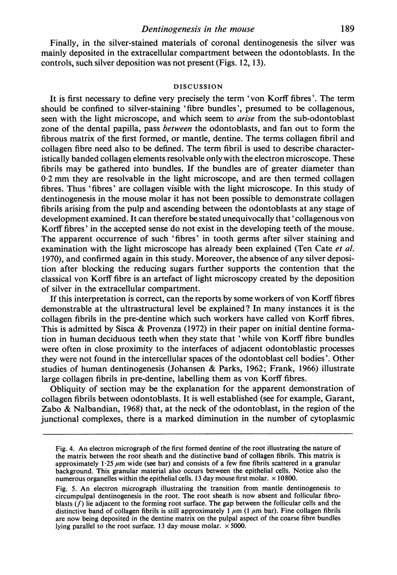
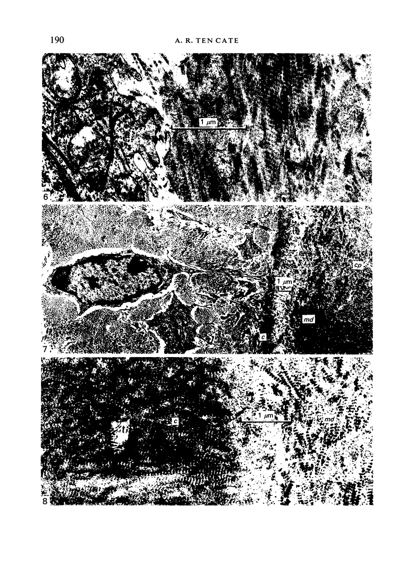
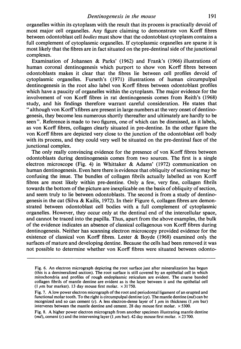
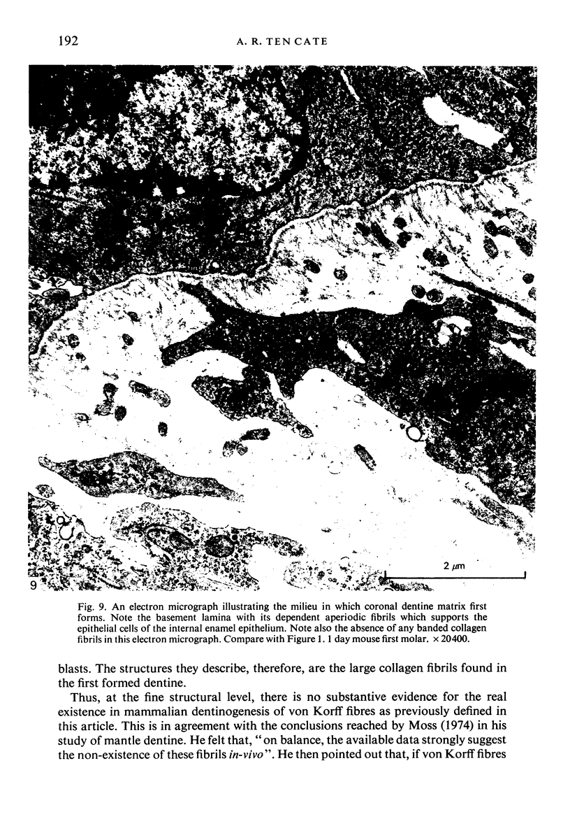
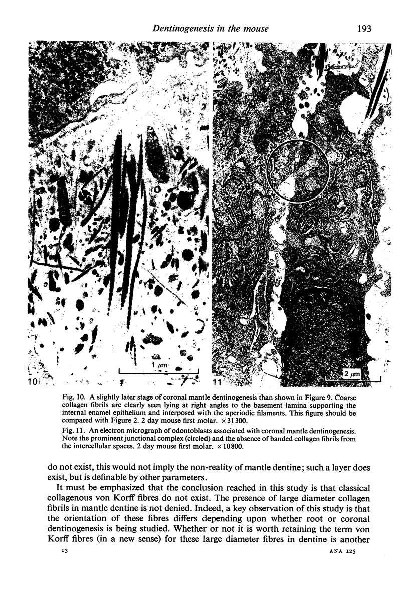
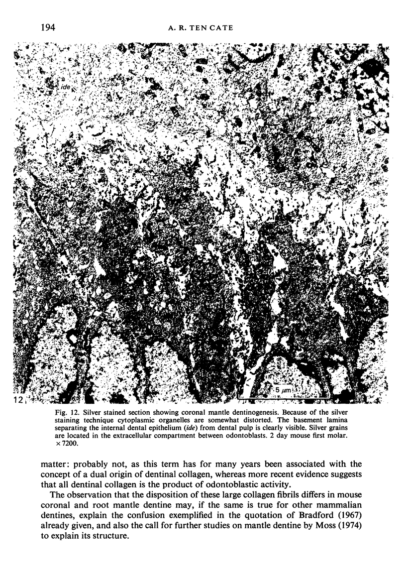
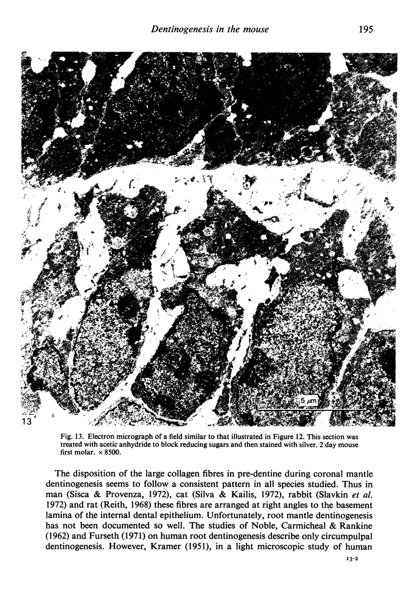
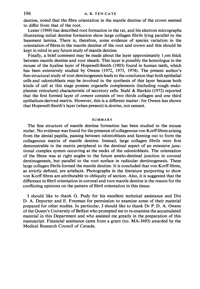
The fine structure of mantle dentine formation has been studied in the mouse molar. No evidence was found for the presence of collagenous von Korff fibres arising from the dental papilla, passing between odontoblasts and fanning out to form the collagenous matrix of mantle detine. Instead, large collagen fibrils were first demonstrable in the matrix peripheral to the dential aspect of an extensive junctional complex system occurring at the necks of the odontoblasts. The orientation of the fibres was at right angles to the future amelo-dentinal junction in coronal dentinogenesis, but parallel to the root surface in radicular dentinogenesis. These large collagen fibrils formed the mantle dentine. It is concluded that von Korff fibres, as strictly defined, are artefacts. Photographs in the literature purporting to show von Korff fibres are attributable to obliquity of section. Also, is suggested that the difference in fibril orientation in coronal and root mantle dentine is the reason for the conflicting opinions on the pattern of fibril orientation in this tissue.
Full text
PDF
Images in this article
Selected References
These references are in PubMed. This may not be the complete list of references from this article.
- Frank R. M. Etude au microscope électronique de l'odontoblaste et du canalicule dentinaire humain. Arch Oral Biol. 1966 Feb;11(2):179–199. doi: 10.1016/0003-9969(66)90186-5. [DOI] [PubMed] [Google Scholar]
- Furseth R. The fine structure of the odontoblast-predentin area in the root. Scand J Dent Res. 1971;79(3):141–150. [PubMed] [Google Scholar]
- Garant P. R., Szabo G., Nalbandian J. The fine structure of the mouse odontoblast. Arch Oral Biol. 1968 Aug;13(8):857–876. doi: 10.1016/0003-9969(68)90002-2. [DOI] [PubMed] [Google Scholar]
- JOHANSEN E., PARKS H. F. Electron-microscopic observations on sound human dentine. Arch Oral Biol. 1962 Mar-Apr;7:185–193. doi: 10.1016/0003-9969(62)90006-7. [DOI] [PubMed] [Google Scholar]
- KRAMER I. R. H. The distribution of collagen fibrils in the dentine matrix. Br Dent J. 1951 Jul 3;91(1):1–7. [PubMed] [Google Scholar]
- Lester K. S., Boyde A. The question of von Korff fibres in mammalian dentine. Calcif Tissue Res. 1968 Mar 27;1(4):273–287. doi: 10.1007/BF02008099. [DOI] [PubMed] [Google Scholar]
- Lester K. S. The unusual nature of root formation in molar teeth of the laboratory rat. J Ultrastruct Res. 1969 Sep;28(5):481–506. doi: 10.1016/s0022-5320(69)80035-3. [DOI] [PubMed] [Google Scholar]
- Moss M. L. Studies on dentin. I. Mantle dentin. Acta Anat (Basel) 1974;87(4):481–507. [PubMed] [Google Scholar]
- NOBLE H. W., CARMICHAEL A. F., RANKINE D. M. Electron microscopy of human developing dentine. Arch Oral Biol. 1962 May-Jun;7:395–399. doi: 10.1016/0003-9969(62)90033-x. [DOI] [PubMed] [Google Scholar]
- Owens P. D. A light microscopic study of the development of the roots of premolar teeth in dogs. Arch Oral Biol. 1974 Jul;19(7):525–538. doi: 10.1016/0003-9969(74)90068-5. [DOI] [PubMed] [Google Scholar]
- Owens P. D. Mineralization in the roots of human deciduous teeth demonstrated by tetracycline labelling. Arch Oral Biol. 1973 Jul;18(7):889–897. doi: 10.1016/0003-9969(73)90059-9. [DOI] [PubMed] [Google Scholar]
- Reith E. J. Collagen formation in developing molar teeth of rats. J Ultrastruct Res. 1967 Dec;21(5):383–414. doi: 10.1016/s0022-5320(67)80148-5. [DOI] [PubMed] [Google Scholar]
- Silva D. G., Kailis D. G. Ultrastructural studies on the cervical loop and the development of the amelo-dentinal junction in the cat. Arch Oral Biol. 1972 Feb;17(2):279–289. doi: 10.1016/0003-9969(72)90211-7. [DOI] [PubMed] [Google Scholar]
- Sisca R. F., Provenza D. V. Initial dentin formation in human deciduous teeth. An electron microscope study. Calcif Tissue Res. 1972;9(1):1–16. doi: 10.1007/BF02061941. [DOI] [PubMed] [Google Scholar]
- Ten Cate A. R., Melcher A. H., Pudy G., Wagner D. The non-fibrous nature of the von Korff fibres in developing dentine. A light and electron microscope study. Anat Rec. 1970 Dec;168(4):491–523. doi: 10.1002/ar.1091680404. [DOI] [PubMed] [Google Scholar]
- Whittaker D. K., Adams D. Electron microscopic studies on Von Korff fibers in the human developing tooth. Anat Rec. 1972 Oct;174(2):175–189. doi: 10.1002/ar.1091740204. [DOI] [PubMed] [Google Scholar]
- Yoshiki S., Kurahashi Y. A light and electron microscopic study of alkaline phosphatase activity in the early stage of dentinogenesis in the young rat. Arch Oral Biol. 1971 Oct;16(10):1143–1154. doi: 10.1016/0003-9969(71)90043-4. [DOI] [PubMed] [Google Scholar]


