Abstract
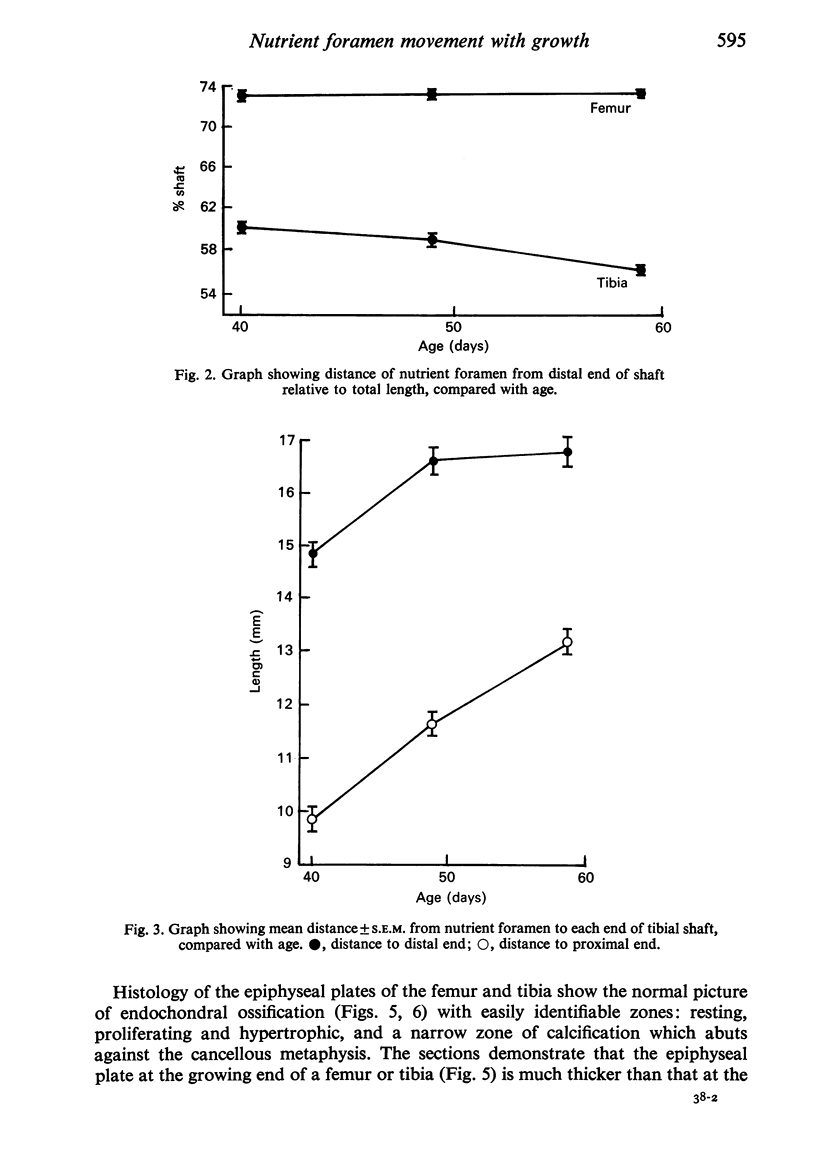
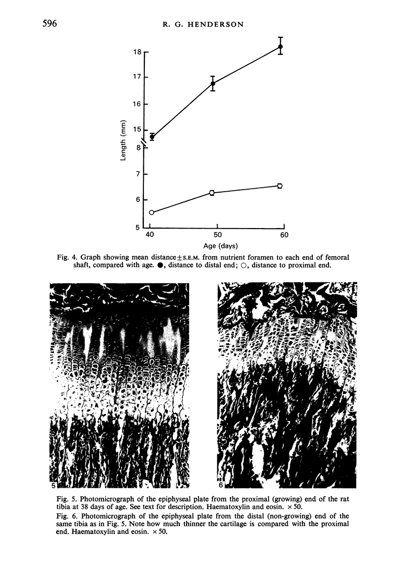
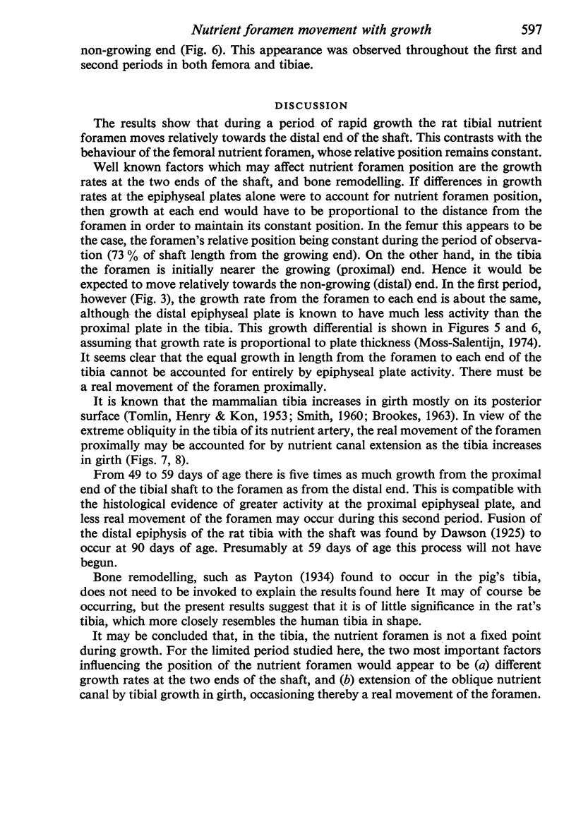
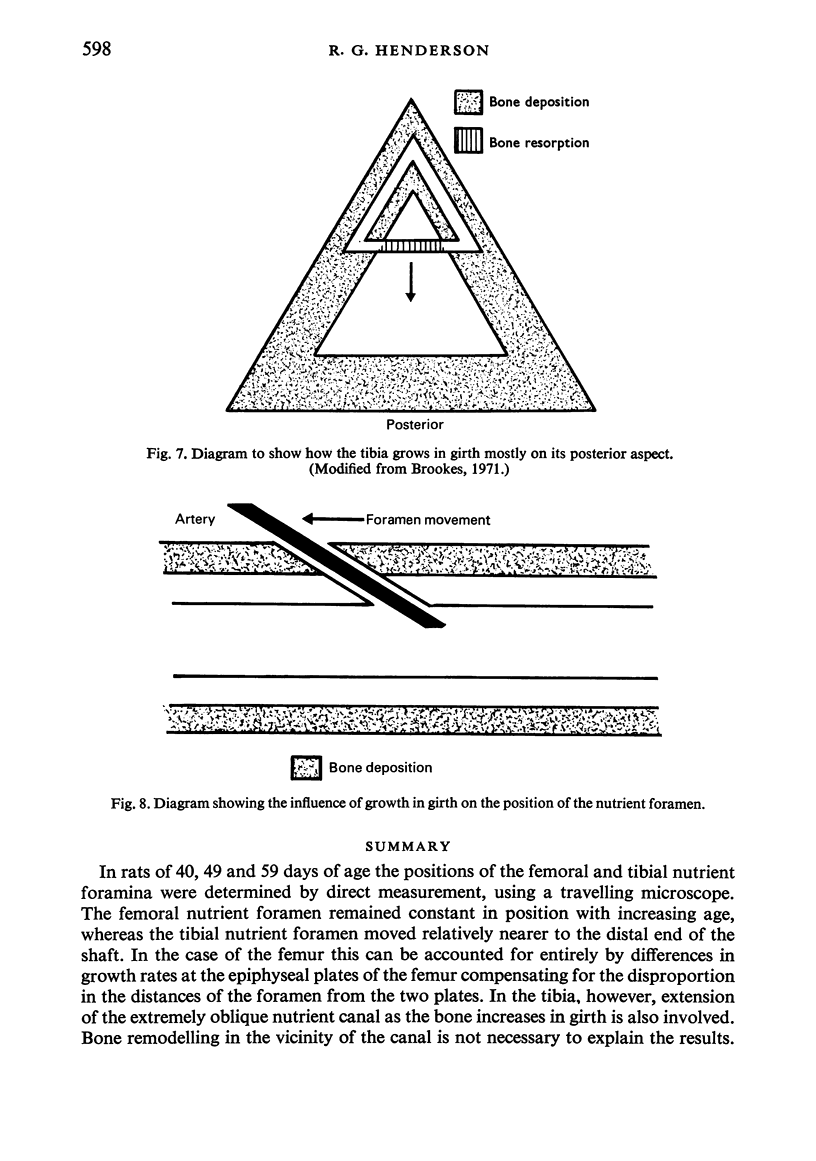
In rats of 40, 49 and 59 days of age the positions of the femoral and tibial nutrient foramina were determined by direct measurement, using a travelling microscope. The femoral nutrient foramen remained constant in position with increasing age, whereas the tibial nutrient foramen moved relatively nearer to the distal end of the shaft. In the case of the femur this can be accounted for entirely by differences in growth rates at the epiphyseal plates of the femur compensating for the disproportion in the distances of the foramen from the two plates. In the tibia, however, extension of the extremely oblique nutrient canal as the bone increases in girth is also involved. Bone remodelling in the vicinity of the canal is not necessary to explain the results.
Full text
PDF
Images in this article
Selected References
These references are in PubMed. This may not be the complete list of references from this article.
- BROOKES M. CORTICAL VASCULARIZATION AND GROWTH IN FOETAL TUBULAR BONES. J Anat. 1963 Oct;97:597–609. [PMC free article] [PubMed] [Google Scholar]
- Moss-Salentijn L. Studies of long bone growth. I. Determination of differential elongation in paired growth plates of the rat. Acta Anat (Basel) 1974;90(1):145–160. [PubMed] [Google Scholar]
- Payton C. G. The Position of the Nutrient Foramen and Direction of the Nutrient Canal in the Long Bones of the Madder-Fed Pig. J Anat. 1934 Jul;68(Pt 4):500–510. [PMC free article] [PubMed] [Google Scholar]
- Smith J. W. Collagen fibre patterns in mammalian bone. J Anat. 1960 Jul;94(Pt 3):329–344. [PMC free article] [PubMed] [Google Scholar]
- TOMLIN D. H., HENRY K. M., KON S. K. Autoradiographic study of growth and calcium metabolism in the long bones of the rat. Br J Nutr. 1953;7(3):235–252. doi: 10.1079/bjn19530029. [DOI] [PubMed] [Google Scholar]


