Abstract
The structure of amniotic papillae in sheep was investigated by light transmission and scanning electron microscopy. Papillae were found on the amnion near the umbilical cord in a majority of the sheep examined, from early mid-term onwards. The papillae possessed a basic connective tissue core with a varying degree of vascularity, the whole being sheathed in squamous epithelial cells in the earlier stages of development; but in larger, and presumably older, papillae, squamous epithelium was absent over the tips. The blood supply to these papillae was shown to originate from the chorion and to pass into the amnion at sites where the two members were closely applied to each other. Tentatively we conclude that amniotic papillae are complex organized structures which develop near amniotic plaques, or, in some instances, from the plaques themselves. The stimuli responsible for their growth and development are unknown.
Full text
PDF
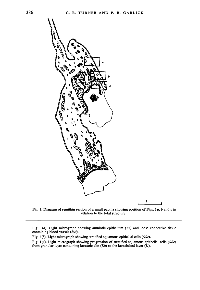
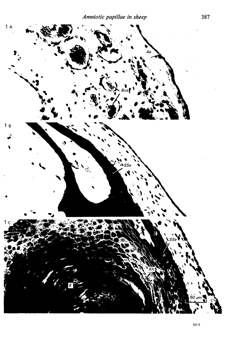
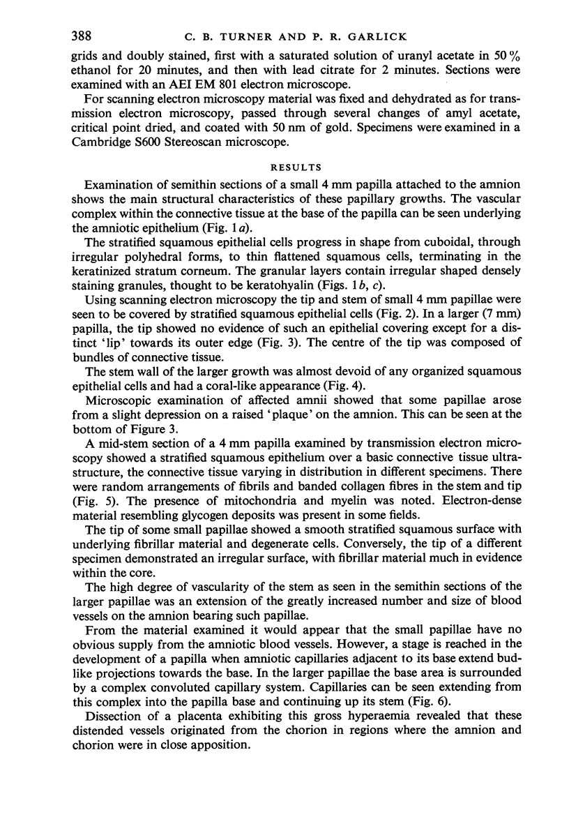
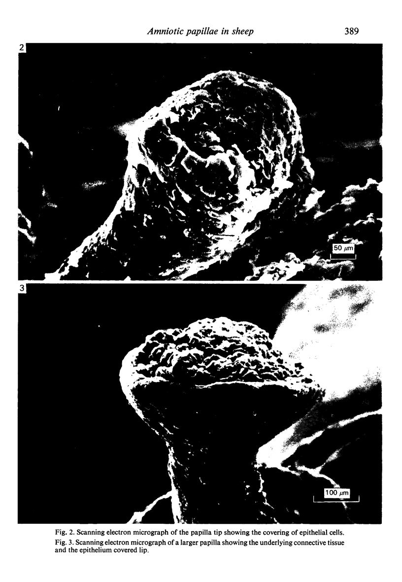
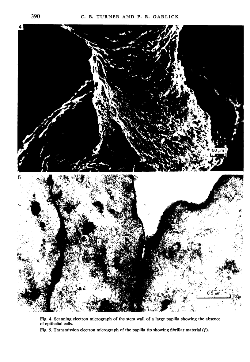
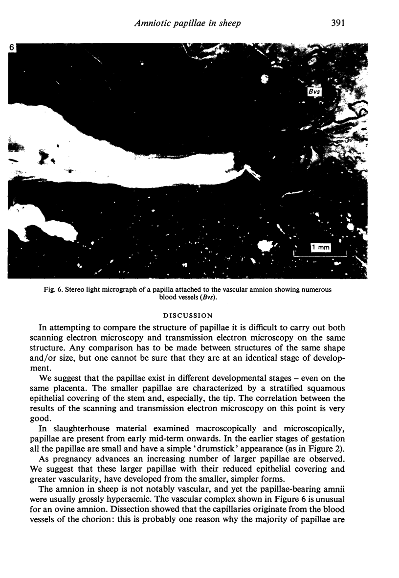
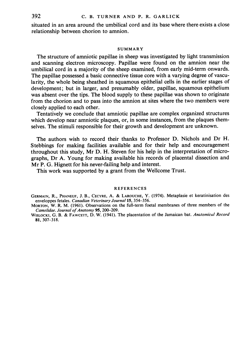
Images in this article
Selected References
These references are in PubMed. This may not be the complete list of references from this article.
- Germain R., Phaneuf J. B., Cécyre A., Larouche Y. Métaplasie et dératinisation des enveloppes foetales. Can Vet J. 1974 Dec;15(12):354–356. [PMC free article] [PubMed] [Google Scholar]
- MORTON W. R. Observations on the full-term foetal membranes of three members of the Camelidae (Camelus dromedarius L., Camelus bactrianus L. and Lama glama L.). J Anat. 1961 Apr;95:200–209. [PMC free article] [PubMed] [Google Scholar]


