Abstract
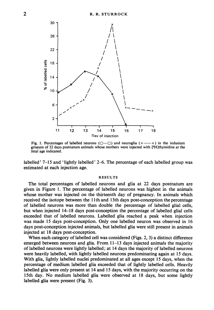
An autoradiographic study of neuronal and glial production was carried out in the indusium griseum of mice. Most neurons were produced between 13 and 15 days post-conception. One part of the glial population underwent its last or second-last divisions between 14 and 16 days post-conception, while the other continued to undergo a number of divisions into postnatal life. It is suggested the former were astrocytes and the latter oligodendrocytes.
Full text
PDF

Selected References
These references are in PubMed. This may not be the complete list of references from this article.
- Atlas M., Bond V. P. The cell generation cycle of the eleven-day mouse embryo. J Cell Biol. 1965 Jul;26(1):19–24. doi: 10.1083/jcb.26.1.19. [DOI] [PMC free article] [PubMed] [Google Scholar]
- Biesold D., Brückner G., Mares V. An autoradiographic study of gliogenesis in the rat lateral geniculate nucleus (LGN). Brain Res. 1976 Mar 12;104(2):295–301. doi: 10.1016/0006-8993(76)90621-1. [DOI] [PubMed] [Google Scholar]
- Blunt M. J., Baldwin F., Wendell-Smith C. P. Gliogenesis and myelination in kitten optic nerve. Z Zellforsch Mikrosk Anat. 1972;124(3):293–310. doi: 10.1007/BF00355032. [DOI] [PubMed] [Google Scholar]
- Dobbing J., Sands J. Timing of neuroblast multiplication in developing human brain. Nature. 1970 May 16;226(5246):639–640. doi: 10.1038/226639a0. [DOI] [PubMed] [Google Scholar]
- Haas R. J., Werner J., Fliedner T. M. Cytokinetics of neonatal brain cell development in rats as studied by the 'complete 3H-thymidine labelling' method. J Anat. 1970 Nov;107(Pt 3):421–437. [PMC free article] [PubMed] [Google Scholar]
- Henrikson C. K., Vaughn J. E. Fine structural relationships between neurites and radial glial processes in developing mouse spinal cord. J Neurocytol. 1974 Dec;3(6):659–675. doi: 10.1007/BF01097190. [DOI] [PubMed] [Google Scholar]
- Korr H., Schultze B., Maurer W. Autoradiographic investigations of glial proliferation in the brain of adult mice. I. The DNA synthesis phase of neuroglia and endothelial cells. J Comp Neurol. 1973 Jul 15;150(2):169–175. doi: 10.1002/cne.901500205. [DOI] [PubMed] [Google Scholar]
- Kraus-Ruppert R., Laissue J., Bürki H., Odartchenko N. Proliferation and turnover of glial cells in the forebrain of young adult mice as studied by repeated injections of 3 H-thymidine over a prolonged period of time. J Comp Neurol. 1973 Mar 15;148(2):211–216. doi: 10.1002/cne.901480206. [DOI] [PubMed] [Google Scholar]
- Mori S., Leblond C. P. Electron microscopic features and proliferation of astrocytes in the corpus callosum of the rat. J Comp Neurol. 1969 Oct;137(2):197–225. doi: 10.1002/cne.901370206. [DOI] [PubMed] [Google Scholar]
- Paterson J. A., Privat A., Ling E. A., Leblond C. P. Investigation of glial cells in semithin sections. 3. Transformation of subependymal cells into glial cells, as shown by radioautography after 3 H-thymidine injection into the lateral ventricle of the brain of young rats. J Comp Neurol. 1973 May 1;149(1):83–102. doi: 10.1002/cne.901490106. [DOI] [PubMed] [Google Scholar]
- Skoff R. P., Price D. L., Stocks A. Electron microscopic autoradiographic studies of gliogenesis in rat optic nerve. II. Time of origin. J Comp Neurol. 1976 Oct 1;169(3):313–334. doi: 10.1002/cne.901690304. [DOI] [PubMed] [Google Scholar]
- Sturrock R. R. A light and electron microscopic study of proliferation and maturation of fibrous astrocytes in the optic nerve of the human embryo. J Anat. 1975 Apr;119(Pt 2):223–234. [PMC free article] [PubMed] [Google Scholar]
- Sturrock R. R. Development of the indusium griseum. I. A quantitative light microscopic study of neurons and glia. J Anat. 1978 Feb;125(Pt 2):293–298. [PMC free article] [PubMed] [Google Scholar]
- Sturrock R. R. Development of the indusium griseum. II. A semithin light microscopic and electron microscopic study. J Anat. 1978 Mar;125(Pt 3):433–445. [PMC free article] [PubMed] [Google Scholar]
- Sturrock R. R. Histogenesis of the anterior limb of the anterior commissure of the mouse brain. II. A quantitative study of pre- and postnatal mitosis. J Anat. 1974 Feb;117(Pt 1):27–35. [PMC free article] [PubMed] [Google Scholar]
- Sturrock R. R. Histogenesis of the anterior limb of the anterior commissure of the mouse brain. II. A quantitative study of pre- and postnatal mitosis. J Anat. 1974 Feb;117(Pt 1):27–35. [PMC free article] [PubMed] [Google Scholar]
- Sturrock R. R. Neurons in the mouse anterior commissure. A light microscopic, electron microscopic and autoradiographic study. J Anat. 1977 Jul;123(Pt 3):751–762. [PMC free article] [PubMed] [Google Scholar]
- Sturrock R. R. Quantitative changes in neuroglia in the white matter of the mouse brain following hypoxic stress. J Anat. 1976 Feb;121(Pt 1):7–13. [PMC free article] [PubMed] [Google Scholar]
- Vaughn J. E. An electron microscopic analysis of gliogenesis in rat optic nerves. Z Zellforsch Mikrosk Anat. 1969;94(3):293–324. doi: 10.1007/BF00319179. [DOI] [PubMed] [Google Scholar]


