Abstract
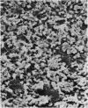
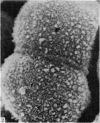
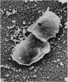
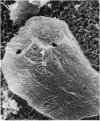
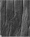
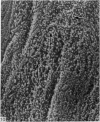
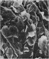
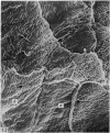
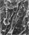
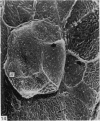
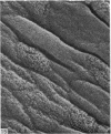
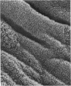
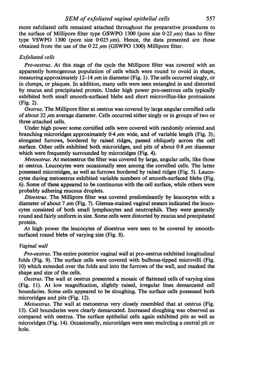
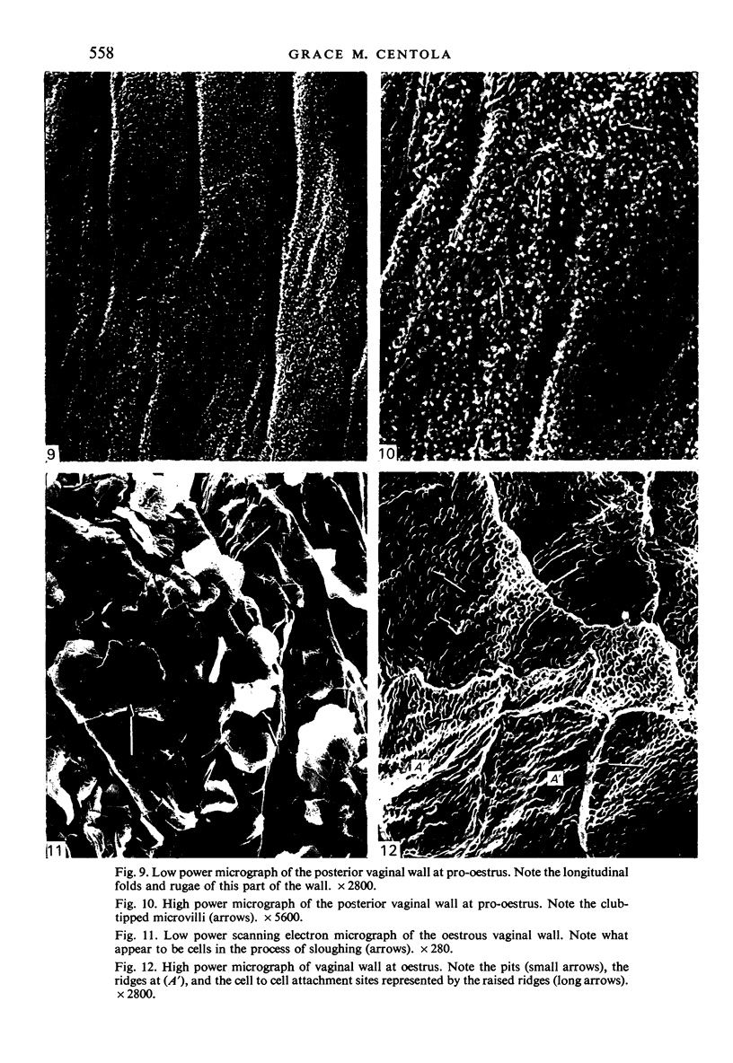
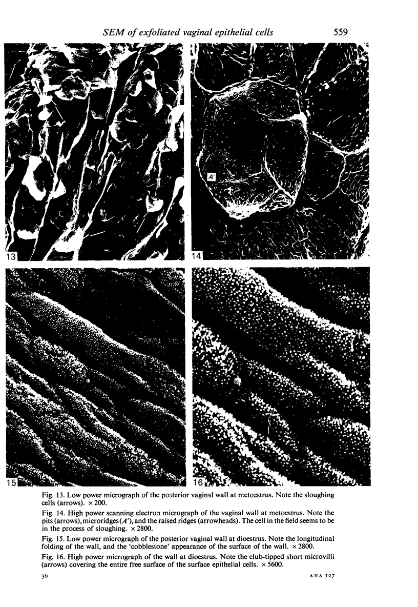
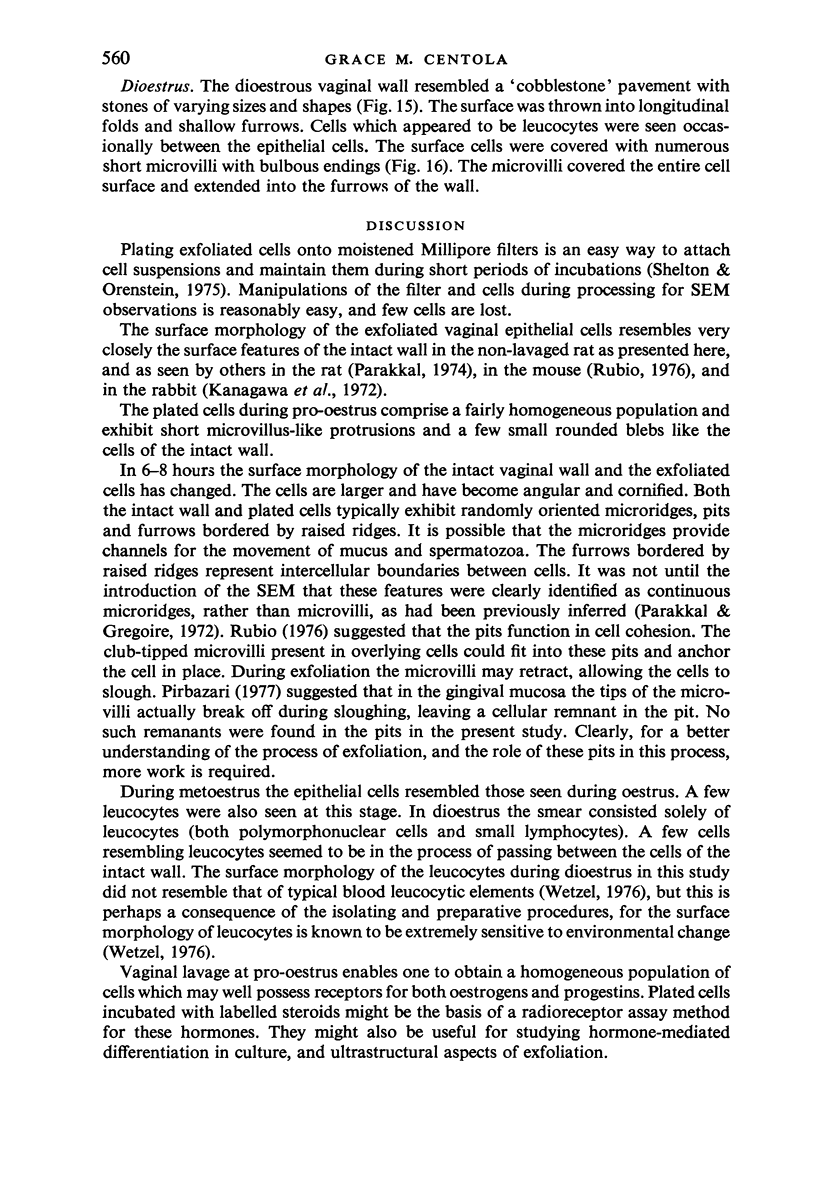
The purpose of this study was to document vaginal exfoliative cytology (smears) as seen with the scanning electron microscope during the first, hormonally induced oestrous cycle of PMS-LH treated immature rats and to compare it with the surface morphology of the intact vaginal wall of similarly treated rats. Exfoliated cells were obtained by vaginal lavage 7--9 hours (pro-oestrus), 18--20 hours (oestrus), 28--34 hours (metoestrus) and 60--64 hours (dioestrus) after administration of luteinizing hormone. Cell suspensions were plated upon moistened Millipore filters according to the method of Shelton & Orenstein (1975). The pro-oestrous stage was characterized by round to ovoid cells with numerous short microvillus-like protrusions and blebs. The vaginal wall exhibited folds lined by cells with many short microvilli. Oestrus was characterized by flattened angular cells with either blebs, microridges or both. The intact wall exhibited 'flattened mosaics' of cells, some in the process of desquamation, with ridges and pits. Metoestrus resembled oestrus, except for the presence of a few leucocytes in the plated specimens. Dioestrus was characterized by an abundance of pleomorphic leucocytes with blebs of varying sizes. The vaginal wall appeared as a 'cobblestone' pavement with cells of varying sizes and shapes possessing numerous short, microvilli with bulbous endings.
Full text
PDF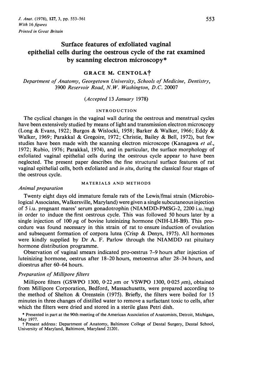
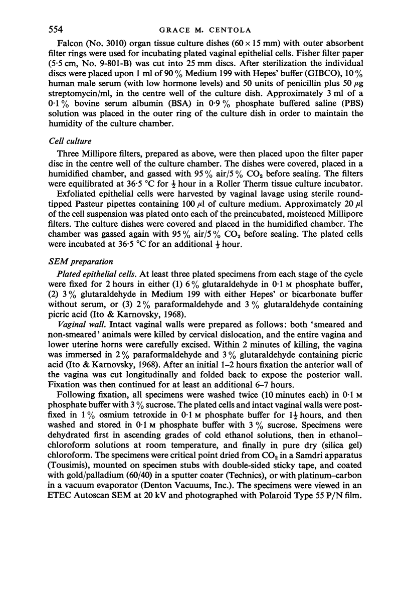
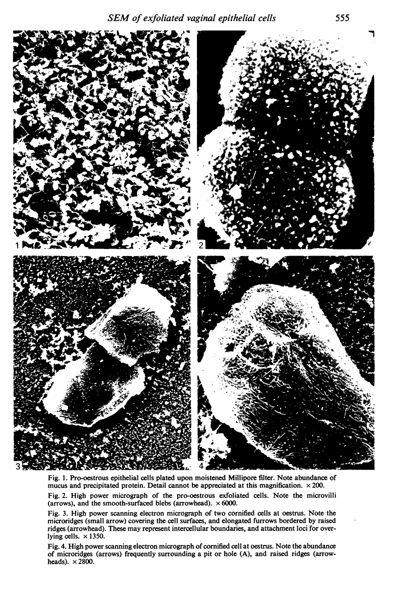
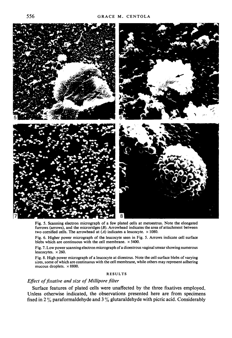
Images in this article
Selected References
These references are in PubMed. This may not be the complete list of references from this article.
- BURGOS M. H., WISLOCKI G. B. The cyclical changes in the mucosa of the guinea pig's uterus, cervix and vagina and in the sexual skin, investigated by the electron microscope. Endocrinology. 1958 Jul;63(1):106–121. doi: 10.1210/endo-63-1-106. [DOI] [PubMed] [Google Scholar]
- Barker T. E., Walker B. E. Initiation of irreversible differentiation in vaginal epithelium. Anat Rec. 1966 Jan;154(1):149–159. doi: 10.1002/ar.1091540112. [DOI] [PubMed] [Google Scholar]
- Christie D. W., Bailey J. B., Bell E. T. Classification of cell types in vaginal smears during the canine oestrous cycle. Br Vet J. 1972 Jun;128(6):301–310. doi: 10.1016/s0007-1935(17)36935-x. [DOI] [PubMed] [Google Scholar]
- Eddy E. M., Walker B. E. Cytoplasmic fine structure during hormonally controlled differentiation in vaginal epithelium. Anat Rec. 1969 Jun;164(2):205–217. doi: 10.1002/ar.1091640207. [DOI] [PubMed] [Google Scholar]
- Kanagawa H., Hafez E. S., Pitchford W. C., Baechler C. A., Barnhart M. I. Surface patterns in the reproductive tracts of the rabbit observed by scanning electron microscopy. Anat Rec. 1972 Oct;174(2):205–225. doi: 10.1002/ar.1091740206. [DOI] [PubMed] [Google Scholar]
- Parakkal P. F. Cyclical changes in the vaginal epithelim of the rat seen by scanning electron microscopy. Anat Rec. 1974 Mar;178(3):529–537. doi: 10.1002/ar.1091780302. [DOI] [PubMed] [Google Scholar]
- Parakkal P. F., Gregoire A. T. Differentiation of vaginal epithelium in the normal and hormone-treated rhesus monkey. Biol Reprod. 1972 Feb;6(1):117–130. doi: 10.1093/biolreprod/6.1.117. [DOI] [PubMed] [Google Scholar]
- Rubio C. A. The exfoliating cervico-vaginal surface. II. Scanning electron microscopical studies during the estrous cycle in mice. Anat Rec. 1976 Jul;185(3):359–372. doi: 10.1002/ar.1091850308. [DOI] [PubMed] [Google Scholar]
- Shelton E., Orenstein J. M. A plating method for preparation of cells for culture and for observation by light or electron microscopy. Exp Mol Pathol. 1975 Oct;23(2):220–227. doi: 10.1016/0014-4800(75)90020-9. [DOI] [PubMed] [Google Scholar]


