Abstract
Foot-and-mouth disease virus (FMDV) initiates infection by binding to integrin receptors via an Arg-Gly-Asp (RGD) sequence found in the G-H loop of the structural protein VP1. Following serial passages of a type A24 Cruzeiro virus (A24Cru) in bovine, via tongue inoculation, a virus was generated which contained an SGD sequence in the cell receptor-binding site and expressed a turbid plaque phenotype in BHK-21 cells. Propagation of this virus in these cells resulted in the rapid selection of viruses that grew to higher titers, produced clear plaques, and now contained an RGD sequence in place of the original SGD. To study the role of the SGD sequence in FMDV receptor recognition and bovine virulence, we assembled an infectious cDNA clone of an RGD-containing A24Cru and derived mutant clones containing either SGD with a single nucleotide substitution in the R144 codon or double substitutions at this position to prevent mutation of the S to an R. The SGD viruses grew poorly in BHK-21 cells and stably maintained the sequence during propagation in BHK-21 cells expressing the bovine αVβ6 integrin (BHK3-αVβ6), as well as in experimentally infected and contact steers. While all the SGD-containing viruses used only the bovine αVβ6 integrin as a cellular receptor with relatively high efficiency, the revertant RGD viruses utilized either the αVβ1 or αVβ3 bovine integrins with higher efficiency than αVβ6 and grew well in BHK-21 cells. Replacing the R at the −1 SGD position with either K or E showed that this residue did not contribute to integrin utilization in vitro. These results illustrate the rapid evolution of FMDV with alteration in receptor specificity and suggest that viruses with sequences other than RGD, but closely related to it, can still infect via integrin receptors and induce and transmit the disease to susceptible animals.
Foot-and-mouth disease virus (FMDV) is the causative agent of foot-and-mouth disease (FMD), a highly contagious disease of cloven-hoofed animals, including cattle, swine, goats, sheep, and other species of wild ruminants. The virus exhibits a remarkable adaptation and serological diversity exemplified by the existence of a large number of subtypes within each serotype and the fact that infected animals can also become persistent carriers (2, 3, 27, 49). In infected animals the virus replicates very rapidly and spreads among in-contact susceptible animals by aerosol or direct contact. Clinical signs of FMD, including vesicles on the feet and the mouth, can appear as early as 2 days after virus exposure (reference 20 and references therein).
FMDV is the type species of the Aphthovirus genus of the family Picornaviridae. The viral genome, consisting of a single open reading frame, is flanked by a long 5′-nontranslated region (NTR) linked to a small viral peptide, VPg, and a short 3′NTR followed by a poly(A) tail (Fig. 1a). Four structural proteins (VP1 to VP4) constitute the viral capsid, and a number of essential nonstructural proteins, including the RNA-dependent RNA polymerase (3Dpol), are involved in the replication of viral RNA (reviewed in references 7 and 45).
FIG. 1.
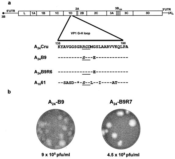
Schematic description of the FMDV genomic RNA. (a) Map of the FMDV genome; the 5′NTR is linked to the viral protein VPg and consists of the S-fragment, the poly(C), the pseudoknots, cis-acting replication element, and internal ribosomal entry site element. The 3′NTR contains a heteropolymeric region and is followed by poly(A). The viral proteins are indicated in the open boxed area, and the amino acid sequence of the VP1 G-H loop of A24Cru is also displayed. For the Colombian (A24-B9 and A24-B9R6; see Results for details) and Argentinian A1061 (GenBank V01130) viruses, only the mutated residue(s) is shown. (b) Titers and plaques formed in BHK-21 cell monolayers by the bovine-derived A24-B9 virus and its cell culture-grown derivative, A24-B9R7 virus. Viruses were titrated on BHK-21 cell monolayers, stained with crystal violet at 48 h postinfection, and photographed.
The first step in FMDV infection involves the recognition of a cell surface receptor. An Arg-Gly-Asp (RGD) amino acid sequence located within the βG-βH (G-H) loop of the capsid protein VP1 (1, 28, 30) is known to bind to several integrins of the αV subgroup, including αVβ1, αVβ3, αVβ6, and αVβ8, and is involved in virus attachment to cells and interaction with neutralizing antibodies (8, 18, 21, 24-26, 34, 36, 39, 40, 52). The generation of genetically engineered virions carrying deletions or mutations of the RGD motif leads to virus which is unable to bind to cultured cells or cause disease in susceptible animals (29, 34, 37, 43).
In cell culture, FMDV binds preferentially to one or more of the above-mentioned integrins with different specificities, and integrin utilization varies between serotypes (7, 17, 18). The reason for this specificity is not clearly understood; however, it is known that sequences surrounding the RGD motif, or located in other regions of the viral capsid, could induce structural changes in the G-H loop of VP1 and modulate binding specificity (14, 42). These changes may occur with the replacement of only one or a few amino acids at the surface of the virus (reviewed in reference 7). Viruses propagated in vitro can exploit alternative mechanisms to bind and enter the host cell independent of integrin binding. Among them, the use of heparan sulfate has been correlated with the acquisition of positively charged amino acids on the virus capsid surface (19, 22, 48). It has also been suggested that there might be other, still-undefined FMDV receptors (5, 31, 53). Despite these observations, there is no evidence of utilization of these alternative receptors in vivo, and the information currently available indicates that FMDV utilizes integrins for entry in the natural host (6, 40; reviewed in references 23 and 33).
The RGD motif is highly conserved among FMDV field isolates, and changes occurring within the G-H loop appear to be the result of antigenic or environmental selective pressures, including cell culture propagation (9, 15, 42, 46, 51). As a result of serial experimental infection with a prototype strain of A24 Cruzeiro (A24Cru) in naïve cattle using tongue inoculation, a variant (A24-B9) was isolated in Colombia with an SGD sequence in the cell receptor recognition site of VP1 instead of the usual RGD sequence. This virus was characterized in our laboratory by a distinctive turbid plaque phenotype and low affinity for BHK-21 cells. Interestingly, a type A FMDV (A1061) field isolate from Argentina with an SGD in this position has been documented (FMDV-A10, Argentina/61; GenBank accession number V01130). Passage of the A24-B9 virus in BHK-21 cells in our laboratory resulted in the rapid selection of viruses that grew to higher titers, produced clear plaques, and contained an RGD sequence. Immunization of cattle with an inactivated vaccine prepared from tissue culture-grown derivatives of A24-B9 has been associated with reduced vaccine protection following challenge with the SGD-containing virus. In the present study we report the phenotypic and genetic characterization of the A24-B9 isolate and analyze the receptor utilization and in vitro replication of molecularly cloned SGD mutant viruses containing additional changes in a conserved amino acid upstream of this motif. Furthermore, we have compared the pathogenesis of SGD- and RGD-containing viruses by direct inoculation and contact transmission in cattle.
MATERIALS AND METHODS
Viruses and virus passages in vitro.
The FMDV type A24Cru used to construct the infectious cDNA clone, pA24Cru, was recovered from the vesicular fluid of a foot lesion of a bovine inoculated with A24Cru virus intradermally into the tongue (38) and provided by Marvin Grubman (Foreign Animal Disease Research Unit, USDA Agricultural Research Service, Plum Island Animal Disease Center, Greenport, NY). The virus was used directly for RNA extraction and first-strand cDNA synthesis without any passage in tissue culture. The variant A24-B9 strain, obtained from the Empresa Colombiana de Productos Veterinarios (Vecol) in Bogota, Colombia, was recovered from the lesions of the ninth serial passage of A24Cru in bovine tongue. A24-B9R7 virus is a tissue culture derivative of the A24-B9 virus, also provided by Vecol, following seven passages of the B9 virus in BHK-21 cells. Accession numbers for the complete sequence of A24Cru and the P1 region of A1061 viruses are AY593768 and V01130, respectively.
Construction of genome-length infectious cDNA of A24Cru and derivation of G-H loop VP1 mutants.
Plasmid pA24Cru is an infectious cDNA clone of FMDV A24Cru constructed by using a long reverse transcription-PCR (RT-PCR) method previously described (32). Briefly, total viral RNA was extracted from vesicular fluid of an infected animal using TRIzol (Life Technologies) and used as a template for first-strand cDNA synthesis with SuperScript II polymerase (Life Technologies) by using specific oligonucleotide primers (P15, 5′-GGCGGCCGCTTTTTTTTTTTTTTT-3′; P665, 5′-TTACGTCTCGGGGGGGGGGGGGGGGGGGGGG-3′). Amplicons, corresponding to the S-fragment and a 920-nucleotide fragment of A24Cru that follows the poly(C), were obtained by amplification of homopolymer-tailed cDNAs and primer pairs P722 (5′-TATCCCGGGTTCTTGAAAGGGGGCGCTAGGG-3′) with P665 (5′-TTACGTCTCGGGGGGGGGGGGGGGGGGGGGG-3′) and P724 (5′-TTACGTCTCCCCCCTAAGTTTTACCG-3′) with P725 (5′-CGATAGCTTCAAGAGTGAGGTTCTCG-3′) with Herculase high-fidelity polymerase (Stratagene, La Jolla, CA). Following endonuclease digestion with EspI/SmaI and EspI/(partial) XbaI, respectively, the fragments were inserted into the SmaI and XbaI sites in a vector derived from the type A12 infectious cDNA plasmid, pRMC35, as previously described (32, 44). This vector contains a T7 promoter sequence upstream of a hammerhead ribozyme at the 5′ end of the FMDV genome (32) and was modified by EcoRI digestion and religation to eliminate most of the viral open reading frame. Construction of pA24Cru was accomplished by replacement of SmaI-to-XbaI fragments derived from the A24Cru isolate into this acceptor vector. The final step in the construction of pA24Cru consisted of the insertion of a 7.5-kb PCR product utilizing the oligonucleotide P741 (5′-ACTCAAGCACTGGTGACAGGCTAAGG-3′) and P15 (see above), which was engineered to add a NotI site following the viral poly(A) tail sequence. Following amplification, the DNA fragment was purified from agarose gels with QIAGEN columns (Valencia, CA), digested with NotI and XbaI restriction enzymes, and inserted into pA24 (SmaI-XbaI) digested with XbaI and EagI enzymes to create pA24Cru.
The nucleotide sequence of pA24Cru was determined using selected oligonucleotides and the ABI PRISM Big Dye Terminator Cycle Sequencing Ready Reaction kit v3.0 (Applied Biosystems), followed by resolution on an ABI PRISM 310 genetic analyzer (Applied Biosystems). PCR site-directed mutagenesis within the G-H loop of VP1 was carried out by mixing PCR-amplified fragments using mutagenic oligonucleotides, reamplifying the purified products with flanking oligonucleotides, and then digesting and cloning into the NdeI/BglII sites of pA24Cru. In all cases, the resulting cDNA clones were sequenced through the entire amplified regions to confirm the presence of expected modifications and absence of unwanted substitutions.
Cell lines and plaque assays.
Baby hamster kidney, strain 21, clone 13 (BHK-21) cells were maintained in Eagle's basal medium containing 10% calf serum (HyClone, South Logan, UT) and 10% tryptose phosphate broth. BHK3 cells are a derivative of BHK-21 cells, obtained from Vecol, which have been adapted to grow in either suspension or monolayer culture and were maintained in the same medium as BHK-21 cells but containing 10% fetal bovine serum. BHK3-αVβ6 is a stable cell line expressing the bovine αVβ6 integrin, propagated in Eagle's basal medium containing 10% calf serum, with the addition of G418 and zeocin (Invitrogen) and has been previously described (18). These cells were periodically monitored for integrin expression by immunohistochemistry (18). COS-1 cells were maintained in Dulbecco's minimal essential medium containing 10% fetal bovine serum and an additional 2 mM l-glutamine and 1 mM sodium pyruvate. Secondary cultures of fetal bovine kidney cells (FBK) were maintained as previously described (41). All tissue culture reagents were obtained from Life Technologies, Gaithersburg, MD. Plaque assays were performed using tragacanth overlay and crystal violet staining as previously described (44). Virus titers were determined on BHK-21 and BHK3-αVβ6 cells and are expressed as either PFU/ml or by calculating the 50% tissue culture infectious dose (TCID50) as described elsewhere (41).
In vitro RNA synthesis and transfection.
Plasmids containing genome-length cDNAs were linearized at the SwaI site in the vector sequence following the poly(A) tract and used as templates for RNA synthesis using the MegaScript T7 kit (Ambion, Austin, TX), following the manufacturer's protocols. BHK-21 cells were transfected with these synthetic RNAs using Lipofectin (Life Technologies) as previously described (44).
Transient expression of bovine integrins in COS-1 cells and infectivity assays.
Expression of integrin subunits in COS-1 cells and virus replication assays were performed as previously described (17, 39). Briefly, cells were transfected with 2 μg each of cDNAs encoding the bovine αV subunit and either the β1, β3, or β6 subunits using Fugene 6 (Boehringer Mannheim, Indianapolis, IN) according to the manufacturer's protocol. Cells were infected 1 day after transfection and labeled with [35S]methionine from 4 to 18 h postinfection. Equal numbers of trichloroacetic acid-precipitable counts were immunoprecipitated with type-specific polyclonal antibodies, and viral protein synthesis in infected cells was analyzed by sodium dodecyl sulfate-polyacrylamide gel electrophoresis as described elsewhere (39).
Animal studies.
Experiments were performed following protocols approved by the PIADC Animal Care and Use Committee in secure facilities at the Plum Island Animal Disease Center. Two 200- to 250-kg Holstein steers, housed in separate rooms, were inoculated intradermally in the tongue (IDL) at five sites per each of a series of 10-fold dilutions containing 2.1 × 10⁴ to 2.1 PFU/site of RGD/A24Cru (bovine 21) or 2.4 × 10⁴ to 2.4 PFU/site of SGD/A24Cru (bovine 22) in 50 μl of tissue culture medium. Bovine tongue titers, expressed as 50% bovine infectious IDL doses (BID50), were determined on inoculated animals as described previously (48). Two days postinoculation (dpi), naïve contact steers (bovines 15 and 18) were moved into each room. All animals were monitored daily for rectal temperature and sedated for clinical examination and sample collection. Lesion scores were based on sites affected that were clearly distinct from inoculation sites and determined by the following criteria: mouth, nostril, or tongue lesion beyond inoculation site = 1; one or more lesions per foot = 1. The maximum score is 5.
Serology and virus isolation.
Virus in blood, vesicles, and swab samples (collected on selected days) was titrated by standard plaque assay in BHK-21 and BHK3-αVβ6 cells, and viral RNA was extracted from these samples, reverse transcribed, and sequenced through the entire P1 region as described above. Serum samples were collected at 0, 7, 14, and 21 days in inoculated (dpi) and contact animals and tested for the presence of neutralizing antibodies against FMDV in a serum microneutralization assay according to the method of Rweyemamu et al. (47). Neutralizing titers were reported as the reciprocal of the last serum dilution to neutralize 100 TCID50 of homologous FMDV in 50% of the wells.
Plasma samples (heparinized blood) were collected at 2 dpi in inoculated animals and aliquots were frozen at −70°C. In contact animals, plasma and nasal swabs were collected the day the animals were placed in contact (day zero) and daily for seven consecutive days. The presence of virus in blood and vesicle samples was examined by duplicate inoculation of monolayers of BHK-21 and BHK3-αVβ6 cells in six-well plates. The cell monolayers were incubated at 37°C and examined at 24, 48, and 72 h for cytopathic effect (CPE). Negative samples were frozen and assayed again on a second passage in the same cells.
RESULTS
Replication of FMDV A24-B9 in different cell lines.
Experimental serial infection of cattle in Colombia with an A24Cru FMDV prototype strain, using tongue inoculations, resulted in a virus isolated from the ninth serial passage (A24-B9) which was characterized by poor growth on BHK-21 cells. In contrast, a vaccine seed-derived virus, generated by seven passages of the A24-B9 virus on BHK-21 cells (A24-B9R7), had titers 100 to 500 times higher than A24-B9 and grew well on these cells. Bovines, immunized in Colombia, with vaccines derived from the A24-B9 virus, cultured in BHK-21 cells, had reduced efficiency in vaccine tests when the animals were challenged with the parental A24-B9 virus (G. Restrepo, Vecol, Colombia, personal communication). Sequence data collected in our laboratory from PCR amplicons encoding the virus P1 (capsid) regions revealed the presence of only one amino acid difference between these two viruses (Fig. 1a). An Arg at position 144 (R144) in the VP1 R144GD motif was found in the cell-adapted A24-B9R7 virus, while an S144 at that position, which changed the receptor-binding motif to an S144GD, characterized the parental A24-B9 virus. Examination of A24-B9 on BHK-21 cells revealed turbid plaque morphology (Fig. 1b), low plaquing efficiency, and poor ability to cause CPE. A second passage of the SGD virus in these cells resulted in the rapid development of CPE and the concomitant enrichment of a virus subpopulation exhibiting a clear plaque phenotype and a mix of S and R at position 144 of VP1 (data not shown). Consistently, the RGD-containing A24-B9R7 virus caused CPE within 24 h postinfection, generated high titers, and formed clear plaques on BHK-21 cultures.
Initial experiments were designed to evaluate the susceptibility of secondary FBK, BHK-21, and BHK3-αVβ6 cells to infection by the bovine A24-B9 virus isolate. Each of these cell lines was infected and subjected to consecutive passages with the resulting virus progeny. As shown in Table 1, the plaque phenotype, titers, and capsid-coding sequences of viruses derived from passages on FBK and BHK3-αVβ6 cells were similar to the corresponding properties of the parental A24-B9 used to initiate the infections. One to two passages of the SGD virus on BHK-21 cells, however, selected a virus that shared the RGD genotype and tissue culture phenotype of A24-B9R7 virus, suggesting the existence of a competitive advantage of the RGD over the SGD genotype during propagation in these cells. Mutations correlating with adaptation in different cell lines all mapped to the G-H loop of VP1 at positions S144→R, and E148→K and following 10 passages on BHK cells a V155→A accompanied the acquisition of the S144→R change. However, this A by itself did not appear to be the main determinant for virus growth in cells. To produce virus stocks and to determine virus titers of SGD-containing viruses, we utilized BHK3-αVβ6 cells, since they were equally sensitive in determining PFU/ml values (by showing defined plaque morphologies in these cells) as using immunohistochemical staining assays in BHK-21 cells (data not shown).
TABLE 1.
Characteristics of A24-B9 FMDV in cell culture
| A24Cru-B9 virus passage history | Amino acid sequence at GH loop of VP1a | Plaque phenotype in BHK-21 cell monolayers | Virus titer (PFU/ml) |
|---|---|---|---|
| No passages | RSGDMESLAARVVK | Turbid | 9.0 × 10⁵ |
| 6× BHK3-αvβ6 | -------------- | Turbid | 2.5 × 10⁵ |
| 4× BHK21 | -R------------ | Clear | 5.0 × 10⁷ |
| 6× BHK3-αvβ6, 4× BHK21 | -R-------A---- | Clear | 1.0 × 10⁷ |
| 3× FBK | -----K-------- | Turbid | 4.4 × 10⁵ |
| 3× FBK, 4× BHK21 | -R---K-------- | Clear | 1.2 × 10⁷ |
The A24-B9 virus amino acid residues 143 to 156 in VP1, with the SGD triplet in boldface letters, is displayed. The dashes represent conserved residues, and the emergent mutations from the original virus are indicated.
Construction and in vitro characterization of wild-type and G-H loop mutants of A24Cru FMDVs.
To examine the abilities of RGD- and SGD-containing viruses to infect cells and cause disease in susceptible animals, we produced full-length cDNA molecules by substitution of the RRGDMGS G-H loop sequence of an infectious A24Cru cDNA clone (pA24Cru [see Materials and Methods]) by an RSGDMES sequence found in the corresponding region of the A24-B9 virus. Two mutants were designed to contain either a single AGA→AGU codon change at position 144 of VP1 (A24-B9-like, designated pA24RSGD) or an AGA→UCA double mutation (designated pA24R*SGD) that imposes a restriction for reversion to the RGD sequence found in the parental virus. We were also interested to determine if the conserved R143 residue at the −1 SGD position found in the two type A “SGD” variants described so far (Fig. 1, A1061 virus, and A24-B9 in the text) influenced cell receptor recognition. Accordingly, we constructed two additional mutant cDNAs (Table 2) to either preserve the basic nature of the residue at position 143 (AGA AGA→AAA UCA, designated pK*SGDMES) or to change it to an acidic residue (AGA AGA→GAA UCA, designated p*E*SGDMES). Transcript RNAs derived from the plasmids listed in Table 2 were checked for their ability to cause CPE and produce plaques following transfection into BHK-21 and BHK3-αVβ6 cells in two independent experiments. Data in Table 2 show that A24-RRGD RNA produced CPE on both transfected cell types, and their progeny viruses formed clear plaques with a similar efficiency on monolayers of either cell type. Large clear plaques were noted on BHK-21 cells transfected with RSGDMES RNA, and after two passages the SGD appeared to be completely replaced by virus encoding the RGD motif and expressing a clear plaque phenotype. Thus, the SGD→RGD reversion allowed for improvement in cell culture growth. When the same genomic RNA was transfected and subsequently passed on BHK3-αVβ6 cells, the turbid plaque phenotype and G-H loop sequence were preserved (Table 2). Significant low affinity for BHK-21 cells and a turbid plaque phenotype clearly distinguished the R*SGDMES, K*SGDMES, and *E*SGDMES double mutants following passages on BHK3-αVβ6 and BHK-21 cells (Table 2); however, the acquisition of a change at position 147 (E→A) was observed following passages in BHK-21 cells. Virus derived from *E*SGDMES RNA acquired additional changes in BHK3-αVβ6 cells but only in one out of two experiments performed, including VP3 (E196→V) and VP4 (N41→K). Similarly, in one experiment an additional change in VP2 (D85→H) was incorporated in the K*SGDMES virus during propagation in BHK-21 cells.
TABLE 2.
Growth properties and sequences of A24Cruz viruses recovered from BHK transfected cells
| RNA sourcea | Encoded amino acid sequenceb | Transfected cell line | Virus growth, plaque phenotypec | Emergent amino acid sequenced | Additional P1 amino acid changes (expt 1/expt 2)e |
|---|---|---|---|---|---|
| A24-RRGD (wt) | RRGDMGS | BHK-21 | +++, C | RRGDMGS | None/none |
| BHK3αvβ6 | +++, C | ------- | |||
| A24-RSGD (B9-like) | SGDMES | BHK-21 | +++, C | -----E- | None/none |
| BHK3αvβ6 | +++, T | -S---E- | |||
| A24-R*SGD | RSGDMES | BHK-21 | ++, T | -S---A- | None/none |
| BHK3αvβ6 | +++, T | -S---E- | |||
| A24-K*SGD | KSGDMES | BHK-21 | ++, T | KS---A- | None/VP2 (D85H) |
| BHK3αvβ6 | +++, T | KS---E- | None/none | ||
| A24-*E*SGD | ESGDMES | BHK-21 | ++, T | ES---A- | None/none |
| BHK3αvβ6 | +++, T | ES---A- | None/VP3 (E196V), VP4 (N41K) |
Transfections with transcript RNAs derived from the indicated sources were carried out as described in Materials and Methods. Transfections were repeated twice in each cell line. The * indicates that a double mutation was introduced in the coding sequence of the amino acid just downstream of the symbol relative to the sequence found in the wild-type (RGD) virus. wt, wild type.
Sequence of amino acids 143 to 149 of VP1.
Growth properties of wild-type and mutant FMDVs. +++, wild-type growth; ++, slower than wild-type growth. Plaque phenotype on BHK-21 cell monolayers stained at 48 hours postinoculation was scored as turbid (T) or clear (C).
Virus derived from two independent transfections in BHK-21 or BHK3αvβ6 cells was passaged two additional times in the same cell line and the P1 sequences were determined. The wild-type A24Cruz virus 143-149 VP1 sequence encoding the RGD triplet (underlined) is displayed; the dashes represent conserved residues, and the emergent mutations from the original cDNA are indicated in boldface letters.
The result of two transfection experiments is indicated.
Receptor specificity of RGD and SGD A24Cru viruses.
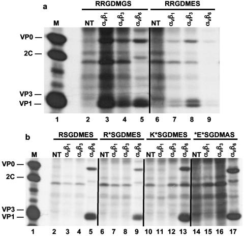
The in vitro characteristics for the A24B9 virus described above appear to be related to alterations on the virion's surface within the cell receptor-binding site. In order to determine preferences for integrin recognition by SGD and RGD A24 viruses, infection/labeling assays in COS-1 cells, previously transfected with specific bovine integrins (see Materials and Methods) were performed. The results of these experiments are shown in Fig. 2. Under these conditions, A24-RGD viruses replicated in cells expressing the αVβ1, αVβ3, and αVβ6 bovine integrins (Fig. 2a), a result consistent with previous studies on A24Cru (17). Interestingly, a different pattern emerged from the SGD viruses (Fig. 2b), including the original A24-B9 virus and R*SGDMES, K*SGDMES, *E*SGDMES, and E*SGDMAS mutant viruses derived from full-length clones. All of these SGD viruses only utilized the bovine αVβ6 integrin receptor, and this effect was independent of mutations at the −1 and +2 positions relative to the SGD motif. The specificity for the αVβ6 bovine integrins by the A24 RGD or SGD viruses has been also demonstrated by blocking the entry of these viruses in transfected COS-1 cells in the presence of soluble αVβ6 bovine integrins or by using a receptor-specific antibody (results not shown). Taken together these results indicate that acquisition of RGD during tissue culture adaptation increased the efficiency of the type A24Cru virus for integrins carrying β1 and β3 integrin subunits.
FIG. 2.
Viral replication in COS-1 cells expressing bovine integrins. Cells were cotransfected with cDNA plasmids encoding the bovine αV subunit and either the β1, β3, or β6 subunits and infected with viruses carrying parental or mutated G-H loop sequences in VP1. Transfected cells were infected with viruses containing RGD (a) or SGD (b) as noted in the figure and labeled with [35S]methionine. Viral protein synthesis was analyzed by radioimmunoprecipitation followed by sodium dodecyl sulfate-polyacrylamide gel electrophoresis separation as described in Materials and Methods. Lanes: M, location of viral structural proteins from lysates prepared from A24Cru-infected BHK-21 cells; NT, immunoprecipitated proteins from nontransfected infected cell lysates.
Pathogenicity of RGD and SGD A24Cru viruses in cattle.
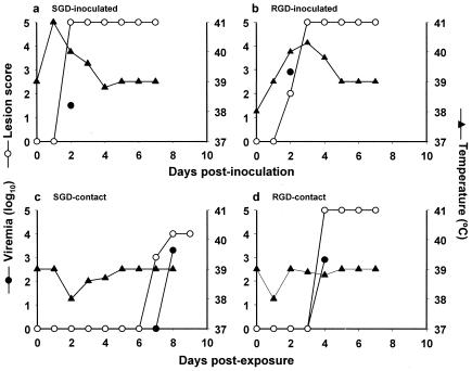
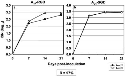
To examine the influence of the receptor specificity on pathogenicity and the ability to transmit disease in cattle, we performed direct inoculation of recombinant RGD and SGD A24Cru viruses in bovines, followed by contact exposure of naïve steers to determine disease transmission. After inoculation, a number of disease parameters were analyzed, including fever, clinical score, viremia, and neutralizing antibodies. Samples of nasal swabs were also collected daily until 7 days postcontact in cohoused animals to measure viral excretion. Plasmid pA24RSGDMES was used to produce transcript RNAs in vitro and then introduced into either BHK-21 or BHK3-αVβ6 cells, and viruses (designated A24-RGD and A24-SGD) were recovered and amplified in the same cell type (Table 2). Virus stocks were prepared following amplification in these cells (passage 3), and the sequences of their P1 regions were compared to the original cDNA. As shown in Table 2, A24SGD viruses propagated in BHK3-αVβ6 cells maintained the nucleic acid capsid sequence of the parental cDNA, while passages in BHK-21 selected viruses carrying reversion to an RGD sequence in this region. A24-RSGDMES virus (A24B9-like) was inoculated into bovine number 22 by IDL injections of serial 10-fold virus dilutions (approximately 1.1 × 10⁵ PFU total). By 24 h, this animal showed fever and extensive tissue damage at the inoculation sites. By day 2 the animal developed a measurable viremia, and lesions had formed on all four feet and beyond the sites of inoculation in the mouth, giving the maximal score achievable (Fig. 3a) and establishing a PFU/BID50 ratio of 3.5 (see Materials and Methods). Vesicular fluid was collected from foot lesions on the second and third days postinoculation, and each sample was separately processed for RT-PCR and sequencing (Table 3). These fluids contained virus that produced turbid plaques on BHK-21 cells and were indistinguishable from the inoculated virus in their capsid sequences, further indicating that the SGD virus had not changed during bovine growth. A contact bovine (number 15) housed together with bovine 22 on day 2 postinoculation developed lesions characteristic of FMD in three feet on day 7 and all four feet by day 8 postcontact which correlated with a high level of viremia (Fig. 3c), but no fever was detected in this animal during the length of the experiment. Virus recovered from blood and vesicular lesions from bovine 15 showed the same SGD virus genotype as the inoculated animal (bovine 22) with no other changes in the capsid region and a turbid plaque phenotype (Table 3). Virus A24-RGD was inoculated into bovine 21 with a total dose of 1.05 × 10⁵ PFU in a similar experimental design as described above. This animal developed clinical manifestations of FMD and viremia by day 2 and achieved the maximal score of lesions on day 3 (Fig. 3b). The specific infectivity, determined as the PFU/BID50 ratio, was 3.1, a result similar to that observed with the SGD virus. However, the virus with the RGD sequence transmitted the disease to the contact animal more rapidly, as judged by viremia and the appearance of vesicles in all feet by day 4 postexposure in bovine 18 (Fig. 3d). This result might be attributed to a higher load of virus present in infected bovine 21 at the time when the contact animal was moved into the room (Fig. 3b, day 2 postinfection). Table 3 also shows that all of the samples collected from directly inoculated bovine 21 and contact bovine 18 contained an RGD motif, no changes in the capsid region from the inoculated virus (A24-RGD), and clear plaque phenotypes on BHK-21 cell monolayers. Figure 4 shows the results of virus neutralization assays performed with the SGD or RGD viruses using sera collected prior to inoculation and at days 7, 14, and 21 postinoculation from bovines 22 (Fig. 4a) and 21 (Fig. 4b). The profile of neutralizing titers and determination of the bilateral relationship (see Materials and Methods), which was established as R = 97%, further indicate that these viruses are very closely related.
FIG. 3.
Evaluation of disease induced in cattle by RGD and SGD (A24-B9-like) viruses. For this experiment, bovine 22 was inoculated intradermally in the tongue with approximately 110,000 PFU of A24-SGD virus (a), and 2 days later a contact steer (bovine 18) was moved to the room (c). In a separate room, bovine 21 was inoculated by the same route with approximately 105,000 PFU of A24-RGD virus (b) and bovine 15 was placed in contact 2 days postinfection (d). The amount of virus detected in the blood was determined as described in Materials and Methods. Lesion scores were calculated by adding the number of locations away from the inoculation site that were affected by the disease (the maximum value achievable was 5; see Materials and Methods for details). Rectal temperatures (in °C) are also shown.
TABLE 3.
Properties and sequence data of A24Cruz viruses recovered from infected or contact cattle
| Sample source | Emergent amino acid sequencea | Virus titer (PFU/ml)b | Plaque phenotypeb |
|---|---|---|---|
| RGD/infected, no. 21 vesiclec | RRGDMES | 1.2 × 10⁷ | Clear |
| RGD/infected, no. 21 blood | RRGDMES | 1.0 × 10³ | Clear |
| RGD/contact, no. 18 vesiclec | RRGDMES | 2.5 × 10⁷ | Clear |
| RGD/contact, no. 18 blood | RRGDMES | 2.0 × 10³ | Clear |
| SGD (A24-B9 like)/infected, no. 22 vesiclec | RSGDMES | 5.0 × 10⁷ | Turbid |
| SGD (A24-B9 like)/infected, no. 22 blood | RSGDMES | 5.0 × 101 | Turbid |
| SGD (A24-B9 like)/contact, no. 15 vesiclec | RSGDMES | 2.3 × 10³ | Turbid |
| SGD (A24-B9 like)/contact, no. 15 blood | RSGDMES | 7.9 × 10² | Turbid |
Sequence data were obtained by RT-PCR of the entire viral capsid region.
Plaque phenotypes and titers were determined on BHK-21 cells after 48 h of incubation postinfection and stained with crystal violet as described in Material and Methods.
Virus recovered from vesicular lesions, away from the inoculation site.
FIG. 4.
Detection of neutralizing antibodies against FMDV in cattle sera. Neutralizing antibody titers (ISN) were determined on bovine sera collected at day zero and at different times postinoculation with A24-RGD (a) and A24-SGD (A24-B9-like) (b) viruses (see Materials and Methods). The antigenic bilateral relationship (R value) between these viruses was determined by a cross-neutralization assay using polyclonal antisera obtained at 21 dpi from bovines 21 and 22.
DISCUSSION
FMDV, like other RNA viruses, has been shown to adapt to new environments through rapid selection of mutants from a quasispecies population (16). The intrinsic genetic variability of FMDV, which has been manifested by changes in antigenicity in FMDV escape mutants (14, 35, 50, 51), and diversity in cellular receptor usage (5, 8, 21, 24, 26, 31, 34, 40) pose concerns for control strategies based on vaccination. The fact that current FMDV vaccines are prepared from virus grown in large amounts in cell cultures generates potential problems in vaccine manufacturing, due to the occasional outgrowth and dominance of mutant viruses. As we, and others, have shown, adaptive changes during passages in cell culture result in animal-derived FMDVs either increasing the efficiency of integrin utilization in vitro or acquiring alternative pathways of entry into the cells (for reviews, see references 7 and 23), such as acquisition of attachment to heparan sulfate (22, 48), or the possible usage of an alternative, as-yet-unidentified receptor in vitro (5, 31, 53). The specific virus (A24-B9) used in the present study was derived from a Colombian A24 Cruzeiro reference strain, following nine serial passages in naïve cattle using tongue inoculations. This virus exhibited low fitness to BHK-21 cells, exemplified by a turbid plaque phenotype and overall low virus yields. Upon passage in vitro, variants with apparent growth advantages in cell culture were selected which exhibited a clear plaque phenotype and yields about two orders of magnitude higher in BHK-21 cells than the original A24-B9 virus. Sequence analysis revealed the presence of only one amino acid change in the viral capsid region that distinguished the A24-B9 virus from its tissue culture derivative (A24-B9R7), mapping to the conserved RGD motif thought to be essential for integrin recognition. Remarkably, the A24-B9 virus carried an atypical SGD sequence in this position that was maintained during passages on secondary FBK cells or BHK3-αVβ6, a cell line engineered to express the bovine αVβ6 integrin (18). Although one additional mutation (E148→K) was selected on third passage on FBK cells and maintained throughout subsequent passages on BHK-21 cells, it did not covary with the S144→R alteration in the cell receptor-binding site, as judged by it absence on viruses recovered from six passages in either BHK3-αVβ6 or BHK-21 cells (Table 1). Additional amino acid substitutions relative to the sequence of the A24Cru infectious cDNA clone were observed in the Colombian A24-B9 virus at position 148 (G148→E) of VP1 and in 3Cpro (not shown), although the latter appeared to be unlikely to influence adaptation to cell culture.
The mechanism by which cell culture-adapted variants are selected by BHK-21 cells is of interest, since it may mimic other adaptation processes leading to alteration in virulence. The evidence presented here strongly suggests a role for the early interaction between the virus and the cell surface as the major selective force in amplifying variant virus populations. This is based upon the observed alteration of receptor specificity from BHK-21 passages. Using a transient integrin expression assay system in COS-1 cells (17, 39) and several FMDV mutants, all derived from a clone of A24Cru, we demonstrated that the efficiency by which different bovine integrins mediate infection differed between FMDVs containing either an RGD or SGD motif. In particular, mutated R*SGDMES, K*SGDMES, and *E*SGDMAS viruses exhibited the same αVβ6-specific receptor utilization as the original RSGDMES (A24-B9) virus. Consistently, for all SGD viruses generated, including those with double mutations in the *SGD sequence, a low fitness to BHK-21 cells and turbid plaque phenotypes were observed (Table 2). In contrast, and in agreement with previous evidence for type A FMDVs (17), RGDMES and RGDMGS viruses replicated in COS-1 cells expressing bovine integrins αVβ1, and αVβ3 and, to a lesser extent, on those cells expressing αVβ6. Furthermore, the results also indicate that the residue immediately preceding the SGD motif (R143→K/E at the −1 SGD position) or the changes acquired at the +2 position (G, A, or E) are not the main determinant of integrin binding specificity in vitro, but they could modulate the affinity to the specific cell receptor. Although our assay does not detect subtle differences in receptor binding affinity, it is possible to speculate that the adaptive mutations (residues 144, 148, and 155 in VP1, 85 in VP2, and 196 in VP3) (Table 2) acquired during virus successive passages in FBK or BHK-21 cells (Tables 1 and 2) could account for affinity gain to the specific cell receptor. This is supported by early reports showing that, depending upon the serotype of the virus, amino acids surrounding the RGD motif or located in other regions of the viral capsid could also influence the virus-receptor interaction (14, 17, 31, 42).
To analyze the effect that a change in the viral receptor-binding site had on FMDV virulence and transmission in an animal naturally susceptible to the disease, we designed tongue inoculation and contact transmission experiments in cattle using the A24-SGD and A24-RGD viruses. Regardless of their differences in receptor utilization and ability to grow in BHK-21 cell monolayers, both SGD and RGD recombinant viruses were highly virulent to bovines (PFU/BID50 ratios of 3.5 and 3.1, respectively), caused rapid onset of clinical disease upon exposure, and transmitted FMDV to cohoused animals. The A24-SGD virus produced more extensive tissue damage at the injected sites and induced fever and vesicles a day earlier than in the A24-RGD-inoculated animal, which indicate a different degree of disease severity. However, a larger number of test animals will be required to confirm this trend. The apparent high virulence of the SGD virus was also supported by the maintenance of an SGD sequence (only one nucleotide change is required to mutate to an RGD motif) in blood and vesicle samples obtained from infected and contact animals. This further suggests that viruses with sequences other than RGD, but closely related to it, can still infect via integrin receptors and induce and transmit the disease to susceptible animals. These observations are particularly interesting since the presence of an RGD motif is believed to be the main determinant to direct FMDV to integrin-containing target tissues during infection in the natural host (2, 6, 7). While conservation of the RGD sequence in the protruding VP1 G-H loop in FMDV field isolates has been extensively reported (see introduction), a type C virus containing a mutated RGGD sequence has been isolated from bovine which was not protected from virus challenge following experimental immunization with a peptide vaccine (50, 51). Moreover, a cell-adapted type C virus, engineered to contain an RGG sequence, was able to infect cells in both an integrin- and heparin-independent manner (4, 5). Although these findings suggest the possibility of a nonintegrin receptor being involved in FMD, there has been no demonstration of the ability of these viruses to produce disease in susceptible animals.
It is not clear why amplification of type A24Cru in cattle selected an SGD variant (via a single point mutation) that utilizes only the αVβ6 integrin in vitro, while the original RGD virus utilizes multiple (αVβ1, αVβ3, >αVβ6) integrin receptors. It is possible that the route of inoculation and the availability of appropriate integrin receptor molecules on the cells at the portal of entry may be the determining factor. Specifically, Breuss and coworkers (10) have reported that αVβ6 is expressed exclusively in epithelial cells, the cell type preferentially infected by FMDV in the susceptible host (2, 11-13), and αVβ6 appears to be the predominant integrin expressed in bovine tissues which also express viral antigen in infected animals (V. O'Donnell and B. Baxt, unpublished data). What is clear, however, is that once the SGD sequence has been established, there does not appear to be any selective pressure to change it to an RGD by infecting the animal either via IDL inoculation or direct contact. Given the evidence that the immune response to the SGD and RGD viruses, measured by levels of neutralizing antibodies, was very similar and cross-neutralization assays suggested that these viruses are very closely related (R = 97%), one could speculate that the SGD virus with enhanced utilization of αVβ6 integrin in bovine epithelium could induce an imbalance favoring virus replication over the host immune defense, other than neutralizing antibodies, which could explain the reduced efficiency of an A24-RGD vaccine in cattle when using A24-SGD challenge virus.
Acknowledgments
We thank Michael Larocco for excellent technical assistance and Guillermo Restrepo, Judith Parraga, and Ricardo Avalo (Vecol, Colombia) for providing A24-B9, A24-B9R6, and A24-B9R7 viruses.
This work was supported by the U.S. Department of Agriculture, Agricultural Research Service through CRIS project no. 1940-32000-035-00D.
REFERENCES
- 1.Acharya, R., E. Fry, D. Stuart, G. Fox, D. Rowlands, and F. Brown. 1989. The three-dimensional structure of foot-and-mouth disease virus at 2.9 Å resolution. Nature 337:709-716. [DOI] [PubMed] [Google Scholar]
- 2.Alexandersen, S., Z. Zhang, A. I. Donaldson, and A. J. Garland. 2003. The pathogenesis and diagnosis of foot-and-mouth disease. J. Comp. Pathol. 129:1-36. [DOI] [PubMed] [Google Scholar]
- 3.Bachrach, H. L. 1968. Foot-and-mouth disease. Annu. Rev. Microbiol. 22:201-244. [DOI] [PubMed] [Google Scholar]
- 4.Baranowski, E., C. M. Ruiz-Jarabo, and E. Domingo. 2001. Evolution of cell recognition by viruses. Science 292:1102-1105. [DOI] [PubMed] [Google Scholar]
- 5.Baranowski, E., C. M. Ruiz-Jarabo, N. Sevilla, D. Andreu, E. Beck, and E. Domingo. 2000. Cell recognition by foot-and-mouth disease virus that lacks the RGD integrin-binding motif: flexibility in aphthovirus receptor usage. J. Virol. 74:1641-1647. [DOI] [PMC free article] [PubMed] [Google Scholar]
- 6.Baxt, B., S. Neff, E. Rieder, and P. Mason. 2002. Foot-and-mouth disease virus-receptor interactions: role in pathogenesis and tissue culture adaptation, p. 115-123. In B. L. Semler and E. Wimmer (ed.), Molecular biology of picornaviruses. ASM Press, Washington, D.C.
- 7.Baxt, B., and E. Rieder. 2004. Molecular aspects of foot-and-mouth disease virus virulence and host range: role of host cell receptors and viral factors, p. 145-172. In F. Sobrino and E. Domingo (ed.), Foot and mouth disease: current perspectives. Horizon Bioscience, Norfolk, England.
- 8.Berinstein, A., M. Roivainen, T. Hovi, P. W. Mason, and B. Baxt. 1995. Antibodies to the vitronectin receptor (integrin αVβ3) inhibit binding and infection of foot-and-mouth disease virus to cultured cells. J. Virol. 69:2664-2666. [DOI] [PMC free article] [PubMed] [Google Scholar]
- 9.Bolwell, C., A. L. Brown, P. V. Barnett, R. O. Campbell, B. E. Clarke, N. R. Parry, E. J. Ouldridge, F. Brown, and D. J. Rowlands. 1989. Host cell selection of antigenic variants of foot-and-mouth disease virus. J. Gen. Virol. 70:45-57. [DOI] [PubMed] [Google Scholar]
- 10.Breuss, J. M., N. Gillett, L. Lu, D. Sheppard, and R. Pytela. 1993. Restricted distribution of integrin β6 mRNA in primate epithelial tissues. J. Histochem. Cytochem. 41:1521-1527. [DOI] [PubMed] [Google Scholar]
- 11.Brown, C. C., R. F. Meyer, H. J. Olander, C. House, and C. A. Mebus. 1992. A pathogenesis study of foot-and-mouth disease in cattle, using in situ hybridization. Can. J. Vet. Res. 56:189-193. [PMC free article] [PubMed] [Google Scholar]
- 12.Brown, C. C., H. J. Olander, and R. F. Meyer. 1995. Pathogenesis of foot-and-mouth disease in swine, studied by in-situ hybridization. J. Comp. Pathol. 113:51-58. [DOI] [PubMed] [Google Scholar]
- 13.Burrows, R., J. A. Mann, A. J. Garland, A. Greig, and D. Goodridge. 1981. The pathogenesis of natural and simulated natural foot-and-mouth disease infection in cattle. J. Comp. Pathol. 91:599-609. [DOI] [PubMed] [Google Scholar]
- 14.Curry, S., E. Fry, W. Blakemore, R. Abu-Ghazaleh, T. Jackson, A. King, S. Lea, J. Newman, D. Rowlands, and D. Stuart. 1996. Perturbations in the surface structure of A22 Iraq foot-and-mouth disease virus accompanying coupled changes in host cell specificity and antigenicity. Structure 4:135-145. [DOI] [PubMed] [Google Scholar]
- 15.Diez, J., M. G. Mateu, and E. Domingo. 1989. Selection of antigenic variants of foot-and-mouth disease virus in the absence of antibodies, as revealed by an in situ assay. J. Gen. Virol. 70:3281-3289. [DOI] [PubMed] [Google Scholar]
- 16.Domingo, E., and J. J. Holland. 1988. High error rates, population equilibrium and evolution of RNA replication systems, p. 3-36. In E. Domingo, J. J. Holland, and P. Ahlquist (ed.), RNA genetics, vol. III: variability of RNA genomes. CRC Press, Boca Raton, Fla.
- 17.Duque, H., and B. Baxt. 2003. Foot-and-mouth disease virus receptors: comparison of bovine αV integrin utilization by type A and O viruses. J. Virol. 77:2500-2511. [DOI] [PMC free article] [PubMed] [Google Scholar]
- 18.Duque, H., M. LaRocco, W. T. Golde, and B. Baxt. 2004. Interactions of foot-and-mouth disease virus with soluble bovine αVβ3 and αVβ6 integrins. J. Virol. 78:9773-9781. [DOI] [PMC free article] [PubMed] [Google Scholar]
- 19.Fry, E. E., S. M. Lea, T. Jackson, J. W. Newman, F. M. Ellard, W. E. Blakemore, R. Abu-Ghazaleh, A. Samuel, A. M. King, and D. I. Stuart. 1999. The structure and function of a foot-and-mouth disease virus-oligosaccharide receptor complex. EMBO J. 18:543-554. [DOI] [PMC free article] [PubMed] [Google Scholar]
- 20.Grubman, M. J., and B. Baxt. 2004. Foot-and-mouth disease. Clin. Microbiol. Rev. 17:465-493. [DOI] [PMC free article] [PubMed] [Google Scholar]
- 21.Jackson, T., S. Clark, S. Berryman, A. Burman, S. Cambier, D. Mu, S. Nishimura, and A. M. King. 2004. Integrin αVβ8 functions as a receptor for foot-and-mouth disease virus: role of the β-chain cytodomain in integrin-mediated infection. J. Virol. 78:4533-4540. [DOI] [PMC free article] [PubMed] [Google Scholar]
- 22.Jackson, T., F. M. Ellard, R. A. Ghazaleh, S. M. Brookes, W. E. Blakemore, A. H. Corteyn, D. I. Stuart, J. W. Newman, and A. M. King. 1996. Efficient infection of cells in culture by type O foot-and-mouth disease virus requires binding to cell surface heparan sulfate. J. Virol. 70:5282-5287. [DOI] [PMC free article] [PubMed] [Google Scholar]
- 23.Jackson, T., A. M. King, D. I. Stuart, and E. Fry. 2003. Structure and receptor binding. Virus Res. 91:33-46. [DOI] [PubMed] [Google Scholar]
- 24.Jackson, T., A. P. Mould, D. Sheppard, and A. M. King. 2002. Integrin αVβ1 is a receptor for foot-and-mouth disease virus. J. Virol. 76:935-941. [DOI] [PMC free article] [PubMed] [Google Scholar]
- 25.Jackson, T., A. Sharma, R. A. Ghazaleh, W. E. Blakemore, F. M. Ellard, D. L. Simmons, J. W. Newman, D. I. Stuart, and A. M. King. 1997. Arginine-glycine-aspartic acid-specific binding by foot-and-mouth disease viruses to the purified integrin αVβ3 in vitro. J. Virol. 71:8357-8361. [DOI] [PMC free article] [PubMed] [Google Scholar]
- 26.Jackson, T., D. Sheppard, M. Denyer, W. Blakemore, and A. M. King. 2000. The epithelial integrin αVβ6 is a receptor for foot-and-mouth disease virus. J. Virol. 74:4949-4956. [DOI] [PMC free article] [PubMed] [Google Scholar]
- 27.Knowles, N. J., and A. R. Samuel. 2003. Molecular epidemiology of foot-and-mouth disease virus. Virus Res. 91:65-80. [DOI] [PubMed] [Google Scholar]
- 28.Lea, S., J. Hernandez, W. Blakemore, E. Brocchi, S. Curry, E. Domingo, E. Fry, R. Abu-Ghazaleh, A. King, J. Newman, et al. 1994. The structure and antigenicity of a type C foot-and-mouth disease virus. Structure 2:123-139. [DOI] [PubMed] [Google Scholar]
- 29.Leippert, M., E. Beck, F. Weiland, and E. Pfaff. 1997. Point mutations within the βG-βH loop of foot-and-mouth disease virus O1K affect virus attachment to target cells. J. Virol. 71:1046-1051. [DOI] [PMC free article] [PubMed] [Google Scholar]
- 30.Logan, D., R. Abu-Ghazaleh, W. Blakemore, S. Curry, T. Jackson, A. King, S. Lea, R. Lewis, J. Newman, N. Parry, D. Rowlands, D. Stuart, and E. Fry. 1993. Structure of a major immunogenic site on foot-and-mouth disease virus. Nature 362:566-568. [DOI] [PubMed] [Google Scholar]
- 31.Martinez, M. A., N. Verdaguer, M. G. Mateu, and E. Domingo. 1997. Evolution subverting essentiality: dispensability of the cell attachment Arg-Gly-Asp motif in multiply passaged foot-and-mouth disease virus. Proc. Natl. Acad. Sci. USA 94:6798-6802. [DOI] [PMC free article] [PubMed] [Google Scholar]
- 32.Mason, P. W., S. V. Bezborodova, and T. M. Henry. 2002. Identification and characterization of a cis-acting replication element (cre) adjacent to the internal ribosome entry site of foot-and-mouth disease virus. J. Virol. 76:9686-9694. [DOI] [PMC free article] [PubMed] [Google Scholar]
- 33.Mason, P. W., M. J. Grubman, and B. Baxt. 2003. Molecular basis of pathogenesis of FMDV. Virus Res. 91:9-32. [DOI] [PubMed] [Google Scholar]
- 34.Mason, P. W., E. Rieder, and B. Baxt. 1994. RGD sequence of foot-and-mouth disease virus is essential for infecting cells via the natural receptor but can be bypassed by an antibody-dependent enhancement pathway. Proc. Natl. Acad. Sci. USA 91:1932-1936. [DOI] [PMC free article] [PubMed] [Google Scholar]
- 35.Mateu, M. G. 1995. Antibody recognition of picornaviruses and escape from neutralization: a structural view. Virus Res. 38:1-24. [DOI] [PubMed] [Google Scholar]
- 36.Mateu, M. G., M. L. Valero, D. Andreu, and E. Domingo. 1996. Systematic replacement of amino acid residues within an Arg-Gly-Asp-containing loop of foot-and-mouth disease virus and effect on cell recognition. J. Biol. Chem. 271:12814-12819. [DOI] [PubMed] [Google Scholar]
- 37.McKenna, T. S., J. Lubroth, E. Rieder, B. Baxt, and P. W. Mason. 1995. Receptor binding site-deleted foot-and-mouth disease (FMD) virus protects cattle from FMD. J. Virol. 69:5787-5790. [DOI] [PMC free article] [PubMed] [Google Scholar]
- 38.Moraes, M. P., G. A. Mayr, P. W. Mason, and M. J. Grubman. 2002. Early protection against homologous challenge after a single dose of replication-defective human adenovirus type 5 expressing capsid proteins of foot-and-mouth disease virus (FMDV) strain A24. Vaccine 20:1631-1639. [DOI] [PubMed] [Google Scholar]
- 39.Neff, S., P. W. Mason, and B. Baxt. 2000. High-efficiency utilization of the bovine integrin αVβ3 as a receptor for foot-and-mouth disease virus is dependent on the bovine β3 subunit. J. Virol. 74:7298-7306. [DOI] [PMC free article] [PubMed] [Google Scholar]
- 40.Neff, S., D. Sa-Carvalho, E. Rieder, P. W. Mason, S. D. Blystone, E. J. Brown, and B. Baxt. 1998. Foot-and-mouth disease virus virulent for cattle utilizes the integrin αVβ3 as its receptor. J. Virol. 72:3587-3594. [DOI] [PMC free article] [PubMed] [Google Scholar]
- 41.Pacheco, J. M., T. M. Henry, V. K. O'Donnell, J. B. Gregory, and P. W. Mason. 2003. Role of nonstructural proteins 3A and 3B in host range and pathogenicity of foot-and-mouth disease virus. J. Virol. 77:13017-13027. [DOI] [PMC free article] [PubMed] [Google Scholar]
- 42.Rieder, E., B. Baxt, and P. W. Mason. 1994. Animal-derived antigenic variants of foot-and-mouth disease virus type A12 have low affinity for cells in culture. J. Virol. 68:5296-5299. [DOI] [PMC free article] [PubMed] [Google Scholar]
- 43.Rieder, E., A. Berinstein, B. Baxt, A. Kang, and P. W. Mason. 1996. Propagation of an attenuated virus by design: engineering a novel receptor for a noninfectious foot-and-mouth disease virus. Proc. Natl. Acad. Sci. USA 93:10428-10433. [DOI] [PMC free article] [PubMed] [Google Scholar]
- 44.Rieder, E., T. Bunch, F. Brown, and P. W. Mason. 1993. Genetically engineered foot-and-mouth disease viruses with poly(C) tracts of two nucleotides are virulent in mice. J. Virol. 67:5139-5145. [DOI] [PMC free article] [PubMed] [Google Scholar]
- 45.Rueckert, R. R. 1996. Picornaviridae: the viruses and their replication, p. 609-654. In B. N. Fields, D. M. Knipe, and P. H. Howley (ed.), Field's virology. Lippincott-Raven, Philadelphia, Pa.
- 46.Ruiz-Jarabo, C. M., N. Sevilla, M. Davila, G. Gomez-Mariano, E. Baranowski, and E. Domingo. 1999. Antigenic properties and population stability of a foot-and-mouth disease virus with an altered Arg-Gly-Asp receptor-recognition motif. J. Gen. Virol. 80:1899-1909. [DOI] [PubMed] [Google Scholar]
- 47.Rweyemamu, M. M., T. W. Pay, and M. J. Parker. 1977. Serological differentiation of foot-and-mouth disease virus strains in relation to selection of suitable vaccine viruses. Dev. Biol. Stand. 35:205-214. [PubMed] [Google Scholar]
- 48.Sa-Carvalho, D., E. Rieder, B. Baxt, R. Rodarte, A. Tanuri, and P. W. Mason. 1997. Tissue culture adaptation of foot-and-mouth disease virus selects viruses that bind to heparin and are attenuated in cattle. J. Virol. 71:5115-5123. [DOI] [PMC free article] [PubMed] [Google Scholar]
- 49.Sutmoller, P., J. W. McVicar, and G. E. Cottral. 1968. The epizootiological importance of foot-and-mouth disease carriers. I. Experimentally produced foot-and-mouth disease carriers in susceptible and immune cattle. Arch. Gesamte Virusforsch. 23:227-235. [DOI] [PubMed] [Google Scholar]
- 50.Taboga, O., C. Tami, E. Carrillo, J. I. Nunez, A. Rodriguez, J. C. Saiz, E. Blanco, M. L. Valero, X. Roig, J. A. Camarero, D. Andreu, M. G. Mateu, E. Giralt, E. Domingo, F. Sobrino, and E. L. Palma. 1997. A large-scale evaluation of peptide vaccines against foot-and-mouth disease: lack of solid protection in cattle and isolation of escape mutants. J. Virol. 71:2606-2614. [DOI] [PMC free article] [PubMed] [Google Scholar]
- 51.Tami, C., O. Taboga, A. Berinstein, J. I. Nunez, E. L. Palma, E. Domingo, F. Sobrino, and E. Carrillo. 2003. Evidence of the coevolution of antigenicity and host cell tropism of foot-and-mouth disease virus in vivo. J. Virol. 77:1219-1226. [DOI] [PMC free article] [PubMed] [Google Scholar]
- 52.Verdaguer, N., M. G. Mateu, D. Andreu, E. Giralt, E. Domingo, and I. Fita. 1995. Structure of the major antigenic loop of foot-and-mouth disease virus complexed with a neutralizing antibody: direct involvement of the Arg-Gly-Asp motif in the interaction. EMBO J. 14:1690-1696. [DOI] [PMC free article] [PubMed] [Google Scholar]
- 53.Zhao, Q., J. M. Pacheco, and P. W. Mason. 2003. Evaluation of genetically engineered derivatives of a Chinese strain of foot-and-mouth disease virus reveals a novel cell-binding site which functions in cell culture and in animals. J. Virol. 77:3269-3280. [DOI] [PMC free article] [PubMed] [Google Scholar]


