Abstract
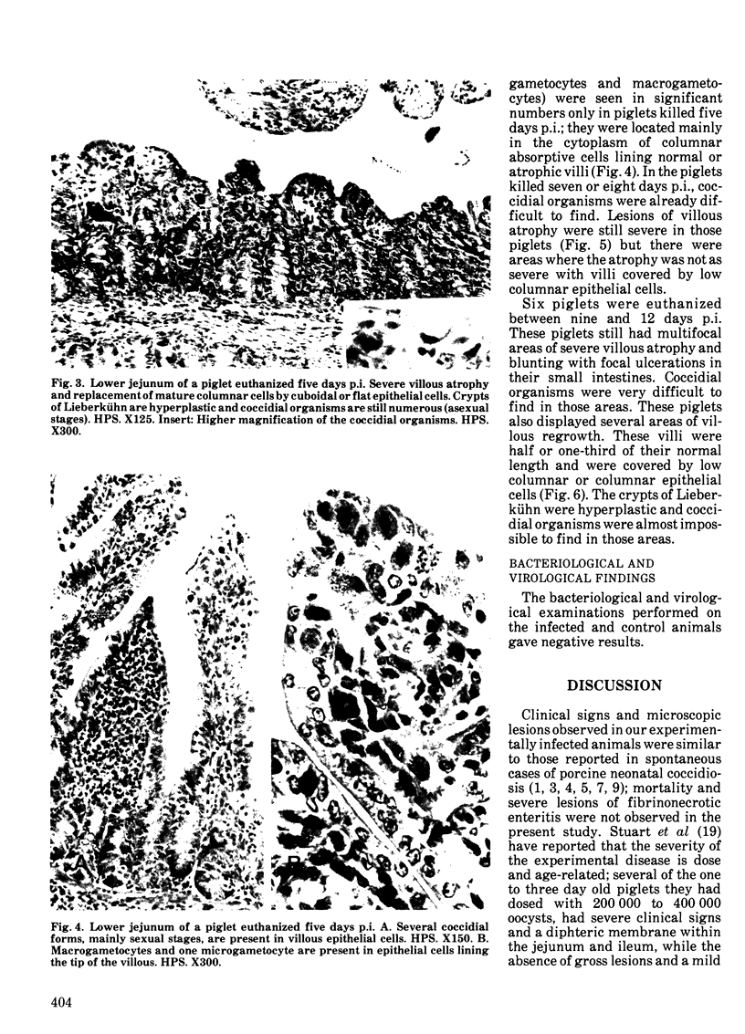
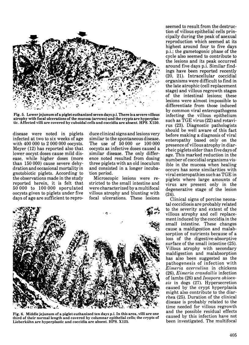
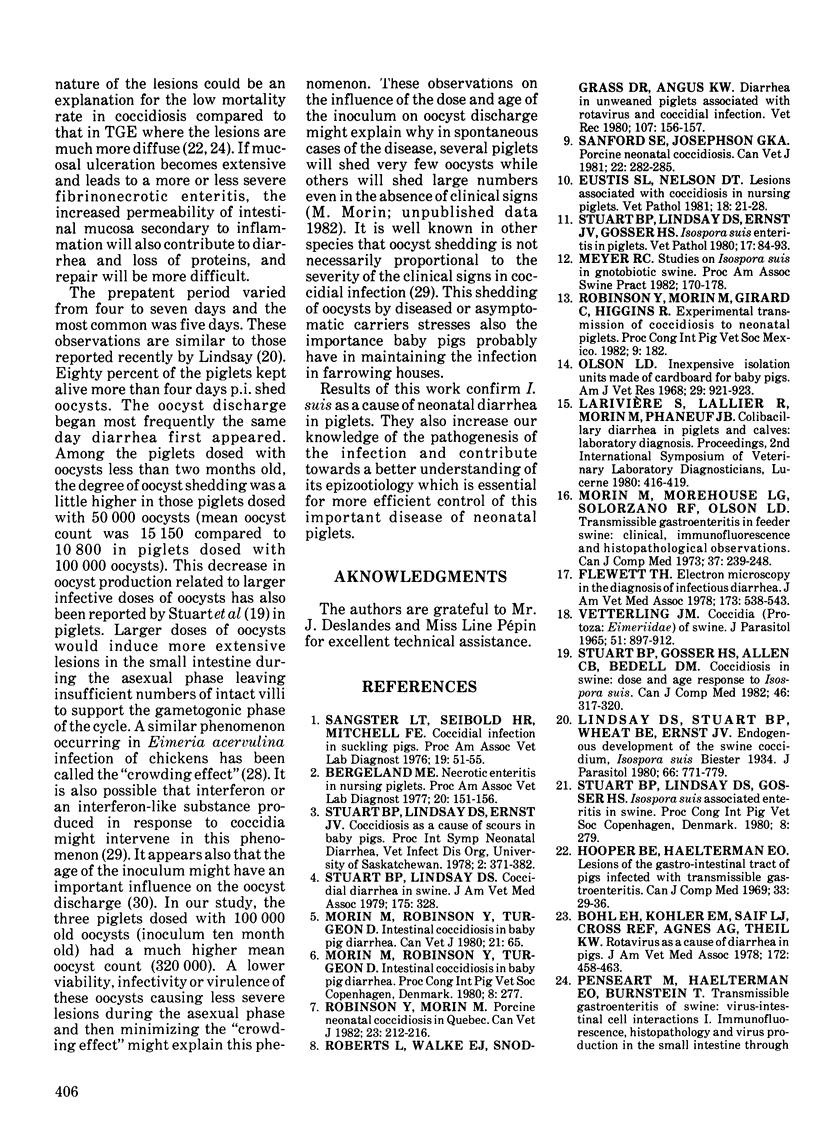
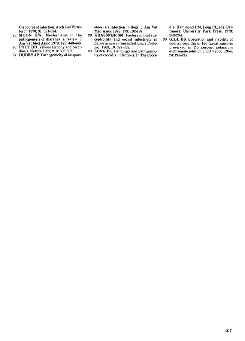
Twenty-eight piglets coming from a "specific pathogen free" herd were inoculated at three days of age with 50 000 or 100 000 sporulated oocysts of Isospora suis. Fecal samples were examined for oocyst shedding daily and several clinical parameters were recorded. Ten piglets were used as normal controls. Groups of piglets were euthanized from three days to 12 days postinoculation and routine necropsies were performed. Bacteriological, virological, parasitological and histopathological examinations were made on the intestinal tracts. The incubation period was four to five days. Clinical signs and microscopic intestinal lesions observed in the experimentally infected animals were similar to those reported in spontaneous cases of porcine neonatal coccidiosis. Lesions of villous atrophy in the small intestine seemed to result from the destruction of villous epithelial cells mainly during the peak of asexual reproduction which occurred around four to five days postinoculation. Intracellular coccidial organisms were difficult to find during the late atrophic and villous regrowth stages of the intestinal lesions. The prepatent period varied from four to seven days and the most common was five days. Eighty percent of the piglets kept alive more than four days postinoculation have shed oocysts. Piglets dosed with old sporulated oocysts (ten months old) shed many more oocysts than those infected with a fresh inoculum (less than two months old). The patent period was not determined precisely with the design of the experiment but some of the infected piglets shed oocysts for at least five days.
Full text
PDF
Images in this article
Selected References
These references are in PubMed. This may not be the complete list of references from this article.
- Bohl E. H., Kohler E. M., Saif L. J., Cross R. F., Agnes A. G., Theil K. W. Rotavirus as a cause of diarrhea in pigs. J Am Vet Med Assoc. 1978 Feb 15;172(4):458–463. [PubMed] [Google Scholar]
- Dubey J. P. Pathogenicity of Isospora ohioensis infection in dogs. J Am Vet Med Assoc. 1978 Jul 15;173(2):192–197. [PubMed] [Google Scholar]
- Eustis S. L., Nelson D. T. Lesions associated with coccidiosis in nursing piglets. Vet Pathol. 1981 Jan;18(1):21–28. doi: 10.1177/030098588101800103. [DOI] [PubMed] [Google Scholar]
- Flewett T. H. Electron microscopy in the diagnosis of infectious diarrhea. J Am Vet Med Assoc. 1978 Sep 1;173(5 Pt 2):538–543. [PubMed] [Google Scholar]
- Hooper B. E., Haelterman E. O. Lesions of the gastrointestinal tract of pigs infected with transmissible gastroenteritis. Can J Comp Med. 1969 Jan;33(1):29–36. [PMC free article] [PubMed] [Google Scholar]
- KRASSNER S. M. FACTORS IN HOST SUSCEPTIBILITY AND OOCYST INFECTIVITY IN EIMERIA ACERVULINA INFECTIONS. J Protozool. 1963 Aug;10:327–333. doi: 10.1111/j.1550-7408.1963.tb01684.x. [DOI] [PubMed] [Google Scholar]
- Lindsay D. S., Stuart B. P., Wheat B. E., Ernst J. V. Endogenous development of the swine coccidium, Isospora suis Biester 1934. J Parasitol. 1980 Oct;66(5):771–779. [PubMed] [Google Scholar]
- Moon H. W. Mechanisms in the pathogenesis of diarrhea: a review. J Am Vet Med Assoc. 1978 Feb 15;172(4):443–448. [PubMed] [Google Scholar]
- Morin M., Morehouse L. G., Solorzano R. F., Olson L. D. Transmissible gastroenteritis in feeder swine: clinical, immunofluorescence and histopathological obervations. Can J Comp Med. 1973 Jul;37(3):239–248. [PMC free article] [PubMed] [Google Scholar]
- Olson L. D. Inexpensive isolation units made of cardboard for baby pigs. Am J Vet Res. 1968 Apr;29(4):921–923. [PubMed] [Google Scholar]
- Pensaert M., Haelterman E. O., Burnstein T. Transmissible gastroenteritis of swine: virus-intestinal cell interactions. I. Immunofluorescence, histopathology and virus production in the small intestine through the course of infection. Arch Gesamte Virusforsch. 1970;31(3):321–334. doi: 10.1007/BF01253767. [DOI] [PubMed] [Google Scholar]
- Pout D. D. Villous atrophy and coccidiosis. Nature. 1967 Jan 21;213(5073):306–307. doi: 10.1038/213306b0. [DOI] [PubMed] [Google Scholar]
- Roberts L., Walker E. J., Snodgrass D. R., Angus K. W. Diarrhoea in unweaned piglets associated with rotavirus and coccidial infections. Vet Rec. 1980 Aug 16;107(7):156–157. doi: 10.1136/vr.107.7.156. [DOI] [PubMed] [Google Scholar]
- Robinson Y., Morin M. Porcine neonatal coccidiosis in quebec. Can Vet J. 1982 Jul;23(7):212–216. [PMC free article] [PubMed] [Google Scholar]
- Sanford S. E., Josephson G. K. Porcine neonatal coccidiosis. Can Vet J. 1981 Sep;22(9):282–285. [PMC free article] [PubMed] [Google Scholar]
- Stuart B. P., Gosser H. S., Allen C. B., Bedell D. M. Coccidiosis in swine: dose and age response to Isospora suis. Can J Comp Med. 1982 Jul;46(3):317–320. [PMC free article] [PubMed] [Google Scholar]
- Stuart B. P., Lindsay D. S., Ernst J. V., Gosser H. S. Isospora suis enteritis in piglets. Vet Pathol. 1980 Jan;17(1):84–93. doi: 10.1177/030098588001700109. [DOI] [PubMed] [Google Scholar]
- Stuart B. P., Lindsay D. Coccidial diarrhea in swine. J Am Vet Med Assoc. 1979 Aug 15;175(4):328–329. [PubMed] [Google Scholar]
- Vetterling J. M. Coccidia (Protozoa: Eimeriidae) of swine. J Parasitol. 1965 Dec;51(6):897–912. [PubMed] [Google Scholar]


