

Abstract
In our continuing effort to characterize the metal cation binding in bacteriorhodopsin (bR) using Ca(2+)-specific electrodes, potentiometric titration was carried out on deionized solubilized bR (containing monomeric units) and deionized bacterioopsin (bR with its retinal removed). Scatchard plots were analyzed. The monomer was found to have plots similar to those of the trimer, suggesting that the binding sites in bR are localized within the protein monomer unit and not between the molecules within the trimer structure. This also supports the previous assumption that the curvature in the Scatchard plot of regenerated bR is not due to cooperativity of metal cation within the trimer, but rather due to multiple sites. Recent studies further support the finding that the curved Scatchard plot is not due to the cooperativity between the metal ions in the two high affinity sites, wherever they are. The results of the analysis of the Scatchard plot for deionized bacterioopsin have shown a change in the binding characteristics of the high affinity but not the low affinity sites from that observed in bR. This result supports previous conclusions that metal cations in the high affinity sites are not far from the retinal cavity.
Full text
PDF
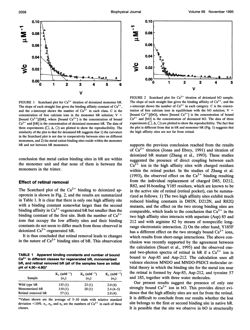
Selected References
These references are in PubMed. This may not be the complete list of references from this article.
- Ariki M., Lanyi J. K. Characterization of metal ion-binding sites in bacteriorhodopsin. J Biol Chem. 1986 Jun 25;261(18):8167–8174. [PubMed] [Google Scholar]
- Bayley H., Radhakrishnan R., Huang K. S., Khorana H. G. Light-driven proton translocation by bacteriorhodopsin reconstituted with the phenyl analog of retinal. J Biol Chem. 1981 Apr 25;256(8):3797–3801. [PubMed] [Google Scholar]
- Becher B. M., Cassim J. Y. Improved isolation procedures for the purple membrane of Halobacterium halobium. Prep Biochem. 1975;5(2):161–178. doi: 10.1080/00327487508061568. [DOI] [PubMed] [Google Scholar]
- Cao Y., Váró G., Chang M., Ni B. F., Needleman R., Lanyi J. K. Water is required for proton transfer from aspartate-96 to the bacteriorhodopsin Schiff base. Biochemistry. 1991 Nov 12;30(45):10972–10979. doi: 10.1021/bi00109a023. [DOI] [PubMed] [Google Scholar]
- Chang C. H., Jonas R., Melchiore S., Govindjee R., Ebrey T. G. Mechanism and role of divalent cation binding of bacteriorhodopsin. Biophys J. 1986 Mar;49(3):731–739. doi: 10.1016/S0006-3495(86)83699-2. [DOI] [PMC free article] [PubMed] [Google Scholar]
- Corcoran T. C., Ismail K. Z., El-Sayed M. A. Evidence for the involvement of more than one metal cation in the Schiff base deprotonation process during the photocycle of bacteriorhodopsin. Proc Natl Acad Sci U S A. 1987 Jun;84(12):4094–4098. doi: 10.1073/pnas.84.12.4094. [DOI] [PMC free article] [PubMed] [Google Scholar]
- Dencher N. A., Heyn M. P. Formation and properties of bacteriorhodopsin monomers in the non-ionic detergents octyl-beta-D-glucoside and Triton X-100. FEBS Lett. 1978 Dec 15;96(2):322–326. doi: 10.1016/0014-5793(78)80427-x. [DOI] [PubMed] [Google Scholar]
- Fischer U., Oesterhelt D. Chromophore equilibria in bacteriorhodopsin. Biophys J. 1979 Nov;28(2):211–230. doi: 10.1016/S0006-3495(79)85172-3. [DOI] [PMC free article] [PubMed] [Google Scholar]
- Jonas R., Ebrey T. G. Binding of a single divalent cation directly correlates with the blue-to-purple transition in bacteriorhodopsin. Proc Natl Acad Sci U S A. 1991 Jan 1;88(1):149–153. doi: 10.1073/pnas.88.1.149. [DOI] [PMC free article] [PubMed] [Google Scholar]
- Jonas R., Koutalos Y., Ebrey T. G. Purple membrane: surface charge density and the multiple effect of pH and cations. Photochem Photobiol. 1990 Dec;52(6):1163–1177. doi: 10.1111/j.1751-1097.1990.tb08455.x. [DOI] [PubMed] [Google Scholar]
- Kimura Y., Ikegami A., Stoeckenius W. Salt and pH-dependent changes of the purple membrane absorption spectrum. Photochem Photobiol. 1984 Nov;40(5):641–646. doi: 10.1111/j.1751-1097.1984.tb05353.x. [DOI] [PubMed] [Google Scholar]
- Mathies R. A., Lin S. W., Ames J. B., Pollard W. T. From femtoseconds to biology: mechanism of bacteriorhodopsin's light-driven proton pump. Annu Rev Biophys Biophys Chem. 1991;20:491–518. doi: 10.1146/annurev.bb.20.060191.002423. [DOI] [PubMed] [Google Scholar]
- Moore T. A., Edgerton M. E., Parr G., Greenwood C., Perham R. N. Studies of an acid-induced species of purple membrane from Halobacterium halobium. Biochem J. 1978 May 1;171(2):469–476. doi: 10.1042/bj1710469. [DOI] [PMC free article] [PubMed] [Google Scholar]
- Mowery P. C., Lozier R. H., Chae Q., Tseng Y. W., Taylor M., Stoeckenius W. Effect of acid pH on the absorption spectra and photoreactions of bacteriorhodopsin. Biochemistry. 1979 Sep 18;18(19):4100–4107. doi: 10.1021/bi00586a007. [DOI] [PubMed] [Google Scholar]
- Oesterhelt D., Stoeckenius W. Rhodopsin-like protein from the purple membrane of Halobacterium halobium. Nat New Biol. 1971 Sep 29;233(39):149–152. doi: 10.1038/newbio233149a0. [DOI] [PubMed] [Google Scholar]
- Papadopoulos G., Dencher N. A., Zaccai G., Büldt G. Water molecules and exchangeable hydrogen ions at the active centre of bacteriorhodopsin localized by neutron diffraction. Elements of the proton pathway? J Mol Biol. 1990 Jul 5;214(1):15–19. doi: 10.1016/0022-2836(90)90140-h. [DOI] [PubMed] [Google Scholar]
- Sweetman L. L., el-Sayed M. A. The binding site of the strongly bound Eu3+ in Eu(3+)-regenerated bacteriorhodopsin. FEBS Lett. 1991 May 6;282(2):436–440. doi: 10.1016/0014-5793(91)80531-7. [DOI] [PubMed] [Google Scholar]
- Szundi I., Stoeckenius W. Effect of lipid surface charges on the purple-to-blue transition of bacteriorhodopsin. Proc Natl Acad Sci U S A. 1987 Jun;84(11):3681–3684. doi: 10.1073/pnas.84.11.3681. [DOI] [PMC free article] [PubMed] [Google Scholar]
- Szundi I., Stoeckenius W. Purple-to-blue transition of bacteriorhodopsin in a neutral lipid environment. Biophys J. 1988 Aug;54(2):227–232. doi: 10.1016/S0006-3495(88)82951-5. [DOI] [PMC free article] [PubMed] [Google Scholar]
- Szundi I., Stoeckenius W. Surface pH controls purple-to-blue transition of bacteriorhodopsin. A theoretical model of purple membrane surface. Biophys J. 1989 Aug;56(2):369–383. doi: 10.1016/S0006-3495(89)82683-9. [DOI] [PMC free article] [PubMed] [Google Scholar]
- Wu S., Awad E. S., El-Sayed M. A. Circular dichroism and photocycle kinetics of partially detergent solubilized and partially retinal regenerated bacteriorhodopsin. Biophys J. 1991 Jan;59(1):70–75. doi: 10.1016/S0006-3495(91)82199-3. [DOI] [PMC free article] [PubMed] [Google Scholar]
- Zhang N. Y., el-Sayed M. A. The C-terminus and the Ca2+ low-affinity binding sites in bacteriorhodopsin. Biochemistry. 1993 Dec 28;32(51):14173–14175. doi: 10.1021/bi00214a015. [DOI] [PubMed] [Google Scholar]
- Zhang Y. N., Sweetman L. L., Awad E. S., El-Sayed M. A. Nature of the individual Ca binding sites in Ca-regenerated bacteriorhodopsin. Biophys J. 1992 May;61(5):1201–1206. doi: 10.1016/S0006-3495(92)81929-X. [DOI] [PMC free article] [PubMed] [Google Scholar]
- Zhang Y. N., el-Sayed M. A., Bonet M. L., Lanyi J. K., Chang M., Ni B., Needleman R. Effects of genetic replacements of charged and H-bonding residues in the retinal pocket on Ca2+ binding to deionized bacteriorhodopsin. Proc Natl Acad Sci U S A. 1993 Feb 15;90(4):1445–1449. doi: 10.1073/pnas.90.4.1445. [DOI] [PMC free article] [PubMed] [Google Scholar]