Abstract
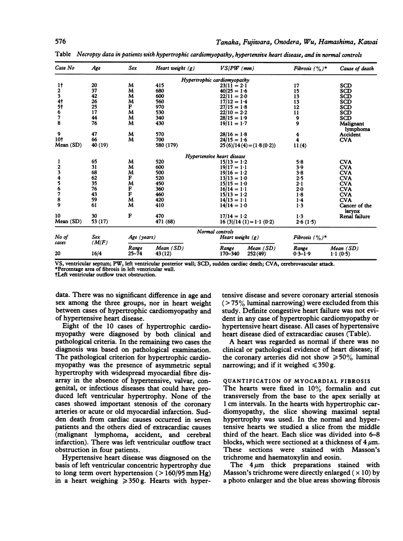
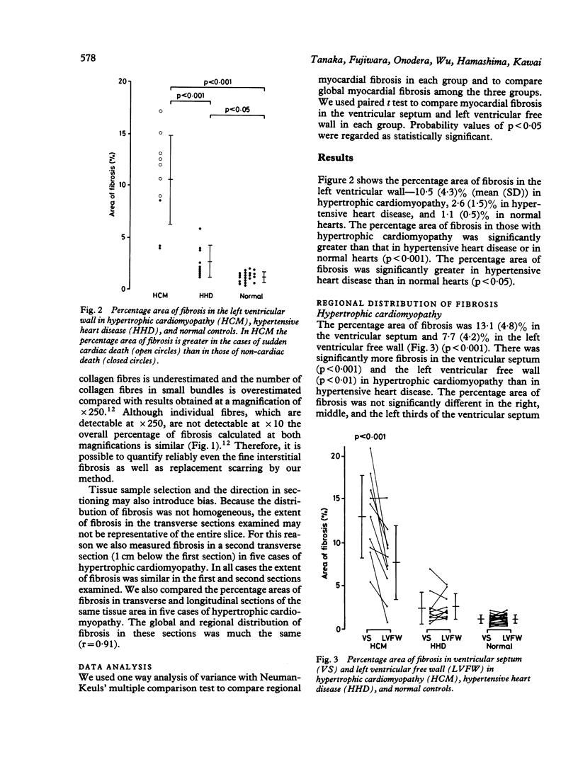
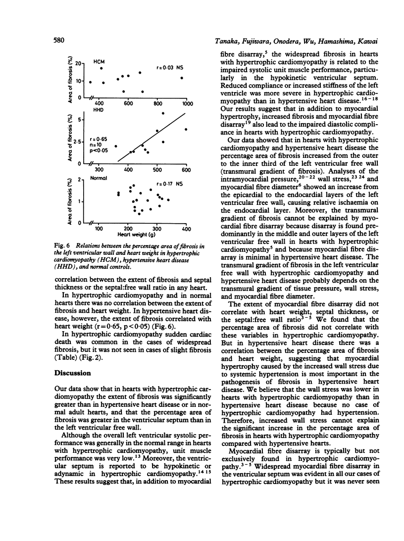
The distribution of fibrosis was studied quantitatively in the entire left ventricular wall of a transverse slice of the heart from 10 necropsy cases of hypertrophic cardiomyopathy, 10 cases of hypertensive heart disease, and 20 normal adults. The percentage area (mean (SD)) of fibrosis in the left ventricular wall in hypertrophic cardiomyopathy (10.5 (4.3)%) was significantly greater than that in hypertensive heart disease (2.6 (1.5)%) or in normal hearts (1.1 (0.5)%). In hypertrophic cardiomyopathy the percentage area of fibrosis was greater (13.1 (4.8)%) in the ventricular septum than in the left ventricular free wall (7.7 (4.2)%) whereas in hypertensive heart disease and normal hearts values in these two areas were similar. The percentage area of fibrosis in the left ventricular free wall (where myocardial fibre disarray was not extensive even in hypertrophic cardiomyopathy) was greater in hypertrophic cardiomyopathy than in hypertensive heart disease. The percentage area of fibrosis correlated with heart weight in hypertensive heart disease, but not in hypertrophic cardiomyopathy. These results suggest that widespread fibrosis in hypertrophic cardiomyopathy cannot be explained by cardiac hypertrophy alone, and that disarray and other factors are also important in pathogenesis. The increase in the percentage area of fibrosis from the outer to the inner third of the left ventricular free wall in hypertrophic cardiomyopathy and in hypertension probably reflected transmural gradients of wall stress and myocardial fibre diameter. Although fibrosis is not specific to hypertrophic cardiomyopathy, its quantification and analysis of its regional distribution provide information that is useful in investigating the pathophysiology of the disorder.
Full text
PDF

Images in this article
Selected References
These references are in PubMed. This may not be the complete list of references from this article.
- Anderson K. R., Sutton M. G., Lie J. T. Histopathological types of cardiac fibrosis in myocardial disease. J Pathol. 1979 Jun;128(2):79–85. doi: 10.1002/path.1711280205. [DOI] [PubMed] [Google Scholar]
- Armour J. A., Randall W. C. Canine left ventricular intramyocardial pressures. Am J Physiol. 1971 Jun;220(6):1833–1839. doi: 10.1152/ajplegacy.1971.220.6.1833. [DOI] [PubMed] [Google Scholar]
- Baandrup U., Olsen E. G. Critical analysis of endomyocardial biopsies from patients suspected of having cardiomyopathy. I: Morphological and morphometric aspects. Br Heart J. 1981 May;45(5):475–486. doi: 10.1136/hrt.45.5.475. [DOI] [PMC free article] [PubMed] [Google Scholar]
- Baird R. J., Manktelow R. T., Shah P. A., Ameli F. M. Intramyocardial pressure. A study of its regional variations and its relationship to intraventricular pressure. J Thorac Cardiovasc Surg. 1970 Jun;59(6):810–823. [PubMed] [Google Scholar]
- Cohen M. V., Cooperman L. B., Rosenblum R. Regional myocardial function in idiopathic hypertrophic subaortic stenosis. An echocardiographic study. Circulation. 1975 Nov;52(5):842–847. doi: 10.1161/01.cir.52.5.842. [DOI] [PubMed] [Google Scholar]
- Fujiwara H., Hoshino T., Fujiwara T., Kawai C., Hamashima Y. Classification and distribution of myocardial fascicle and fiber disarray in 14 hearts with hypertrophic cardiomyopathy in 25 mu thick sections. Jpn Circ J. 1982 Mar;46(3):225–234. doi: 10.1253/jcj.46.225. [DOI] [PubMed] [Google Scholar]
- Fujiwara H., Hoshino T., Yamana K., Fujiwara T., Furuta M., Hamashima Y., Kawai C. Number and size of myocytes and amount of interstitial space in the ventricular septum and in the left ventricular free wall in hypertrophic cardiomyopathy. Am J Cardiol. 1983 Oct 1;52(7):818–823. doi: 10.1016/0002-9149(83)90421-6. [DOI] [PubMed] [Google Scholar]
- Fujiwara H., Onodera T., Tanaka M., Fujiwara T., Kawai C., Hamashima Y. The reliability of the tracing-method of fine cardiac fibrosis at a magnification of x10--preliminary study for quantitative analysis of fibrosis in large tissue sections of hearts with cardiomyopathy. Heart Vessels. 1985 Aug;1(3):182–184. doi: 10.1007/BF02066416. [DOI] [PubMed] [Google Scholar]
- Gaasch W. H., Battle W. E., Oboler A. A., Banas J. S., Jr, Levine H. J. Left ventricular stress and compliance in man. With special reference to normalized ventricular function curves. Circulation. 1972 Apr;45(4):746–762. doi: 10.1161/01.cir.45.4.746. [DOI] [PubMed] [Google Scholar]
- Hirota Y. A clinical study of left ventricular diastolic properties. Jpn Circ J. 1982 Jan;46(1):49–57. doi: 10.1253/jcj.46.49. [DOI] [PubMed] [Google Scholar]
- Hirota Y. A clinical study of left ventricular relaxation. Circulation. 1980 Oct;62(4):756–763. doi: 10.1161/01.cir.62.4.756. [DOI] [PubMed] [Google Scholar]
- Hirota Y., Furubayashi K., Kaku K., Shimizu G., Kino M., Kawamura K., Takatsu T. Hypertrophic nonobstructive cardiomyopathy: a precise assessment of hemodynamic characteristics and clinical implications. Am J Cardiol. 1982 Nov;50(5):990–997. doi: 10.1016/0002-9149(82)90407-6. [DOI] [PubMed] [Google Scholar]
- Hoshino T., Fujiwara H., Kawai C., Hamashima Y. Myocardial fiber diameter and regional distribution in the ventricular wall of normal adult hearts, hypertensive hearts and hearts with hypertrophic cardiomyopathy. Circulation. 1983 May;67(5):1109–1116. doi: 10.1161/01.cir.67.5.1109. [DOI] [PubMed] [Google Scholar]
- Maron B. J., Anan T. J., Roberts W. C. Quantitative analysis of the distribution of cardiac muscle cell disorganization in the left ventricular wall of patients with hypertrophic cardiomyopathy. Circulation. 1981 Apr;63(4):882–894. doi: 10.1161/01.cir.63.4.882. [DOI] [PubMed] [Google Scholar]
- Maron B. J., Roberts W. C. Quantitative analysis of cardiac muscle cell disorganization in the ventricular septum of patients with hypertrophic cardiomyopathy. Circulation. 1979 Apr;59(4):689–706. doi: 10.1161/01.cir.59.4.689. [DOI] [PubMed] [Google Scholar]
- Matsubara O., Tanaka M., Kasuga T., Ishii T., Chino M. Idiopathic cardiomyopathy: the pathologic roles of arteriolopathy. Hum Pathol. 1984 Jan;15(1):39–47. doi: 10.1016/s0046-8177(84)80328-7. [DOI] [PubMed] [Google Scholar]
- Mirsky I. Left ventricular stresses in the intact human heart. Biophys J. 1969 Feb;9(2):189–208. doi: 10.1016/S0006-3495(69)86379-4. [DOI] [PMC free article] [PubMed] [Google Scholar]
- Roberts W. C., Ferrans V. J. Pathologic anatomy of the cardiomyopathies. Idiopathic dilated and hypertrophic types, infiltrative types, and endomyocardial disease with and without eosinophilia. Hum Pathol. 1975 May;6(3):287–342. [PubMed] [Google Scholar]
- Sanderson J. E., Gibson D. G., Brown D. J., Goodwin J. F. Left ventricular filling in hypertrophic cardiomyopathy. An angiographic study. Br Heart J. 1977 Jun;39(6):661–670. doi: 10.1136/hrt.39.6.661. [DOI] [PMC free article] [PubMed] [Google Scholar]
- St John Sutton M. G., Lie J. T., Anderson K. R., O'Brien P. C., Frye R. L. Histopathological specificity of hypertrophic obstructive cardiomyopathy. Myocardial fibre disarray and myocardial fibrosis. Br Heart J. 1980 Oct;44(4):433–443. doi: 10.1136/hrt.44.4.433. [DOI] [PMC free article] [PubMed] [Google Scholar]
- Wong A. Y., Rautaharju P. M. Stress distribution within the left ventricular wall approximated as a thick ellipsoidal shell. Am Heart J. 1968 May;75(5):649–662. doi: 10.1016/0002-8703(68)90325-6. [DOI] [PubMed] [Google Scholar]
- ten Cate F. J., Hugenholtz P. G., Roelandt J. Ultrasound study of dynamic behaviour of left ventricle in genetic asymmetric septal hypertrophy. Br Heart J. 1977 Jun;39(6):627–633. doi: 10.1136/hrt.39.6.627. [DOI] [PMC free article] [PubMed] [Google Scholar]


