Abstract
Phytochromes are an important class of chromoproteins that regulate many cellular and developmental responses to light in plants. The model plant species Arabidopsis thaliana possesses five phytochromes, which mediate distinct and overlapping responses to light. Photobiological analyses have established that, under continuous irradiation, phytochrome A is primarily responsible for plant's sensitivity to far-red light, whereas the other phytochromes respond mainly to red light. The present study reports that the far-red light sensitivity of phytochrome A depends on the structure of the linear tetrapyrrole (bilin) prosthetic group. By reconstitution of holophytochrome in vivo through feeding various synthetic bilins to chromophore-deficient mutants of Arabidopsis, the requirement for a double bond on the bilin D-ring for rescuing phytochrome A function has been established. In contrast, we show that phytochrome B function can be rescued with various bilin analogs with saturated D-ring substituents.
Plants perceive light as environmental information for adaptation to fluctuating circumstances using various pigments in nature. We now know that three major classes of chromoproteins—phytochromes (1), cryptochromes (2), and phototoropins (3)—are engaged in the photoregulation of plant life. Among these photoreceptors, phytochromes have been best characterized by their molecular structure and biological function (4). Phytochromes absorb light of wider wavelengths than our vision and sense extremely low fluence light; they are unique pigments that operate through photochromicity, the property of photoreversible absorbance changes between two spectrally distinct forms, a red (R) light-absorbing form and a far-red (FR) light-absorbing form on light irradiations (5). Phytochromes are encoded by a small gene family, and five distinct phytochromes (A to E) were identified in Arabidopsis (6, 7), in which phytochromes A (PhyA) and B (PhyB) are most abundant, principally working throughout the life cycle. The question which phytochrome is responsible for which phytochrome-mediated responses has been addressed in the past decade, using various phytochrome-deficient mutants (4). Phytochrome was first discovered a half century ago as a pigment of photoreceptor for the R/FR-reversible effect on lettuce seed germination on alternate pulse irradiation with R and FR light (8). Then it had been a central dogma for a long time that phytochrome in the FR-absorbing form is physiologically active. We now know that such photoreversible regulation results from PhyB but not from PhyA (9, 10). PhyA triggers seed germination photoirreversibly by a single pulse irradiation with very low fluence light of broad spectral range (300–770 nm) and requires four orders of magnitude less in fluence than is required by PhyB to switch on or off of R/FR reversible responses (9). Further, PhyA mediates the inhibitory effect of continuous FR light on hypocotyl elongation (10). This effect can be replaced by intermittent irradiation with FR light if given every 3 min, and the effect of each FR pulse is reversible by irradiation of R pulses (11). Therefore, the molecular mechanism of photoperception by PhyA appears essentially different from that of PhyB. It thus is an open question what molecular property of phytochromes causes such photosensory specificity of PhyA and PhyB.
Phytochrome molecules have two functional domains: the chromophore-bearing photosensory N-terminal domain and the signaling C-terminal domain (12). In oat phytochrome, isolated from etiolated tissues, phytochromobilin (PΦB), an open linear tetrapyrrole (bilin), covalently binds to a cysteine residue located in the N-terminal domain of phytochrome apoprotein through a thioether linkage (13). Arabidopsis PhyA and PhyB show 52% identity in amino acid sequence (6) and have conserved subdomain structure (14) and similar spectral properties in vitro (15). However, as mentioned above, their modes of photoperception are essentially different. To probe the intramolecular determinants that are responsible for the photosensory specificity of PhyA and PhyB, the physiological consequences of reciprocal PhyA/PhyB generic chimeras were examined under continuous R or FR light conditions (16). The results suggested that the chromophore-bearing N-terminal domain determined the photosensory specificity for hypocotyl responses to R and FR light in Arabidopsis. This work focused on differences in apoproteins between PhyA and PhyB, but little is known of the role of the bilin chromophore in the photobiological specificity of PhyA and PhyB.
Through chemical synthesis of PΦB (17), phycocyanobilin (PCB) (18), and various analogs (19–21), the structural determinants of the bilin precursor on reconstituted phytochrome can now be addressed. Photochromic properties of holophytochromes are influenced by the nature of the side chains of the bilin chromophores, if they are adducted with recombinant phytochrome apoproteins (19–21). A question arises whether different bilin structures affect the photobiological activities of phytochromes in vivo. To elucidate this question, we have incorporated synthetic bilin chromophores into apophytochromes A (PHYA) and B (PHYB) in Arabidopsis hy1 and hy2 mutants, which are deficient in PΦB biosynthesis (22–24). Parks and Quail restored photomorphogenesis in these mutants with exogenously supplied biliverdin IXα, a direct precursor of PΦB (25).
In the present study, by using an analogous approach, synthetic bilins were fed exogenously to hy1 and hy2 seedlings to test whether they restore the photobiological functions of PhyA and PhyB in vivo. Here, we demonstrated the possibility that the structural requirement of bilin chromophore of PhyA and PhyB determines their functional specificity in PhyA- and PhyB-mediated responses in Arabidopsis seedlings.
Materials and Methods
Plant Materials.
Seedlings of wild-type (WT) Arabidopsis thaliana (L.) Heynh., PhyA-deficient mutant (phyA), phyA-201 (26), PhyB-deficient mutant (phyB), phyB-1 (27), and chromophore-deficient mutants (hy1 and hy2), hy1–1 and hy2–1 (27) were used for assay. Chromophore-deficient and phyA-double mutant (hy1/phyA), hy1–1/phyA-201 and chromophore-deficient and phyB-double mutant (hy1/phyB), hy1–1/phyB-1 were generous gifts from J. Clark Lagarias (University of California, Davis) and Jason W. Reed (University of North Carolina). The ecotype of all strains was Ler (Landsberg erecta).
Plant Growth Conditions and Light Treatment.
Each well of a 24-well tissue culture dish plate (Corning) contained 1 ml of agar medium [Murasige–Skoog medium (28) diluted to one-tenth with 0.7% (wt/vol) agar]. Seeds were planted on the agar plate and kept at 4°C for 3 days. Plates were subsequently transferred to 23°C and exposed to white light for 24 h to induce seed germination, then seeds were kept in the dark for 24 h. At the beginning of the light irradiation, 10 μl of bilin stock solutions (generally 2 mM each) in DMSO or DMSO alone as control were added to each seedling well. The germinated seedlings were irradiated with intermittent pulses of monochromatic light or light-emitting diodes (LEDs) for 2 days. Then seedlings were kept in the dark for 5 days at 23°C for further analysis. Monochromatic R light (55 μmol⋅ m−2⋅s−1 for 60 sec) at 4-h cycles was obtained from white fluorescent tubes [FL20SSW/18(G); Hitachi, Tokyo], which was filtered through a 3-mm red acrylic sheet (Shinkolite A102; Mitsubishi Rayon, Tokyo). Alternatively, monochromatic FR light (110 μmol⋅m−2⋅s−1 for 60 sec) at 3-min cycles was obtained from FR fluorescent tubes (FL20S.FR-74; Toshiba, Tokyo), which was filtered through a 3-mm FR acrylic sheet (Deraglass A-900; Asahikasei, Tokyo). We used long wave pass filters: the wavelengths of 50% of peak transmittance are 610 nm for R light and 738 nm for FR light, respectively. R/FR and FR/R were generated by using a custom-built LED irradiation system, as previously reported (11). R light (110 μmol⋅m−2⋅s−1 for 26 sec) immediately followed by FR light (846 μmol⋅m−2⋅s−1 for 8 sec) (R/FR) was delivered at 4-h cycles. FR light (846 μmol⋅m−2⋅s−1 for 8 sec) immediately followed by R light (386 μmol⋅m−2⋅s−1 for 8 sec) (FR/R) was delivered at 3-min cycles.
Bilin Preparation.
PCB (18), PΦB (17), and their analogs (21, 29) were chemically synthesized, as reported. The bilins were stored as 2 mM stock solutions in dimethyl sulfoxide under nitrogen atmosphere at −80°C.
Spectrophotometry.
For measurement of difference spectra of crude protein extract, approximately 1,000 7-day-old seedlings were used. Crude extract was obtained as described with slight modification (30). Frozen seedlings were homogenized in liquid nitrogen to powder by using a mortar and pestle. Extraction buffer A [1,200 μl; 100 mM Tris⋅HCl/5 mM EDTA/1 unit Complete protein inhibitor cocktail (Amersham Pharmacia Biotech), pH 8.3] was added to the powder and allowed to stand for 15 min on ice. After centrifugation (12,000 × g for 30 min at 4°C), 1,200 μl of the supernatant was transferred to a new tube, and 800 μl of saturated ammonium sulfate solution was added before 30-min incubation on ice. The precipitated material was collected by centrifugation (12,000 × g for 30 min at 4°C) and resuspended in 120 μl of extraction buffer B [100 mM Tris⋅HCl/25% (vol/vol) ethylene glycol/2 mM EDTA, pH 8.3]. The resultant extraction fractions were used for spectrophotometry. The difference spectra of extracted fractions were measured as previously described (21) by using custom-built spectrophotometer (Genespec V; Naka, Ibaragi, Japan).
Measurements of Physiological Response.
For each data point, approximately 100 seeds were planted on each well of a 24-well culture dish. Then 50 seedlings were picked randomly, and the hypocotyl lengths of the longest 35 seedlings were measured by using a digimatic caliper (CD-15C, Mitsutoyo, Tokyo). Mean value, SE, and statistical analysis were calculated by using excel Ver. 8.0 (Microsoft).
Results
Both PΦB and PCB Restore Spectrally Active Phytochrome in Chromophore-Deficient hy1 Mutant.
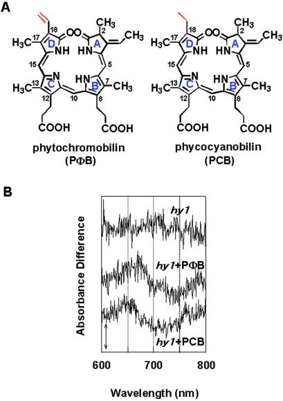
Fig. 1A shows the chemical structure of PΦB and PCB. The only difference between these two bilins is the substitution of vinyl of PΦB for ethyl of PCB in the bilin D-ring. We measured difference spectra in crude extracts of etiolated hy1 mutant seedlings grown on media in the presence or absence of synthetic PΦB and PCB. No photoreversible spectral change was observed in the extract of hy1 seedlings grown without exogenous bilin chromophores, whereas extracts prepared from PCB- or PΦB-treated mutant seedlings showed the characteristic difference absorption spectra of phytochrome on actinic R and FR light irradiations (Fig. 1B). Estimation of the photochemically active holophytochrome is difficult because of a small absorbance difference, ≈0.001 ΔΔA unit. The amount of absorbance difference of hy1 seedlings treated by either PΦB or PCB resembled that of WT (data not shown). These difference absorption spectra could be attributed mainly to the reconstituted PhyA with exogenously supplied PΦB or PCB in vivo, because PhyA is the most abundant phytochrome present in dark grown seedlings (31).
Figure 1.
Spectrally detectable phytochrome in 7-day-old etiolated hy1 seedlings grown with or without PΦB and PCB. (A) Chemical structures of PΦB and PCB. Red indicates the positions of the different substituents between them. (B) Difference spectra of crude extract generated from bilin-supplied seedlings were obtained by subtracting the absorption spectra measured after saturating R light irradiation from those measured after saturating FR light irradiation. (Bar = 0.001 ΔΔA units.)
Effects on Photomorphogenesis of PΦB and PCB.
We fed the synthetic bilin chromophores to germinated hy1 and hy2 mutant seedlings as described in Materials and Methods and exposed the growing seedlings to different light conditions. When these seedlings were intermittently irradiated with FR light for 48 h, the PΦB-treated hy1 and hy2 seedlings showed a significantly different growth phenotype compared with PCB-treated mutants in terms of hypocotyl length, cotyledon opening, and cotyledon expansion. FR-irradiated PCB-treated seedlings resembled dark-grown seedlings, whereas the PΦB-treated seedlings showed a characteristic de-etiolated phenotype similar to that of the FR-irradiated WT seedlings. In contrast, R-irradiated PΦB- or PCB-treated seedlings showed the same de-etiolated phenotype as that of the R-irradiated WT seedlings. Table 1 summarizes the hypocotyl lengths of 7-day-old seedlings that were exposed to R or FR light. Under either R or FR light conditions, the PΦB-treated hy1 and hy2 seedlings possessed hypocotyl lengths as short as WT. FR-irradiated PCB-treated hy1 seedlings showed a statistically significant but very small response of hypocotyl growth inhibition, whereas hy2 seedlings were indistinguishable from the dark-grown controls. Thus, we conclude that PΦB rescued both PhyA and PhyB function, whereas PCB rescued only PhyB function. Similar results were observed for cotyledon opening (data not shown).
Table 1.
Hypocotyl length of WT, phytochrome-deficient, and chromophore-deficient mutants grown with or without PΦB and PCB
| Light treatment | WT | phyA | phyB |
hy1
|
hy2
|
||||
|---|---|---|---|---|---|---|---|---|---|
| − | +PΦB | +PCB | − | +PΦB | +PCB | ||||
| D | 12.4 ± 1.2 | 10.2 ± 1.5 | 13.7 ± 1.7 | 11.3 ± 2.3 | 12.1 ± 0.7 | 12.8 ± 1.0 | 11.1 ± 1.3 | 11.1 ± 1.3 | 10.7 ± 1.5 |
| 10.5 ± 2.4 | 7.8 ± 0.8 | 14.4 ± 1.5 | 10.5 ± 2.0 | 9.6 ± 0.4 | 8.6 ± 0.8 | 10.9 ± 0.4 | 8.9 ± 0.3 | 7.8 ± 1.0 | |
| R | P < 0.01 | P < 0.01 | ns | ns | P < 0.01 | P < 0.01 | ns | P < 0.01 | P < 0.01 |
| (t = 5.9) | (t = 9.5) | (t = −2.3) | (t = 2.5) | (t = 14.2) | (t = 18.6) | (t = 0.9) | (t = 10.4) | (t = 11.0) | |
| 6.7 ± 1.0 | 10.7 ± 1.6 | 8.1 ± 0.6 | 11.6 ± 2.3 | 6.2 ± 0.9 | 10.9 ± 1.1 | 11.2 ± 0.6 | 5.9 ± 2.1 | 10.5 ± 0.3 | |
| FR | P < 0.01 | ns | P < 0.01 | ns | P < 0.01 | P < 0.01 | ns | P < 0.01 | ns |
| (t = 23.3) | (t = −1.6) | (t = 22.1) | (t = −0.7) | (t = 27.8) | (t = 7.7) | (t = −0.6) | (t = 16.7) | (t = 0.9) | |
Germinated Arabidopsis seedlings were irradiated with intermittent pulses of monochromatic light (D, kept in darkness; R, intermittent R pulse at 4-h cycles; FR, intermittent FR pulse at 3-min cycles) for 2 days. Values are means (mm) ± SE of 35 plant materials. Two-tailed t tests of mean differences were calculated in R and FR light-treated samples compared with D samples, respectively. ns, no significant differences. t values are given in parentheses.
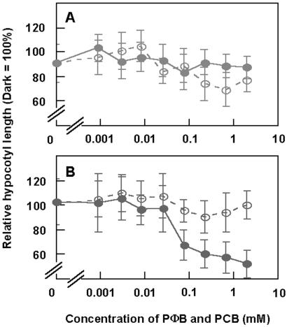
The dose dependence of bilin solution in the concentration range of 0.001–2 mM on R- and FR-mediated growth inhibition is shown in Fig. 2. Under R light, the resultant dose–response curves for PΦB and PCB were identical (Fig. 2A), whereas under FR light they were essentially different from each other (Fig. 2B). It is evident, again, that the PhyA-dependent photoinhibition occurred only when PΦB was supplied, but the PhyB-mediated responses were induced by both PCB and PΦB.
Figure 2.
Relationship between hypocotyl length of hy1 seedlings and concentration of bilin solutions added to agar media. At the beginning of light irradiation, 10 μl of PΦB (●) and PCB (○) in various concentrations, ranging from 0.001 to 2 mM of stock solutions, were added to 1 ml of agar media. If bilins are fully diffused to agar media, the final concentrations are 0.01–20 μM. (A) The seedlings were irradiated by the intermittent R light for 2 days (12 cycles). (B) The seedlings were irradiated by the intermittent FR light for 2 days (960 cycles). Error bars represent SE.
Effects of Bilins on Photoreversible Responses.
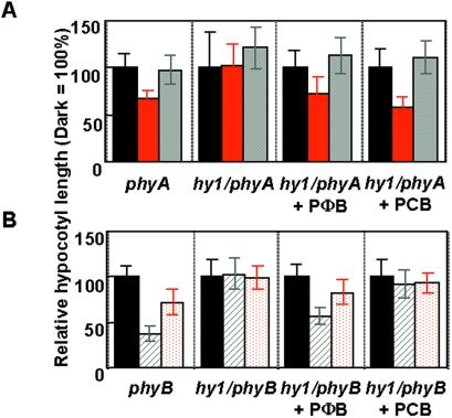
To examine further the role of PΦB and PCB in PhyA- and PhyB-specific responses, we measured the hypocotyl lengths of hy1/phyA and hy1/phyB double mutants in the presence or absence of PΦB and PCB followed by intermittent R, R/FR, FR, and FR/R light treatments. The hy1/phyA double mutant seedlings clearly showed R light-induced growth inhibition and R/FR photoreversibility similar to the phyA mutant when either PΦB or PCB was supplied (Fig. 3A). Because untreated hy1/phyA seedlings are not responsive to R light, the results indicated that PhyB action was restored with both PΦB and PCB. When PΦB was supplied to hy1/phyB double mutant seedlings, intermittently given FR light inhibited hypocotyl growth significantly. Any inhibitory effect was not observed in the seedlings that were exposed to intermittent FR/R (Fig. 3B). In contrast, when PCB was supplied to hy1/phyB seedlings, intermittent FR light did not inhibit hypocotyl elongation. The results, again, indicate that only PΦB restores the PhyA-mediated response in the hy1/phyB double mutant, whereas the supplement of either PΦB or PCB to the hy1/phyA double mutant seedlings rescues PhyB action. These results are consistent with those in Table 1 and Fig. 2.
Figure 3.
Effect of PΦB and PCB adduction on photoreversible response of hy1/phyA and hy1/phyB double mutant seedlings. (A) The phyA and hy1/phyA double mutant seedlings were irradiated with intermittent pulses of light. D (black), kept in darkness; R (red) and R/FR (gray) at 4-h cycles for 2 days. (B) The phyB and hy1/phyB double mutant seedlings were irradiated with intermittent pulses of light. D (black), kept in darkness; FR (shade) and FR/R (red dot) at 3-min cycles for 2 days. Error bars represent SE.
Effect of Bilin Analogs on PhyA- and PhyB-Mediated Responses.
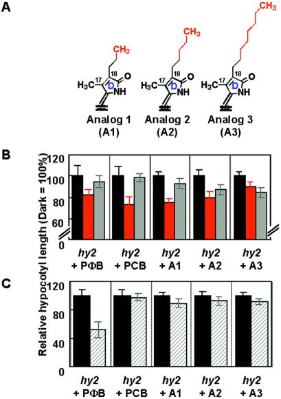
We next examined the effect of exogenously supplied PΦB analogs (Fig. 4A) on the hypocotyl growth of hy2 mutant seedlings that were grown under intermittent R or FR light irradiation. These analogs were substituted at the C18 position of the bilin D-ring (Fig. 1A) with a saturated alkyl group such as ethyl (PCB), n-propyl (A1), n-pentyl (A2), or n-octyl (A3) (Fig. 4A). All analogs rescued the PhyB responsiveness in hy2 seedlings. But the degree of inhibitory effect of R light on hypocotyl elongation and the degree of R/FR reversibility decreased with increasing length of the side chains (Fig. 4B). In contrast to the PhyB-dependent responsiveness, the analogs A1, A2, and A3 failed to restore the PhyA-dependent responsiveness to FR light. Hypocotyl growth inhibition was not as significantly restored by any of these analogs under the condition where PΦB clearly rescued photomorphogenesis in the hy2 mutant (Fig. 4C).
Figure 4.
Effect of exogenously supplied PΦB analogs to hy2 mutant seedlings. (A) The D-ring structures of PΦB analogs. Whole structures are identical to PΦB except where indicated. (B) The seedlings were irradiated with intermittent pulses of light. D (black), kept in darkness; R (red) and R/FR (gray) at 4-h cycles for 2 days. (C) The seedlings were irradiated with intermittent pulses of light. D (black), kept in darkness; FR (shade) at 3-min cycles for 2 days. Error bars represent SE.
Discussion
The structure of PΦB is closely related to PCB, which is a bilin chromophore of the light-harvesting pigment in algae, C-phycocyanin. In the past decade, PCB has been widely used as a chromophore for assembly in vitro with recombinant phytochrome apoproteins (32), and it yields a photoactive holophytochrome with molecular and spectrophotometric properties similar to the natural adduct with PΦB (33–35). Indeed, Murphy and Lagarias (36) reported that the photochemical properties and the molar absorption coefficients of PΦB and PCB adducts of oat PHYA are quite similar. In the present work, similar difference spectra were observed in the extracts from PΦB- and PCB-treated seedlings (Fig. 1B), suggesting that both bilins assembled with apoprotein of phytochrome A (PHYA) in vivo afford spectrophotometrically functional holophytochrome. A similar result was reported for chromophore-deficient oat seedlings many years ago (37). Therefore, one expects that both PCB and PΦB adducts of apophytochrome could be photobiologically active in plants. This hypothesis, however, was valid in the case of PhyB- but not PhyA-mediated response (Table 1, Figs. 2–4). The present study clearly demonstrated that PΦB is required for PhyA responsiveness on FR irradiation, and that PCB is not a functional analog for that. Moreover, PhyA has been known to exhibit two different modes of action: depending on the light wavelength and intensity in environment, one is induced by very low fluence light irreversibly (9) and the other by continuous and intermittent FR light (11). Therefore, further experiments are needed to find out whether PCB restores the PhyA function in irreversible very low fluence response.
The loss of PhyA-mediated response to FR light in PCB-supplied seedlings might be caused by reasons other than the spectrophotometric properties of the PhyA adduct with PCB. One possible explanation is that PΦB (not PCB) might have some direct role as signaling molecules in PhyA signaling pathway. Another possibility is that loss of PhyA function in PCB-supplied chromophore-deficient mutants was caused by a lack of interaction between chromophore and phytochrome apoprotein. Previous studies revealed that determinants of the photosensory specificity of PhyA and PhyB for physiological functions exist in the chromophore-bearing N-terminal domain (16). In particular, the α-helix-forming N-terminal 6-kDa fragment of PhyA is important for physiological activity (12, 38, 39). The present work demonstrated a quite suggestive discovery that a double bond in the vinyl side chain of D-ring of PΦB is crucial for the photosensory function of PhyA. Perhaps some amino acid(s) in the N-terminal domain of PHYA interacting directly would carry out an essential role for direct interaction with this vinyl side chain. One candidate for such an amino acid in PHYA is Ile-80, as Bhoo et al. (40) previously reported that Ile-80 preferentially interacts with the vinyl group of bilin D-ring in a qualitative model. However, Ile-80 is conserved among Arabidopsis PHYA, PHYB, and PHYC, so Ile-80 alone is not likely to be a determinant of the photosensory specificity between PhyA and PhyB. It is more likely that other amino acid(s) of PHYA would directly interact with the vinyl group of the D-ring. It is likely that tight binding with PHYA through the vinyl group at the hydrophobic cavity prevents free movement of PΦB from the binding site even when PhyA is irradiated with FR light. This binding process might be required for PhyA-specific biological activity for photomorphogenesis.
PHYB seems to be more flexible than PHYA in chromophore compatibility, because PhyB accepts both PΦB and PCB as a functional chromophore for induction of physiological response. Relative to PHYA, PHYC, and PHYE (14), PHYB has 37 extra amino acid residues just before the N-terminal 6-kDa motif (12, 38, 39), and it is possible that the extended peptide directly participates in the PHYB–chromophore interaction and results in the PhyB-specific property (12). If this 37-aa extension is important for adduct formation with PCB as a functional bilin precursor, PCB would probably not rescue PhyC- and PhyE-mediated response in chromophore-deficient mutants.
In recent years, the diversity of phytochrome-related chromoproteins discovered in photosynthetic and nonphotosynthetic prokaryotes raises a question whether chromophores other than PΦB are present in phytochromes (for review, see ref. 41). For example, it is known that recombinant bacterial phytochromes were able to bind various bilins autocatalytically in the absence of other factors in vitro (42–44). The difference absorption spectra of phytochromes in vivo determined from several lower organisms exhibited blue-shifted absorption peaks for both R and FR light-absorbing forms relative to those of phytochromes isolated from etiolated higher plants (45–47). These results suggest the possibility of alternative chromophores for these phytochrome-related proteins. Indeed, the phytochrome from Mesotaenium caldariorum utilizes PCB as its chromophore (48). Although it is tempting to speculate on the existence of PCB in higher plants, this has not been identified. Interestingly, the binding efficiency of various bilins to recombinant PHYB was determined in vitro, with the result that PCB bound to PHYB more efficiently than PΦB (21). In the present work, we found that PCB and PΦB restore PhyB-mediated physiological responses. Moreover, it is interesting that the effectiveness of PCB was greater than that of PΦB (Table 1, Figs. 2–4), suggesting that the PHYB–PCB adduct is either more stable than the PHYB–PΦB adduct or more biologically active.
According to phylogenetic analysis of the higher plant phytochrome sequence, the first branch between PhyA/C and PhyB/D/E was generated from the common ancestral phytochrome about the time of the origin of seed plants (49). In their evolutionary pathway, PhyA and PhyB might have gained structural specificity of the bilin prosthetic group as well as a variation of amino acid sequence. As a result of the diversity of structural requirements of bilin, PhyA and PhyB can detect identical environmental signals and regulate photomorphogenesis of plants in a different manner.
Acknowledgments
This work is dedicated to Francesco Lenci and Masamitsu Wada on the occasion of their 60th birthdays. We thank Pill-Soon Song and Jason W. Reed for critical reading of the manuscript and helpful suggestions, J. Clark Lagarias and Jason W. Reed for providing seeds of the hy1/phyA and hy1/phyB double mutants, Kenko Uchida, Sadao Minagawa, and Yoshio Sugiura for developing a newly custom-built spectrophotometer, and Chiemi Hoshina and Hee-Jun Chung for technical assistance. This work was supported by a grant from the Program for Promotion of Basic Research Activities for Innovative Biosciences (to M.F.).
Abbreviations
- PhyA (or -B)
spectrally active holoprotein of phytochrome A (or B)
- PHYA (or -B)
apoprotein of phytochrome A (or B)
- bilin
linear tetrapyrrole
- PΦB
phytochromobilin
- PCB
phycocyanobilin
- R
red light
- FR
far-red light
- WT
wild type
References
- 1.Sage L. Pigment of the Imagination: A History of Phytochrome Responses. San Diego: Academic; 1992. [Google Scholar]
- 2.Lin C. Trends Plant Sci. 2000;5:337–342. doi: 10.1016/s1360-1385(00)01687-3. [DOI] [PubMed] [Google Scholar]
- 3.Briggs W R, Beck C F, Cashmore A R, Christie J M, Hughes J, Jarillo J A, Kagawa T, Kanegae H, Liscum E, Nagatani A, et al. Plant Cell. 2001;13:993–997. doi: 10.1105/tpc.13.5.993. [DOI] [PMC free article] [PubMed] [Google Scholar]
- 4.Fankhauser C. J Biol Chem. 2001;276:11453–11456. doi: 10.1074/jbc.R100006200. [DOI] [PubMed] [Google Scholar]
- 5.Butler W L, Norris K H, Siegelman H W, Hendricks S B. Proc Natl Acad Sci USA. 1959;45:1703–1708. doi: 10.1073/pnas.45.12.1703. [DOI] [PMC free article] [PubMed] [Google Scholar]
- 6.Sharrock R A, Quail P H. Genes Dev. 1989;3:1745–1757. doi: 10.1101/gad.3.11.1745. [DOI] [PubMed] [Google Scholar]
- 7.Clack T, Mathews S, Sharrock R A. Plant Mol Biol. 1994;25:413–427. doi: 10.1007/BF00043870. [DOI] [PubMed] [Google Scholar]
- 8.Borthwick H A, Hendricks S B, Parker M W, Toole E H, Toole V K. Proc Natl Acad Sci USA. 1952;38:662–666. doi: 10.1073/pnas.38.8.662. [DOI] [PMC free article] [PubMed] [Google Scholar]
- 9.Shinomura T, Nagatani A, Hanzawa H, Kubota M, Watanabe M, Furuya M. Proc Natl Acad Sci USA. 1996;93:8129–8133. doi: 10.1073/pnas.93.15.8129. [DOI] [PMC free article] [PubMed] [Google Scholar]
- 10.Quail P H, Boylan M T, Parks B M, Short T W, Xu Y, Wagner D. Science. 1995;268:675–680. doi: 10.1126/science.7732376. [DOI] [PubMed] [Google Scholar]
- 11.Shinomura T, Uchida K, Furuya M. Plant Physiol. 2000;122:147–156. doi: 10.1104/pp.122.1.147. [DOI] [PMC free article] [PubMed] [Google Scholar]
- 12.Park C M, Bhoo S H, Song P-S. Semin Cell Dev Biol. 2000;11:449–456. doi: 10.1006/scdb.2000.0200. [DOI] [PubMed] [Google Scholar]
- 13.Lagarias J C, Rapoport H. J Am Chem Soc. 1980;102:4821–4828. [Google Scholar]
- 14.Quail P H. Plant Cell Environ. 1997;20:657–665. [Google Scholar]
- 15.Eichenberg K, Bäurle I, Paulo N, Sharrock R A, Rüdiger W, Schäfer E. FEBS Lett. 2000;470:107–112. doi: 10.1016/s0014-5793(00)01301-6. [DOI] [PubMed] [Google Scholar]
- 16.Wagner D, Fairchild C D, Kuhn R M, Quail P H. Proc Natl Acad Sci USA. 1996;93:4011–4015. doi: 10.1073/pnas.93.9.4011. [DOI] [PMC free article] [PubMed] [Google Scholar]
- 17.Kakiuchi T, Kinoshita H, Inomata K. Synlett. 1999;S1:901–904. [Google Scholar]
- 18.Kakiuchi T, Kato H, Jayasundera K P, Higashi T, Watabe K, Sawamoto D, Kinoshita H, Inomata K. Chem. Lett. 1998. 1001–1002. [Google Scholar]
- 19.Lindner I, Knipp B, Braslavsky S E, Gärtner W, Schaffner K. Angew Chem Int Ed. 1998;37:1843–1846. [Google Scholar]
- 20.Robben U, Lindner I, Gärtner W, Schaffner K. Angew Chem Int Ed. 2001;40:1048–1050. doi: 10.1002/1521-3773(20010316)40:6<1048::aid-anie10480>3.0.co;2-9. [DOI] [PubMed] [Google Scholar]
- 21.Hanzawa H, Inomata K, Kinoshita H, Kakiuchi T, Jayasundera K P, Sawamoto D, Ohta A, Uchida K, Wada K, Furuya M. Proc Natl Acad Sci USA. 2001;98:3612–3617. doi: 10.1073/pnas.051629698. [DOI] [PMC free article] [PubMed] [Google Scholar]
- 22.Davis S J, Kurepa J, Vierstra R D. Proc Natl Acad Sci USA. 1999;96:6541–6546. doi: 10.1073/pnas.96.11.6541. [DOI] [PMC free article] [PubMed] [Google Scholar]
- 23.Muramoto T, Kohchi T, Yokota A, Hwang I, Goodman H M. Plant Cell. 1999;11:335–347. doi: 10.1105/tpc.11.3.335. [DOI] [PMC free article] [PubMed] [Google Scholar]
- 24.Kohchi T, Mukougawa K, Frankenberg N, Masuda M, Yokota A, Lagarias J C. Plant Cell. 2001;13:425–436. doi: 10.1105/tpc.13.2.425. [DOI] [PMC free article] [PubMed] [Google Scholar]
- 25.Parks B M, Quail P H. Plant Cell. 1991;3:1177–1186. doi: 10.1105/tpc.3.11.1177. [DOI] [PMC free article] [PubMed] [Google Scholar]
- 26.Nagatani A, Reed J W, Chory J. Plant Physiol. 1993;102:269–277. doi: 10.1104/pp.102.1.269. [DOI] [PMC free article] [PubMed] [Google Scholar]
- 27.Koornneef M, Rolff E, Spruit C J P. Z Pflanzenphsiol. 1980;100:147–160. [Google Scholar]
- 28.Murashige T, Skoog F. Physiol Plant. 1962;15:473–497. [Google Scholar]
- 29.Takeda S, Jayasundera K P, Kakiuchi T, Kinoshita H, Inomata K. Chem. Lett. 2001. , 590–591. [Google Scholar]
- 30.Hisada A, Hanzawa H, Weller J L, Nagatani A, Reid J B, Furuya M. Plant Cell. 2000;12:1063–1078. doi: 10.1105/tpc.12.7.1063. [DOI] [PMC free article] [PubMed] [Google Scholar]
- 31.Furuya M. Annu Rev Physiol Plant Mol Biol. 1993;44:617–645. [Google Scholar]
- 32.Furuya M, Song P-S. In: Photomorphogenesis in Plants. Kendrick R E, Kronenberg G H M, editors. Dordrecht, The Netherlands: Kluwer; 1994. pp. 105–140. [Google Scholar]
- 33.Deforce L, Tomizawa K, Ito N, Farrens D, Song P-S, Furuya M. Proc Natl Acad Sci USA. 1991;88:10392–10396. doi: 10.1073/pnas.88.23.10392. [DOI] [PMC free article] [PubMed] [Google Scholar]
- 34.Wahleithner J A, Li L M, Lagarias J C. Proc Natl Acad Sci USA. 1991;88:10387–10391. doi: 10.1073/pnas.88.23.10387. [DOI] [PMC free article] [PubMed] [Google Scholar]
- 35.Li L, Lagarias J C. J Biol Chem. 1992;267:19204–19210. [PubMed] [Google Scholar]
- 36.Murphy J T, Lagarias J C. Photochem Photobiol. 1997;65:750–758. doi: 10.1111/j.1751-1097.1997.tb01920.x. [DOI] [PubMed] [Google Scholar]
- 37.Elich T D, McDonagh A F, Palma L A, Lagarias J C. J Biol Chem. 1989;264:183–189. [PubMed] [Google Scholar]
- 38.Boylan M, Douglas N, Quail P H. Plant Cell. 1994;6:449–460. doi: 10.1105/tpc.6.3.449. [DOI] [PMC free article] [PubMed] [Google Scholar]
- 39.Jordan E T, Marita J M, Clough R C, Vierstra R D. Plant Physiol. 1997;115:693–704. doi: 10.1104/pp.115.2.693. [DOI] [PMC free article] [PubMed] [Google Scholar]
- 40.Bhoo S H, Hirano T, Jeong H-Y, Lee J-G, Furuya M, Song P-S. J Am Chem Soc. 1997;119:11717–11718. [Google Scholar]
- 41.Vierstra R D, Davis S J. Semin Cell Dev Biol. 2000;11:511–521. doi: 10.1006/scdb.2000.0206. [DOI] [PubMed] [Google Scholar]
- 42.Davis S J, Vener A V, Vierstra R D. Science. 1999;286:2517–2520. doi: 10.1126/science.286.5449.2517. [DOI] [PubMed] [Google Scholar]
- 43.Park C M, Shim J Y, Yang S S, Kang J G, Kim J I, Luka Z, Song P-S. Biochemistry. 2000;39:6349–6356. doi: 10.1021/bi992916s. [DOI] [PubMed] [Google Scholar]
- 44.Yeh K C, Wu S H, Murphy J T, Lagarias J C. Science. 1997;277:1505–1508. doi: 10.1126/science.277.5331.1505. [DOI] [PubMed] [Google Scholar]
- 45.Hughes J, Lamparter T. Plant Physiol. 1999;121:1059–1068. doi: 10.1104/pp.121.4.1059. [DOI] [PMC free article] [PubMed] [Google Scholar]
- 46.Kidd D G, Lagarias J C. J Biol Chem. 1990;265:7029–7035. [PubMed] [Google Scholar]
- 47.Oyama H, Yamamoto K T, Wada M. Plant Cell Physiol. 1990;31:1229–1238. [Google Scholar]
- 48.Wu S H, McDowell M T, Lagarias J C. J Biol Chem. 1997;272:25700–25705. doi: 10.1074/jbc.272.41.25700. [DOI] [PubMed] [Google Scholar]
- 49.Smith H. Nature (London) 2000;407:585–591. doi: 10.1038/35036500. [DOI] [PubMed] [Google Scholar]


