Abstract
Background
The single-cone obturation technique with iRoot SP demonstrated strong filling capacity in circular root canals; however, its sealing ability in oval root canals remains underexplored. This study aimed to evaluate the sealing performance of the single-cone obturation technique with iRoot SP in oval root canals, in comparison with warm vertical compaction obturation.
Methods
A total of 129 single-rooted teeth with oval canals were prepared using rotary instruments and randomly divided into three groups (n = 43 per group): warm vertical compaction obturation with AH Plus (WVC + AH Plus), warm vertical compaction obturation with iRoot SP (WVC + iRoot SP), and single-cone obturation with iRoot SP (SC + iRoot SP). Treatment assignment were blinded to researchers. Sealing ability was evaluated based on void volume percentage using micro-computed tomography, sealer penetration depth and percentage of penetration area in dentinal tubules using confocal laser scanning microscopy, and apical dye leakage depth using transparent teeth. Statistical analyses were conducted using one-way analysis of variance and post hoc Tukey’s test with a significance threshold of 5%.
Results
The SC + iRoot SP group exhibited lower void volumes in the middle and apical thirds of the root canal but higher viod volumes in the cervical third (P < 0.05). In both the WVC + iRoot SP and SC + iRoot SP groups, the maximum and mean penetration depths at 2 mm and 5 mm from the apex were greater, and the percentage of penetration area was higher, compared to the WVC + AH Plus group (P < 0.05). The SC + iRoot SP group also displayed the smallest apical dye penetration depth (P < 0.05).
Conclusion
SC + iRoot SP demonstrated superior sealing performance in the middle and apical regions of oval root canals making it a suitable option for sealing these specific areas.
Supplementary Information
The online version contains supplementary material available at 10.1186/s12903-025-06713-9.
Keywords: iRoot SP, Single cone, Oval root canal, Micro-CT, Confocal laser scanning microscopy, Warm vertical compaction, Root canal obturation
Background
The primary objectives of root canal obturation are to maintain the disinfection achieved through chemomechanical preparation and to prevent the infiltration of periradicular exudates or oral fluids into the root canal system [1, 2]. While achieving a perfect airtight or hermetic seal is unrealistic, every effort should be made to approximate this goal, as it is a critical determinant of successful root canal therapy(RCT) [3].
A comprehensive understanding of the morphological characteristics and variations of root canals is essential for treatment success. Obturating oval-shaped root canals remains more challenging than filling those with a circular cross-section. In oval canals, the gap between the canal wall and the gutta-percha, which has a circular cross-section, is significantly larger, complicating efforts to achieve a tightly sealed fill [2, 4]. Additionally, oval root canals are more susceptible to vertical root fractures, which may result from excessive pressure during lateral or vertical compaction [5, 6]. Therefore, tailored obturation techniques are critical for managing oval root canals effectively.
Various techniques are currently employed to achieve a three-dimensional, homogeneous fill of the root canal system. The Cold Lateral Condensation obturation technique, while widely uesd, has limitations, such as the risk of void formation [7] and the potential for vertical root fractures caused by the wedging forces exerted by instruments like spreaders [5, 7]. In contrast, the warm vertical compaction (WVC) obturation technique involves condensing heated gutta-percha within the root canal, allowing it to adapt closely to the canal walls. This method provides superior sealing but is time intensive and can cause apical extrusion of the sealer, potentially leading to postoperative pain [3, 8]. Over the past decade, the single-cone (SC) obturation technique has gained significant attention due to advancement in bioceramic sealers with low shrinkage rates and single-file systems paired with matching gutta-percha points. This technique is valued for its simplicity, efficiency, and reduced stress on the root canal walls [3, 9]. However, as the SC technique relies on larger volume of sealer, the flowability and physicochemical properties of the sealer are crucial to the success of endodontic treatment [1, 4].
The iRoot SP (Innovative BioCeramix Inc., Vancouver, BC, Canada) sealer, composed of calcium silicate, calcium phosphate, calcium hydroxide, zirconium oxide, and other agents [10], demonstrates desirable intratubular penetration [11]. It forms hydroxyapatite in the presence of water [12], enabling a direct bond between the sealer and dentin [13, 14].These properties contribute to its excellent sealing performance. Furthermore, iRoot SP exhibits favorable attributes, including low cytotoxicity, antibacterial effects, biocompatibility, and bioactivity [15–17], making it an.ideal root canal sealer. While the SC obturation technique with iRoot SP has demonstrated good filling capacity in circular root canals [18, 19], research on its sealing performance in oval root canals remians limited.
This study aimed to evaluate the sealing ability of the SC obturation technique with iRoot SP in oval root canals.
Methods
Sample size estimation
The sample size was calculated using SPSS 21.0 software (version 22.0, Chicago, IL, USA). Based on pre-test data, with 80% statistical power and a 95% confidence interval, the required sample sizes were 13 cases for micro-computed tomography (micro-CT), 15 cases for confocal laser scanning microscopy (CLSM), and 15 cases for apical dye leakage assessments in each group.
Extracted teeth collection and selection
The study was approved by the ethics committee (KYKQ2022MEC 003). A total of 129 extracted human teeth with a single root were collected with the owners’ consent. Cone-beam computed tomography scans were performed on all samples. Inclusion criteria included: a long-to-short diameter ratio of 2–4 at 5 mm from the apex, no prior root canal treatment, absence of internal or external resorption, no calcifications, no immature root apices, no caries, cracks, or fractures on the root surface, and a root canal curvature under 25 degrees [20].
Root canal preparation
The crowns were removed using a dental air turbine, and roots were trimmed to a standardized length of 14 mm. Root canals were instrumented using ProTaper Universal files(Dentsply Maillefer, Ballaigues, Switzerland) from size SX to F3 with an X-smart instrument (Dentsply Maillefer, Ballaigues, Switzerland). During instrumentation, canals were irrigated with 2 mL of 3% sodium hypochlorite (NaOCl) after each instrument change. Post-instrumentation, canals were irrigated with 5 mL of 3% NaOCl using an ultrasonic device (P5 Newtron XS, Satelec Acteon Group, Merignac, France) at power level 4 for 1 min, followed by 5 mL of 17% ethylenediaminetetraacetic acid (EDTA) for 1 min, and then a final flush with 5 mL of 0.9% saline.
Root canal obturation
Specimens were randomly divided into three groups (Groups A1/B1/C1, A2/B2/C2, and A3/B3/C3) based on the obturation method. All canals were dried with paper points.
The sealer used for Groups A2/B2/C2 was labeled with rhodamine B (Sigma-Aldrich, St. Louis, MO, USA) at a concentration of 0.1%.
Group A (WVC + AH Plus): A gutta-percha cone (#25/06, #30/06, or #35/06; Dentsply Maillefer, Ballaigues, Switzerland) matched to the canal was lightly coated with 20 µL of AH Plus sealer (Dentsply Detrey GmbH, Konstanz, Germany) and gently inserted to the working length (WL). The cone was then condensed using System B pluggers (BP5506/BP5508; Sybron Endodontics, Orange, CA, USA) at 150 °C for 3 s, up to 4 mm from the apex. Backfilling was completed using the Obtura II device (Obtura Spartan, Fenton, MO, USA).
Group B (WVC + iRoot SP): The procedure was identical to Group A, except that the root canal sealer used was iRoot SP.
Group C (SC + iRoot SP): A single gutta-percha cone (#25/06, #30/06, or #35/06) matched to the canal was placed directly to the WL.
Following root canal filling, the coronal 2 mm of all specimens were sealed with resin. The samples were then immersed in saline at 37 °C for 1 week to ensure complete setting.
Micro computed tomography scanning
For micro-CT analysis (Groups A1, B1, C1), specimences were scanned using micro-CT (Nikon, Tokyo, Japan), and 3D reconstructions were created using Mimics 21.0 software. Measurements included the long and short diameters at 5 mm from the apex, and the percentage of void volume in the entire canal, as well as in the apical, middle, and cervical thirds.
Confocal laser scanning microscopy analysis
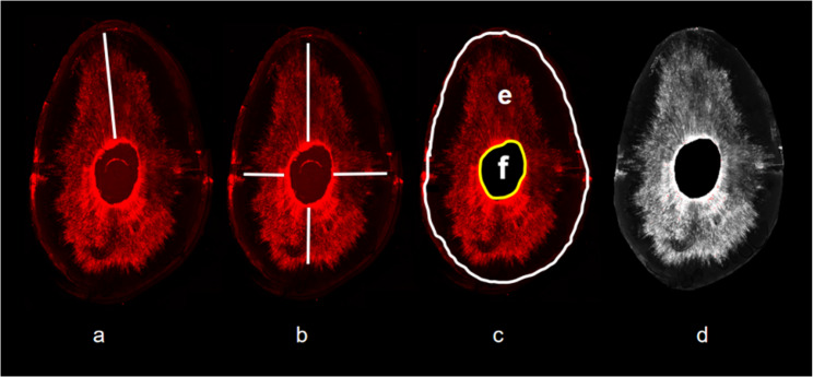
For CLSM analysis (Groups A2, B2, C2), teeth were sectioned into slices at depths of 2 mm, 5 mm, and 8 mm. Using CLSM (Nikon, Tokyo, Japan) at 10× magnification, and a wavelength of 560–600 nm, digital images were analysed with ImageJ program (National Institutes of Health, Bethesda, MD, USA). Maximum and mean sealer penetration depths at four points (3, 6, 9, and 12 o’clock) were calculated along with the percentage of sealer penetration into the dentinal tubules (Fig. 1).
Fig. 1.
Measurement diagram of sealer penetration. Maximum depth of sealer penetration(a); sealer penetration depths at four points (b); area inside the white circle (e); area inside the yellow circle (e); sealer penetration area into the dentin (d) ( Percentage of the sealer penetration area into the dentin=S(d)/S(e-f)*100%)
Apical dye leakage
For dye penetration test (Groups A3, B3, C3), teeth were coated with nail polish, leaving the apical 2 mm exposed, and immersed in 0.2% Indian ink for 7 days. After removal of the nail polish and dye, speciments were demineralized in 10% nitric acid for 48 h, dehydrated in ethanol (70%, 95%, and 100%), and stored in methyl salicylate. Dye penetration was measured under a stereomicroscope at 20× magnification, and images were analysed with ImageJ to determine the linear extent of penetration from the apex to the coronal portion.
Statistical analysis
In statistical analysis, data were analyzed usingc SPSS 21.0 software. One-way analysis of variance and Tukey’s post hoc tests were used for normally distributed data, with the significance level set at 5%. For non-homogeneous variance, the Kruskal–Wallis test was applied (P < 0.05), followed by pairwise comparison using the Bonferroni correction method when necessary, with a 5% significance level. Results were reported as mean ± standard deviation.
Results
Morphological analysis
Pre-instrumentation CBCT morphological parameters (WVC + AH Plus: 2.47 ± 0.53, WVC + iRoot SP: 2.49 ± 0.49, SC + iRoot SP: 2.47 ± 0.39) and post-instrumentation micro-CT parameters (WVC + AH Plus: 1.87 ± 0.2, WVC + iRoot SP: 1.88 ± 0.19, SC + iRoot SP: 1.87 ± 0.23) showed no statistically significant differences between groups (P > 0.05).
Tridimensional reconstructions and the percentage of void volume
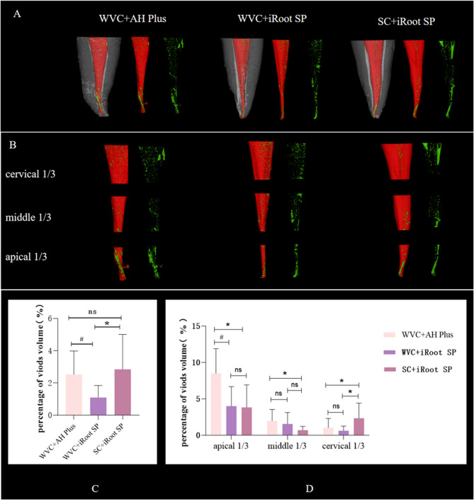
Three-dimensional reconstructions of specimens from the three groups and their respective void volume percentages are depicted in Fig. 2. Among the groups, the SC + iRoot SP group exhibited the lowest void volume percentage across the entire root canal (P < 0.05). In the apical and middle thirds, the SC + iRoot SP group demonstrated significantly lower void volumes than the WVC + AH Plus group (P < 0.05). However, in the cervical third, the void volume was higher than in the WVC + iRoot SP/AH Plus group (P < 0.05).
Fig. 2.
Three-dimensional reconstruction images and the percentage of void volume. Three-dimensional reconstruction images of specimens in the three groups (A); three-dimensional reconstruction images of the apical 1/3, middle 1/3, and cervical 1/3 in the three groups (B) (Gray indicates dental tissue, red indicates the filling material, green indicates voids, and white indicates artifacts); percentage of void volume in the whole root canal in the three groups (C); percentage of void volume in the apical, middle, and cervical thirds of the root canalD. ("*" indicates significant differences between the SC+iRoot SP group and the WVC+AH Plus, WVC+iRoot SP groups; "#" indicates significant differences between the WVC+AH Plus and WVC+iRoot SP groups; "ns" indicates no significant difference)
Penetration of sealers in dentinal tubules
Figure 3 illustrates the penetration situation of sealers into dentinal tubules, along with corresponding images. At 2 mm from the apex, the maximum and mean penetration depths in the WVC + AH Plus group were shorter than in the other groups. At 5 mm from the apex, the WVC + iRoot SP group displayed significantly shorter penetration depths compared to the SC + iRoot SP group (P < 0.05). Conversly, at 8 mm from the apex, the maximum penetration depth in the WVC + iRoot SP group was greater than in the SC + iRoot SP group (P < 0.05). Additionally, at 2 mm, 5 mm, and 8 mm, the percentage of sealer penetration area in the WVC + AH Plus group was significantly lower than in the other two groups (P < 0.05).
Fig. 3.
Penetration of sealers into dentin tubules. Confocal microscopy images of representative sections A. Maximum penetration depth of sealers in the three groups (B); mean penetration depth of sealers in the three groups (C); percentage of the penetration area of sealers in the three groups D. ("*" indicates significant differences between the SC+iRoot SP group and the WVC+AH Plus and WVC+iRoot SP groups; "#" indicates significant differences between the WVC+AH Plus and WVC+iRoot SP groups;"ns" indicates no significant difference)
Apical dye penetration
Figure 4 shows representative demineralized tooth images for each group. The dye penetration depth was significantly lower in the SC + iRoot SP group (750 ± 340 μm) compared to the WVC + AH Plus (1110 ± 490 μm) and WVC + iRoot SP (1090 ± 510 μm) groups. Statistically significant differences were observed between the SC + iRoot SP group and the other two groups (P < 0.05).
Fig. 4.
Representative samples of demineralization teeth images. (In the image, the distance is the length of the line)
Discussion
Considering that sample may influence results, meticulous selection and randomization of the teeth were employed in this study. Consequently, CBCT scanning was utilized for pre-instrumentation, evaluation, while Micro-CT was performed post-instrumentation to define morphological parameters before and after preparation.
The presence of voids in root canal filling material can facilitate the proliferation of residual microorganisms, potentially compromising treatment outcomes [21].
The filling technique significantly impacts the presence of these voids. In this study, the percentage of void volume across in the entire root canal was lower with the WVC technique than with the SC technique. This finding aligns with Santos-Junior et al. [22], who reported superior filling performance with WVC over SC. Similarly, Iglecias et al. [23] demonstrated better obturation quality with WVC particularly in band-shaped isthmuses. These findings suggest that WVC may be more effective than SC in irregular root canals. However, variations in filling effectiveness across different sections of the root canal warrant attention. Santos-Junior AO’s study revealed that while SC resulted in more voids in the middle and cervical thirds of the root canal, it achieved comparable sealing to WVC in the apical third [24]. Another study assessing voids in mesial root canals of mandibular molars showed similar void volumes in the middle and apical thirds between SC and continuous wave of condensation (CWC) techniques [25].In the present study, SC resulted in more voids in the cervical third but demonstrated excellent sealing in the middle and apical thirds, surpassing WVC. These findings indicate that for irregular or oval root canals, different obturation techniques may be suitable for different segments of the canal.
Sealer penetration into dentinal tubules enhances mechanical retention by forming a physical barrier against bacterial microleakage and recontamination of the root canal system [26, 27]. Achieving optimal sealer diffusion into the tubules is therefore critical for ensuring a hermetic seal and improving long-term outcomes [28].
Several studies have reported similar interfacial adaptation for root canals filled with iRoot SP and AH Plus sealers. Amaral et al. [29] found significantly lower maximum and mean penetration depths with AH Plus at 5 mm from the apex compared to iRoot SP. Turkyilmaz and Erdemir [30] also demonstrated a significantly larger penetration area for iRoot SP. In this study, iRoot SP showed superior maximum and mean penetration depths, as well as a greater percentage of penetration area, at both of 2 mm and 5 mm from the apex, compared to AH Plus. The enhanced dentinal tubule penetration of iRoot SP is likely due to its ultrafine particle size (< 2 microns) and high viscosity.
The effect of root canal filling methods on sealer penetration was not evident in this study. No significant differences were observed in penetration depth or percentage of penetration area between WVC + iRoot SP and SC + iRoot SP. Regardless of the obturation technique, the sealer’s properties predominantly determined penetration. Consistent with prior studies [31, 32], extensive sealer penetration into dentinal tubules was observed, independent of the obturation method.
Inadequate sealing in the apical region can permit the infiltration of microorganisms and their byproducts between the root canal system and periradicular tissues, potentially resulting in treatment failure [1]. Our study, found that the SC + iRoot SP group exhibited the lowest apical dye penetration depth, which in SC + iRoot SP was the lowest, associated with the formation of sealer clumps in the apical region, consistent with Ballullaya SV’s findings [33]. The minimal penetration depth and reduced void volume in the apical third of the root canal in the SC + iRoot SP group indicate its superior sealing efficacy in this region.
Caution is warranted when extrapolating these findings to clinical practice, given the limitations of ex vivo studies and the snapshot nature of obturation quality assessment. Long-term stability of these obturations remains speculative. Future laboratory methods should prioritize predicting the long-term clinical performance of root canal fillings rather than merely ranking materials or techniques.
These findings guide clinicians in choosing the most effective, safe, straightforward, and cost-efficient techniques for filling oval root canals.
Conclusions
Despite more voids observed in the cervical third of the root canal with SC + iRoot SP, its sealing performance in the middle and apical segments was excellent, making it the preferred method for these regions.
Supplementary Information
Acknowledgements
Not applicable.
Abbreviations
- RCT
root cnal therapy
- WVC
Warm Vertical Compaction Obturation Technique
- SC
Single-Cone Obturation Technique
- micro-CT
micro-Computed Tomography
- CLSM
Confocal Laser Scanning Microscopy
- WL
Working Length
- NaOCl
sodium hypochlorite
Authors’ contributions
Huo Lijun: Conception, design, investigation, data collection, drafting and critical revision of the article; Zhang Qiaoyun: data collection, data curation, methodology and drafting of the article; Lei Yayan critical revision of the article; Meng Xinhui: writing—review and editing; Zhan JIyuan: statistics. All authors have read and agreed to the published version of the manuscript.
Funding
The authors declare that no funds, grants, or other support were received during the preparation of this manuscript.
Data availability
No datasets were generated or analysed during the current study.
Declarations
Ethics approval and consent to participate
The collected teeth were extracted for therapeutic reasons unrelated to the study were collected, with prior informed consent from healthy individuals who were seeking dental care at The Oral and Maxillofacial Surgery Department Clinic, Affiliated Stomatological Hospital of Kunming Medical University. The patients were voluntary donating the extracted teeth to The Faculty for utilizing in research purpose. This study was previously approved by the ethics committee of the School of Stomatology at Kunming Medical University(KYKQ2022MEC003).
Consent for publication
Not applicable.
Competing interests
The authors declare no competing interests.
Footnotes
Publisher’s Note
Springer Nature remains neutral with regard to jurisdictional claims in published maps and institutional affiliations.
Contributor Information
Qiaoyun Zhang, Email: 2580637941@qq.com.
Lijun Huo, Email: danling430@126.com.
References
- 1.Tomson RM, Polycarpou N, Tomson PL. Contemporary obturation of the root canal system. Br Dent J. 2014;216(6):315–22. 10.1038/sj.bdj.2014.205. [DOI] [PubMed] [Google Scholar]
- 2.Pawar AM, Bhardwaj A, Banga KS, et al. Deficiencies in root canal fillings subsequent to adaptive instrumentation of oval canals. Biology. 2021;10(11): 1074. 10.3390/biology10111074. [DOI] [PMC free article] [PubMed] [Google Scholar]
- 3.Jaha HS. Hydraulic (single cone) versus thermogenic (warm vertical compaction) obturation techniques: a systematic review. Cureus. 2024;16(6):e62925. 10.7759/cureus.62925. [DOI] [PMC free article] [PubMed] [Google Scholar]
- 4.da Penha Silva PJ, Marceliano-Alves MF, Provenzano JC, et al. Quality of root canal filling using a bioceramic sealer in oval canals: a three-dimensional analysis. Eur J Dent. 2021;15:475–80. 10.1055/s-0040-1722095. [DOI] [PMC free article] [PubMed] [Google Scholar]
- 5.Patel S, Bhuva B, Bose R. Present status and future directions: vertical root fractures in root filled teeth. Int Endod J. 2022;55(3):804–26. 10.1111/iej. [DOI] [PMC free article] [PubMed] [Google Scholar]
- 6.Khasnis SA, Kidiyoor KH, Patil AB, et al. Vertical root fractures and their management. J Conserv Dent. 2014;17:103–10. 10.4103/0972-0707.128034. [DOI] [PMC free article] [PubMed] [Google Scholar]
- 7.Liao WC, Chen CH, Pan YH, et al. Vertical root fracture in non-endodontically and endodontically treated teeth: current understanding and future challenge. J Pers Med. 2021;11: 1375. 10.3390/jpm11121375. [DOI] [PMC free article] [PubMed] [Google Scholar]
- 8.Canakci BC, Sungur R, Er O. Comparison of warm vertical compaction and cold lateral condensation of α, β gutta-percha and Resilon on apically extruded debris during retreatment. Niger J Clin Pract. 2019;22(7):926–31. 10.4103/njcp.njcp_663_18. [DOI] [PubMed] [Google Scholar]
- 9.Heran J, Khalid S, Albaaj F, et al. The single cone obturation technique with a modified warm filler. J Dent. 2019;89: 103181. 10.1016/j.jdent.2019.103181. [DOI] [PubMed] [Google Scholar]
- 10.Jafari F, Jafari S. Composition and physicochemical properties of calcium silicate based sealers: a review Article. J Clin Exp Dent. 2017;9:e1249–55. 10.4317/jced.54103. [DOI] [PMC free article] [PubMed] [Google Scholar]
- 11.Vouzara T, Dimosiari G, Koulaouzidou EA, et al. Cytotoxicity of a new calcium silicate endodontic sealer. J Endod. 2018;44:849–52. 10.1016/j.joen.2018.01.015. [DOI] [PubMed] [Google Scholar]
- 12.Ashkar I, Sanz JL, Forner L, et al. Calcium silicate-based sealer dentinal tubule penetration a systematic review of in vitro studies. Mater Basel. 2023;16: 2734. 10.3390/ma16072734. [DOI] [PMC free article] [PubMed] [Google Scholar]
- 13.Yadav S, Nawal RR, Chaudhry S, et al. Assessment of quality of root Canal filling with C point, Guttacore and lateral compaction technique: a confocal laser scanning microscopy study. Eur Endod J. 2020;5:236–41. 10.14744/eej.2020.62534. [DOI] [PMC free article] [PubMed] [Google Scholar]
- 14.Sfeir G, Zogheib C, Patel S, Giraud T, et al. Calcium silicate-based root canal sealers: a narrative review and clinical perspectives. Materials. 2021;14: 3965. 10.3390/ma14143965. [DOI] [PMC free article] [PubMed] [Google Scholar]
- 15.Bukhari S, Karabucak B. The antimicrobial effect of bioceramic sealer on an 8-week matured Enterococcus faecalis biofilm attached to root canal dentinal surface. J Endod. 2019;45(8):1047–52. 10.1016/j.joen.2019.04.004. [DOI] [PubMed] [Google Scholar]
- 16.Rodríguez-Lozano FJ, García-Bernal D, Oñate-Sánchez RE, et al. Evaluation of cytocompatibility of calcium silicate-based endodontic sealers and their effects on the biological responses of mesenchymal dental stem cells. Int Endod J. 2017;50:67–76. 10.1111/iej.12596. [DOI] [PubMed] [Google Scholar]
- 17.Park MG, Kim IR, Kim HJ, et al. Physicochemical properties and cytocompatibility of newly developed calcium silicate-based sealers. Aust Endod J. 2021;47(3):512–9. 10.1111/aej.12515. [DOI] [PubMed] [Google Scholar]
- 18.Wang Y, Liu S, Dong Y. In vitro study of dentinal tubule penetration and filling quality of bioceramic sealer. PLoS ONE. 2018;13:e0192248. 10.1371/journal.pone. [DOI] [PMC free article] [PubMed] [Google Scholar]
- 19.Mohamed EA, Fathieh SM, Farzaneh TA et al. Effect of Different Irrigation Solutions on the Apical Sealing Ability of Different Single-cone Obturation Systems: An In Vitro Study. J Contemp Dent Pract. 2019;20(2):158–165.Wang Y, Liu S, Dong Y. In vitro study of dentinal tubule penetration and filling quality of bioceramic sealer. PLoS One. 2018;13:e0192248. 10.1371/journal.pone [PubMed]
- 20.Schneider SW. A comparison of canal preparations in straight and curved root canals. Oral Surg Oral Med Oral Pathol. 1971;32(2):271–5. 10.1016/0030-4220(71)90230-1. [DOI] [PubMed] [Google Scholar]
- 21.Tabassum S, Khan FR. Failure of endodontic treatment: the usual suspects. Eur J Dent. 2016;10:144–7. 10.4103/1305-7456.175682. [DOI] [PMC free article] [PubMed] [Google Scholar]
- 22.Xu H, Qiu XH, Zhang GD, et al. Evaluation of the filling quality of different root Canal obturation techniques using micro-CT. Shanghai Kou Qiang Yi Xue. 2018;27:349–53. [PubMed] [Google Scholar]
- 23.Zhang P, Yuan K, Jin Q, et al. Presence of voids after three obturation techniques in band-shaped isthmuses: a micro-computed tomography study. BMC Oral Health. 2021;21: 227. 10.1186/s12903-021-01584-2. [DOI] [PMC free article] [PubMed] [Google Scholar]
- 24.Santos-Junior AO, Tanomaru-Filho M, Pinto JC, et al. Effect of obturation technique using a new bioceramic sealer on the presence of voids in flattened root canals. Braz Oral Res. 2021;35: e028. 10.1590/1807-3107bor-2021.vol35.0028. [DOI] [PubMed] [Google Scholar]
- 25.Iglecias EF, Freire LG, de Miranda Candeiro GT, et al. Presence of voids after continuous wave of condensation and Single-cone obturation in mandibular molars: A Micro-computed tomography analysis. J Endod. 2017;43(4):638–42. 10.1016/j.joen.2016. [DOI] [PubMed] [Google Scholar]
- 26.Sfeir G, Zogheib C, Patel S, et al. Calcium silicate-based root canal sealers: a narrative review and clinical perspectives. Materials. 2021;14(14): 3965. 10.3390/ma14143965. [DOI] [PMC free article] [PubMed] [Google Scholar]
- 27.De Sarkar M, Mala K, Shenoy Mala S, et al. Antimicrobial efficacy of Kerr pulp Canal sealer (EWT) in combination with 10% amoxicillin on Enterococcus faecalis: a confocal laser scanning microscopic study. F1000Res. 2023;12:725. 10.12688/f1000research.132047. [DOI] [PMC free article] [PubMed] [Google Scholar]
- 28.Donnermeyer D, Bunne C, Schäfer E, et al. Retreatability of three calcium silicate- containing sealers and one epoxy resin-based root canal sealer with four different root canal instruments. Clin Oral Investig. 2018;22:811–7. 10.1007/s00784-017-2156-5. [DOI] [PubMed] [Google Scholar]
- 29.El Hachem R, Khalil I, Le Brun G, et al. Dentinal tubule penetration of AH plus, BC sealer and a novel tricalcium silicate sealer: a confocal laser scanning microscopy study. Clin Oral Investig. 2019;23:1871–6. 10.1007/s00784-018-2632-6. [DOI] [PubMed] [Google Scholar]
- 30.Akcay M, Arslan H, Durmus N, et al. Dentinal tubule penetration of AH plus, iRoot SP, MTA fillapex, and guttaflow bioseal root Canal sealers after different final irrigation procedures: A confocal microscopic study. Lasers Surg Med. 2016;48(1):70–6. 10.1002/lsm. [DOI] [PubMed] [Google Scholar]
- 31.Girelli CF, Lacerda MF, Lemos CA, et al. The thermoplastic techniques or single-cone technique on the quality of root Canal filling with tricalcium silicate-based sealer: an integrative review. J Clin Exp Dent. 2022;14:e566–72. 10.4317/jced.59387. [DOI] [PMC free article] [PubMed] [Google Scholar]
- 32.Turkyilmaz A, Erdemir A. Comparison of dentin penetration ability of different root canal sealers used with different obturation methods. Microsc Res Tech. 2020;83:1544–51. 10.1002/jemt.23548. [DOI] [PubMed] [Google Scholar]
- 33.Ballullaya SV, Vinay V, Thumu J, et al. Stereomicroscopic dye leakage measurement of six different root Canal sealers. J Clin Diagn Res. 2017;11(6):ZC65–8. 10.7860/JCDR/2017/25780.10077. [DOI] [PMC free article] [PubMed] [Google Scholar]
Associated Data
This section collects any data citations, data availability statements, or supplementary materials included in this article.
Supplementary Materials
Data Availability Statement
No datasets were generated or analysed during the current study.


