Abstract
We report here, for the first time, that bacteria associated with marine snow produce communication signals involved in quorum sensing in gram-negative bacteria. Four of 43 marine microorganisms isolated from marine snow were found to produce acylated homoserine lactones (AHLs) in well diffusion and thin-layer chromatographic assays based on the Agrobacterium tumefaciens reporter system. Three of the AHL-producing strains were identified by 16S ribosomal DNA gene sequence analysis as Roseobacter spp., and this is the first report of AHL production by these α-Proteobacteria. It is likely that AHLs in Roseobacter species and other marine snow bacteria govern phenotypic traits (biofilm formation, exoenzyme production, and antibiotic production) which are required mainly when the population reaches high densities, e.g., in the marine snow community.
Marine snows are millimeter- to centimeter-size aggregates which typically consist of detrital particles, inorganic particles, and diatoms as well as other microorganisms. Marine snows play a significant role in the vertical transport of organic material in the ocean (11) but are also sites of elevated heterotrophic activity (27, 28). Bacteria colonize sinking aggregates rapidly (10), and by means of exoenzymes they solubilize the aggregated organic particulate material (40). Since particulate material is solubilized faster than the attached bacteria can take up the solutes (44), dissolved organics leak (17, 40) and form a plume trailing behind the sinking aggregate (22). The growth of free bacteria may be substantially enhanced in this plume and, in fact, may account for a significant fraction of the bacterial production and remineralization in the water column (22). The balance between the rate at which aggregates form and sink on the one hand and the rate at which they are solubilized and remineralized on the other hand therefore has a major impact on ocean carbon fluxes and thus on atmospheric CO2 and global climate (1).
Regulation of bacterial activity is key to the understanding of marine snow degradation rates. Levels of microorganisms in marine and lake snow are typically 108 to 109 cells ml−1, which are 100- to 10,000-fold higher than those in the surrounding water column (20, 27, 29, 37). Long and Azam (23) recently suggested that bacterial interactions may occur where microbial densities are high, and they showed that approximately 50% of particle-associated bacteria display antagonistic activities towards other bacteria isolated from particles and free water masses. In particular, members of the α-Proteobacteria growing on marine particles showed a much higher production of inhibitory molecules than those in the surrounding water. Bacteria probably have little advantage of antagonistic and extracellular hydrolytic activities in the free-living stage. In contrast, such activities are crucial at the higher densities in the marine snow, and it has therefore been suggested that quorum-sensing mechanisms may be operating in these communities (21). This hypothesis is also supported by the observation that particle-associated bacteria are metabolically more active than free-living bacteria (41) and exhibit a higher extracellular enzymatic hydrolysis rate per cell (17).
In gram-negative bacteria, the quorum-sensing mechanism relies on the ability of the bacteria to communicate by using chemical signals such as acylated homoserine lactones (AHLs) (12). The AHLs allow the bacterial population to sense its own density and to express target genes only at particular (high) cell densities. The AHL-based quorum-sensing mechanism was first studied in the marine symbiotic bacterium, Vibrio fischeri, in which colonization of the light organ crypts (35) and light emission are regulated by AHLs and occur only at high cell densities (26). The list of organisms capable of producing AHLs and the phenotypes regulated has since been growing (7, 45), and these include antibiotic production in Pseudomonas aureofaciens and (up)regulation of hydrolytic enzymatic activity in pseudomonads and strains of Enterobacteriaceae (7, 14, 32; A. B. Christensen, K. Riedel, L. Ravn, L. Eberl, S. Molin, L. Gram, and M. Givskov, submitted for publication).
Due to the involvement of AHLs in phenotypes of importance for bacterial behavior in marine snow and the high bacterial densities present in this environment, it is possible that such coordinated bacterial regulation takes place. We here demonstrate that bacteria isolated from marine snow and marine diatoms appear to be capable of producing AHLs.
Isolation and identification of bacterial strains.
Forty-three strains of bacteria were isolated from particles collected in surface waters of the Øresund (Denmark) and in the German Waddensea (Neuharlingersiel). Bacterial strains were isolated either directly from individual marine particles or from axenic marine diatoms (Thalassiosira rotula or Skeletonema costatum) which had been incubated with natural seawater. Incubations were done in sterile rolling tanks which were kept at in situ temperature and natural light conditions for 1 day or several days. Bacteria were cultivated and isolated on a minimal medium (f/2 diatom medium [19]) which was enriched with 1 mg of a sterile algal hydrolysate as the sole carbon source. This medium selects for growth of bacteria adapted to low nutrient concentrations and reduces growth of bacteria adapted to high nutrient concentrations. Separation of bacteria was done by a dilution series performed directly on agar plates (13-strike method). Isolates were checked for purity by using the denaturing gradient gel electrophoresis method (25). Only isolates with a single band on the denaturing gradient gel were defined as pure and were sequenced thereafter. Following isolation, the strains were grown and pure cultured on agar plates (2%) enriched with marine broth medium (MB 2216; Difco, Detroit, Mich.). All cultures were grown at 15°C (in situ temperature).
Chromosomal DNA was extracted by cycles of freezing and heating the cell suspension to 95°C for 30 min at least three times. An approximately 1,500-bp segment of the 16S rRNA gene was amplified by PCR with a pair of primers. One (GM3F, 5′-AGAGTTTGATC[AC]TGGC-3′; MWG, Ebersberg, Germany) targeted the beginning and the other (GM4R, 5′-TACCTTGTTACGACT T-3′; MWG) targeted the end of the 16S rRNA gene. Two microliters of the cell suspension was added to a 98 μl of PCR mixture containing 1.5 U of Sigma Red Taq polymerase, a 250 μM concentration of each deoxynucleoside triphosphate, 2.1 μM MgCl2, and 25 pmol of each primer. The PCR protocol consisted of a denaturing step of 95°C for 5 min, followed by 30 cycles of denaturing for 1 min at 95°C, annealing for 1 min at 40°C, and a 3-min extension at 72°C. A final extension at 72°C for 10 min was then performed. The PCR products were purified by using the QIAquick 250 purification kit (Qiagen, Hilden, Germany). Purified PCR products were sequenced with the DYEnamic direct cycle sequencing kit (US79535; Amersham Life Science, Inc.) and a Li-Cor Long Redir 4200 automated DNA sequencer (MWG). Sequencing primers were GM 3F (5′-AGAGTTTGATC[AC]TGGC-3′; MWG) and GM 8R (5′-TGGGTATCTAATCCT-3′; MWG) labeled with IRDyeTM800 (binding at base 798 of Escherichia coli in the reverse orientation). Two microliters was added to the reaction mixture containing 1 pmol of the primer and 2 μl of the reaction solution. The sequencing protocol consisted of a denaturing step at 95°C for 5 min, followed by 30 cycles of denaturing for 0.5 min, annealing for 0.5 min at 40°C, and a 1-min extension at 70°C. The reaction mixture was immediately cooled to 4°C and kept at this temperature until the sequence gel was loaded. Electrophoresis was performed at 2,000 V and 45°C for 16 h in 1× Tris-borate-EDTA buffer. Sequences were compared to those in GenBank by using the BLAST function of the National Center for Biotechnology Information server (http://www.ncbi.nlm.nih.gov).
The culturable microflora was dominated by α- and γ-Proteobacteria and organisms belonging to the Cytophaga-Flavobacterium-Bacteroides complex (Table 1). In addition, gram-positive bacteria, mainly from the genera Bacillus and Clostridium, were also culturable. These data are in overall agreement with studies of marine and lake snow done by using direct molecular analysis of the microbial community (fluorescent in situ hybridization or rRNA cloning). Bacteria constitute 80 to 99% of the microbial population of marine and lake snow (18, 37), and within this group, organisms belonging to α- and β-Proteobacteria and to the Cytophaga-Flavobacterium-Bacteroides complex dominate on marine particles (5, 6, 37). Our finding of a significant proportion of γ-Proteobacteria is probably due to the high proportion of culturable organisms in this group (9).
TABLE 1.
Identification of and AHL production by 43 bacterial strains isolated from detrital and algal aggregates, based on partial (800-bp) 16S rRNA gene sequencing
| Phylum or group | Strain | Identification by GenBank alignment | % Homology to GenBank sequence | AHLb |
|---|---|---|---|---|
| α-Proteobacteria | HP39 | AF025321, α-Proteobacterium KAT8 | 99 | − |
| HP40 | AF025321, α-Proteobacterium KAT8 | 98 | − | |
| HP24 | U70680, Rhodobacter-Roseobacter strain OM42 | 94 | − | |
| HP12 | AF359535, Roseobacter strain ATAM407 | 98 | + | |
| HP30 | AJ294355, Roseobacter strain 667-19 (uncultured) | 98 | + | |
| HP32 | AF359535, Roseobacter strain ATAM407 | 97 | + | |
| HP44w | Y15343, Roseobacter strain PRLISTO6 | 98 | − | |
| HP27 | X91814, Sphingobacterium comitans | 92 | − | |
| HP18 | AY007681, Sphingomonas sp. (unknown) | 98 | − | |
| HP22 | AJ318163, uncultured α-Proteobacterium Blri | 94 | − | |
| HP28 | AJ290003, uncultured α-Proteobacterium FukaNS | 100 | − | |
| HP33 | AF345550Rhizobium sp. strain SDW052 | 99 | − | |
| AF388033, A. tumefaciens | 99 | |||
| HP13 | AF359546, marine bacterium SCRIPPS 739 | 96 | − | |
| CFBa | HP34 | AF367847, bacterium KM1 | 96 | − |
| HP25 | AF277514, Cellulophaga strain SIC.834 (uncultured) | 98 | − | |
| HP2 | AF235114, Cytophaga strain KTO2ds22 | 98 | − | |
| HP14 | AF235114, Cytophaga strain KTO2ds22 | 98 | − | |
| HP19 | AF235114, Cytophaga strain KTO2ds22 | 98 | − | |
| HP31 | AF235114, Cytophaga strain KTO2ds22 | 97 | − | |
| HP35 | AF235114, Cytophaga strain KTO2ds22 | 98 | − | |
| HP20 | AF321022, Frigobacterium sp. strain GOB | 98 | − | |
| HP11 | M58792, Microscilla furvescens | 90 | − | |
| HP23 | AF287047, uncultured strain EC-B39 | 92 | − | |
| γ-Proteobacteria | HP3 | AF062642, Alcanivorax borkumensis | 98 | − |
| HP26 | L10938, Alteromonas macleodii | 98 | − | |
| HP1 | AJ002006, Curacaobacter baltica | 98 | − | |
| HP9 | AJ002006, C. baltica | 97 | − | |
| HP41 | AJ302707, Marinobacter strain ME108 | 97 | − | |
| HP6 | AJ000647, Marinobacter strain PCOB-2 | 99 | − | |
| HP15 | AJ000647, Marinobacter strain PCOB-2 | 98 | − | |
| HP36 | AJ000647, Marinobacter strain PCOB-2 | 98 | + | |
| HP4 | AJ295154, Oleiphilus messinensis | 93 | − | |
| Bacillus-Clostridium | HP16 | AJ296095, Bacillus sp. strain OS-5 | 96 | − |
| HP17 | AJ296095, Bacillus sp. strain OS-5 | 98 | − | |
| HP29 | AJ296095, Bacillus sp. strain OS-5 | 98 | − | |
| HP38 | AJ296095, Bacillus sp. strain OS-5 | 98 | ||
| HP21 | AY030327, Bacillus pumilis KL-052 | 99 | − | |
| HP8 | AY038905, marine bacterium SE165 | 97 | − | |
| HP10 | AF275714, Haeler soda lake bacterium Z6 | 99 | − | |
| Actinomycete | HP42 | AJ344143, Citrococcus muralis 1b | 98 | − |
| HP5 | AF321022, Frigobacterium sp. strain GOB | 98 | − | |
| HP7 | AF197036, Arthrobacter sp. strain SMCC G980 | 97 | − | |
| Cyanobacterium | HP43 | D90916, Synechocystis sp. strain PCC6803 | 100 |
CFB, Cytophaga-Flavobacterium-Bacteroides.
Presence (+) or absence (−) of AHL as tested by using the A. tumefaciens monitor system. A positive reaction was not reproducible for strain HP43.
Detection of AHLs from marine snow bacteria.
Isolation and pure culturing are required to determine the ability of bacteria to produce AHLs. Due to the heterogeneity of the AHL synthetase (I) genes, it has not been possible to develop PCR protocols or rRNA probes allowing a direct determination of AHL-producing capability at the molecular level in single cells. Therefore, preliminary screening for AHLs was done by preparing sterile filtered supernatants from cultures grown for 1 1/2 to 2 weeks at 15°C and testing the samples in three AHL monitor systems using Agrobacterium tumefaciens (3) and Chromobacterium violaceum CV026 (24) as described by Ravn et al. (31) and in the E. coli pSB403 LuxR assay (8, 43) as described by Gram et al. (15). Since AHLs are not stable at high pH (above 8), all cultures were also grown in MB in which pH was adjusted to 6.2. In none of these outgrown cultures did the pH increase above 7.5. All cultures eliciting an AHL response were positive both when grown in MB at normal pH (7.8) and when grown at pH 6.2. For the monitor assays, A. tumefaciens strain NT1(pZLR4) was grown with 20 μg of gentamicin ml−1 in Luria-Bertani broth (2) with 5 g of NaCl liter−1 (LB5) for 24 h and inoculated into 50 ml of ABt broth with 0.5% glucose and 0.5% Casamino Acids (4). The outgrown culture was mixed with 100 ml of melted, 45°C ABt agar containing 50 μg of X-Gal (5-bromo-4-chloro-3-indolyl-β-d-galactopyranoside) (Promega 9683801 L) ml−1 and poured into petri dishes. C. violaceum CV026 (24) was grown in LB5 with 20 μg of kanamycin ml−1 for 24 h, inoculated in 50 ml of LB5, and incubated overnight. Plates were poured after the outgrown culture was mixed with 100 ml of 45°C LB5 agar. Wells of 6 mm in diameter were punched in the solidified agars, and samples of 60 μl were pipetted into the wells. Plates with A. tumefaciens or C. violaceum were incubated for 2 days and 1 day, respectively, at 25°C and read for zones of blue color due to AHL-induced β-galactosidase activity or zones of purple pigment due to AHL-induced violacin formation in the agar. E. coli pSB403 was grown in LB5 with 10 μg of tetracycline ml−1 overnight and diluted to an optical density at 450 nm of 0.8. One hundred microliters of this culture was mixed with 100 μl of sterile filtered supernatant from marine bacteria, and luminescence was measured with a MicroBeta 1450 TriLux scintillation and luminescence counter (Wallac).
Sterile filtered supernatants from 4 strains (HP12, HP30, HP32, and HP36) out of 43 tested elicited a reproducible response in the A. tumefaciens monitor system (Fig. 1), indicating the presence of AHLs. No responses were elicited in the C. violaceum or E. coli pSB403 system from any of the 43 strains, while the standard N-hexanoyl-homoserine lactone (C6-HSL) caused induction of a purple zone in the CV026 assay. In principle, it is possible that some of the 38 strains that did not elicit AHL responses in the monitor systems do produce AHLs, since they may cause a reaction in other AHL receptors. However, several authors have reported that the TraR system is broad (31, 38), and it is therefore a good screening system.
FIG. 1.
Presence of acylated homoserine lactones in supernatants on bacteria isolated from marine snow particles. AHLs were determined by the sensor strain A. tumefaciens NT1(pZLR4). Plates 1 and 2, twofold dilutions of N-3-oxo-hexanoyl-homoserine lactone. Plate 3, sterile filtered supernatants from Roseobacter strains (upper left well, HP12; lower left well, HP30; lower right well, HP32).
Three strains producing AHLs were closely related and clustered in the Roseobacter-Ruegeria subgroup of the α-Proteobacteria. This is, to our knowledge, the first report of AHLs in this group (7, 45). However, AHLs have been detected in other members of the α-Proteobacteria, such as Rhodobacter sphaeroides (30), Rhizobium species (16, 34), and A. tumefaciens (46). We tested four Roseobacter strains isolated from similar marine environments and found two, isolated from diatom aggregates in the North Sea, also to be AHL positive. Thus, several but not all marine Roseobacter strains produce AHLs. Many genera of the γ-Proteobacteria (Alteromonadales and Vibrionales) are common in marine particles (23), and although these organisms are common producers of AHLs (7, 45), we detected AHLs only from one Marinobacter strain in this bacterial group (Table 1).
Roseobacter species are important members of the aquatic microbial community (39), and 20 to 30% of bacterial small-subunit ribosomal DNAs isolated from the upper 50 m of Monterey Bay belong to this clade (42). The exact ecological role of Roseobacter species is not known, but they metabolize dimethyl-sulfonio-propionate and other organic sulfur compounds (M. A. Moran, J. M. Gonzáles, R. P. Kiene, R. Simó, and C. Pedrós-Alió, talk presented at the American Society of Limnology and Oceanography, 2001). They display chemotactic behavior towards S and C compounds and are likely to be of major importance for cycling of these compounds in the ocean.
All AHL-positive Roseobacter strains were isolated from living marine diatoms (S. costatum and T. rotula). While AHLs clearly are involved in bacterium-bacterium interactions allowing up-regulation of production of hydrolytic enzymes or antibiotics, these regulatory systems may also be a key to understanding specific bacterium-eucaryote interactions. Thus, the marine alga Delisea pulchra produces a range of halogenated furanones which specifically interfere with the bacterial AHL regulatory systems and eliminate or reduce bacterial surface colonization (13). The possible involvement of AHL regulation in Roseobacter metabolism can have major implications for turnover of organic material in the ocean as well as their colonization of and growth on phytoplankton or organic aggregates.
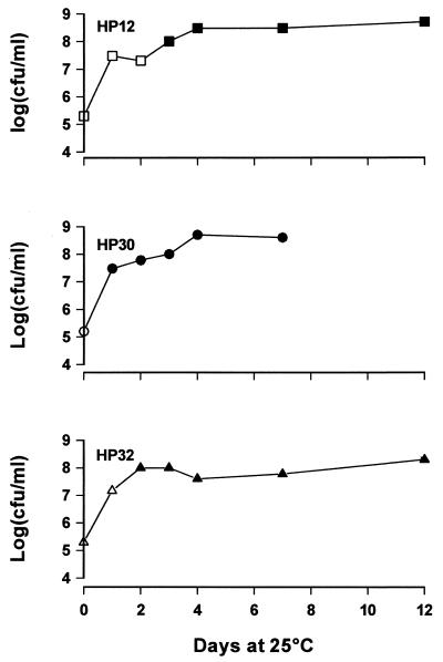
Three Roseobacter strains (HP12, HP30, and HP32) which were positive in the initial screening were inoculated in MB at approximately 105 CFU ml−1 and incubated at 25°C. Samples were withdrawn regularly for 1 week, and sterile filtered supernatants were tested in the AHL assay. The strains grew well in MB at 25°C, and AHL-inducing zones were seen from sterile filtered culture supernatants only when counts exceeded 5 × 10⁷ to 108 CFU ml−1 (Fig. 2). Thus, AHL production in Roseobacter species appears to be similar to that in other organisms in being a phenomenon related to high bacterial densities. AHL production clearly occurs at the bacterial densities typically found in marine snow.
FIG. 2.
Growth of three Roseobacter strains isolated from marine snow in MB at 25°C. Closed symbols, AHLs were not detected in sterile filter supernatants; filled symbols, AHLs were detected in sterile filtered supernatants.
TLC profiling of AHLs.
Ten milliliters of sterile filtered bacterial supernatants was mixed with an equal volume of ethylacetate containing 0.5% formic acid. Following thorough mixing, the ethylacetate phase was removed and the extraction was repeated twice. The three volumes of ethylacetate were combined, evaporated under an N2 atmosphere, and redissolved in 1 ml of ethylacetate. The samples were loaded onto a C18 thin-layer chromatography (TLC) plate (20- by 20-cm TLC aluminum sheets; RP-18 F254 S, 1.05559) (64271; Merck, Darmstadt, Germany), and the plates were developed in 100 ml of 60:40 (vol/vol) methanol-Millipore water (31). The plates were air dried and covered with ABt agar containing the A. tumefaciens monitor strain. Standards included on the plates were N-3-oxo-hexanoyl-homoserine lactone, N-hexanoyl-homoserine lactone (HHL), and N-octanoyl-homoserine lactone (OHL), all from Sigma Chemicals.
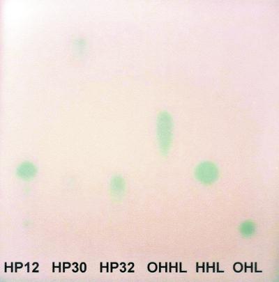
Ethylactetate extracts were prepared from 7-day-old cultures. TLC separation of the extracts revealed different AHL profiles from the three Roseobacter strains (Fig. 3). HP12 induced three spots in the A. tumefaciens monitor system. One was equivalent in size and Rf value to HHL and one was equivalent to OHL, but the other could not be identified. The AHLs of HP30 and HP32 did not correspond to any of the standards used. Tailing of the spots could indicate presence of 3-oxo substitutions on the AHL-inducing compound. Production of HHL and OHL has been detected in another α-Proteobacteria, Rhizobium leguminosarum (33), but the majority of studies of Rhizobium species describe the autoregulation of a hydroxy-C-14-acylated homoserine lactone (36) which is involved in antagonism against other microorganisms (34).
FIG. 3.
Separation of AHLs from ethylacetate extracts of Roseobacter strains isolated from marine snow particles. TLC plates were developed with the sensor strain A. tumefaciens NT1(pZLR4). OHHL, N-3-oxo-hexanoyl-homoserine lactone.
Concluding remarks.
In conclusion, we have shown that several bacterial strains isolated from marine snow particles produce AHL communication signals. To our knowledge this is the first report of AHL production from such communities and the first report of AHL production by Roseobacter-Ruegeria strains. Bacteria attached to marine snow aggregates may spend most of their time freely swimming in the water while searching for particles to colonize. In the pelagic stage, production of extracellular hydrolytic enzymes and antibiotics would presumably be a waste of energy. Due to the common involvement of AHLs in antibiotic production and in hydrolytic enzymatic activity, it is tempting to suggest that the AHL allows the marine snow bacteria to express such phenotypes only when occupying the snow particles and interacting with marine phytoplankton reaching high bacterial cell densities.
Acknowledgments
This work was supported by research grants from the Danish Research Council for the Technical Sciences (9700726) to L.G., from the Danish Ministry for Food and Agriculture to L.G. and T.K., and from the Danish Natural Science Research Council (9801391) to T.K. Isolation and sequencing of bacteria were supported by the Niedersaechsischer Forschungsschwerpunkt Meeresbiotechnologie Themenbereich 5 given to Meinhard Simon by the Volkswagen Foundation.
We thank Meinhard Simon and Thorsten Brinkhoff for numerous discussions and support as well as Jette Melchiorsen for skillful technical assistance.
REFERENCES
- 1.Azam, F., and R. A. Long. 2001. Sea snow microcosms. Nature 414:495-498. [DOI] [PubMed] [Google Scholar]
- 2.Bertani, G. 1951. Studies on lysogenesis. I. The mode of phage liberation by lysogenic Escherichia coli. J. Bacteriol. 62:293-300. [DOI] [PMC free article] [PubMed] [Google Scholar]
- 3.Cha, C., P. Gao, Y.-C. Chen, P. D. Shaw, and S. K. Farrand. 1998. Production of acyl-homoserine lactone quorum-sensing signals by Gram-negative plant-associated bacteria. Mol. Plant-Microbe Interact. 11:1119-1129. [DOI] [PubMed] [Google Scholar]
- 4.Clark, D. J., and O. Maaløe. 1967. DNA Replication and division cycle in Escherichia coli. J. Mol. Biol. 23:99-112. [Google Scholar]
- 5.Cottrell, M. T., and D. L. Kirchman. 2000. Community composition of marine bacterioplankton determined by 16S rRNA gene clone libraries and fluorescence in situ hybridization. Appl. Environ. Microbiol. 66:5116-5122. [DOI] [PMC free article] [PubMed] [Google Scholar]
- 6.DeLong, E. F., D. G. Franks, and A. L. Alldredge. 1993. Phylogenetic diversity of aggregate-attached vs. free-living marine bacterial assemblages. Limnol. Oceanogr. 38:924-934. [Google Scholar]
- 7.Eberl, L. 1999. N-acyl homoserinelactone-mediated gene regulation in Gram-negative bacteria. Syst. Appl. Microbiol. 22:493-506. [DOI] [PubMed] [Google Scholar]
- 8.Eberl, L., M. K. Winson, C. Sternberg, G. S. A. B. Stewart, G. Christiansen, S. R. Chhabra, M. Daykin, P. Williams, S. Molin, and M. Givskov. 1996. Involvement of N-acyl-L-homoserine lactone autoinducers in control of multicellular behavior of Serratia liquefaciens. Mol. Microbiol. 20:127-136. [DOI] [PubMed] [Google Scholar]
- 9.Eilers, H., J. Pernthaler, and R. Amann. 2000. Succession of pelagic marine bacteria during enrichment: a close look at cultivation-induced shifts. Appl. Environ. Microbiol. 66:4634-4640. [DOI] [PMC free article] [PubMed] [Google Scholar]
- 10.Fenchel, T. 2001. Eppur si muove: many water column bacteria are motile. Aquat. Microb. Ecol. 24:197-201. [Google Scholar]
- 11.Fowler, S. W., and G. A. Kanuaer. 1986. Role of large particles in transport of elements and organic compounds through the ocean water column. Proc. Oceanogr. 16:147-194. [Google Scholar]
- 12.Fuqua, C., S. C. Winans, and E. P. Greenberg. 1996. Census and consensus in bacterial ecosystems: the LuxR-LuxI family of quorum-sensing transcriptional regulators. Annu. Rev. Microbiol. 50:727-751. [DOI] [PubMed] [Google Scholar]
- 13.Givskov, M., R. deNys, M. Manefield, L. Gram, R. Maximilien, L. Eberl, S. Molin, P. D. Steinberg, and S. Kjelleberg. 1996. Eucaryotic interference with homoserine lactone-mediated procaryotic signalling. J. Bacteriol. 178:6618-6622. [DOI] [PMC free article] [PubMed] [Google Scholar]
- 14.Givskov, M., L. Eberl, and S. Molin. 1997. Control of exoenzyme production, motility and cell differentiation in Serratia liquefaciens. FEMS Microbiol. Lett. 148:115-122. [Google Scholar]
- 15.Gram, L. A. B. Christiansen, L. Ravn, S. Molin, and M. Givskov. 1999. Production of acylated homoserine lactones by psychrotrophic members of the Enterobacteriaceae isolated from foods. Appl. Environ. Microbiol. 65:3458-3463. [DOI] [PMC free article] [PubMed] [Google Scholar]
- 16.Gray, K. M., J. P. Pearson, J. A. Downie, B. E. A. Boboye, and E. P. Greenberg. 1996. Cell-to-cell singaling in the symbiotic nitrogen-fixing bacterium Rhizobium leguminosarum: autoinduction of stationary phase and rhizosphere-expressed genes. J. Bacteriol. 178:372-376. [DOI] [PMC free article] [PubMed] [Google Scholar]
- 17.Grossart, H. P., and M. Simon. 1998. Bacterial colonization and microbial decomposition of limnetic organic aggregates (lake snow). Aquat. Microb. Ecol. 15:127-140. [Google Scholar]
- 18.Grossart, H. P., and H. Ploug. 2001. Microbial degradation of organic carbon and nitrogen on diatom aggregates. Limnol. Oceanogr. 46:267-277. [Google Scholar]
- 19.Guillard, R. R. L., and J. H. Ryther. 1962. Studies on marine planktonic diatoms. I. Cyclotella nana HUSTEDT and Detonula confercacea (CLEVE) GRAN. Can. J. Microbiol. 8:229-239. [DOI] [PubMed] [Google Scholar]
- 20.Kiørboe, T. 2000. Colonization of marine snow aggregates by invertebrate zooplankton: abundance, scaling, and possible role. Limnol. Oceanogr. 45:479-484. [Google Scholar]
- 21.Kiørboe, T. 2001. Formation and fate of marine snow: small-scale processes with large-scale implications. Sci. Mar. 65:57-71. [Google Scholar]
- 22.Kiørboe, T., and G. A. Jackson. 2001. Marine snow, organic solute plumes and optimal chemosensory behavior of bacteria. Limnol. Oceanogr. 46:1309-1318. [Google Scholar]
- 23.Long, R. A., and F. Azam. 2001. Antagonistic interactions among marine pelagic bacteria. Appl. Environ. Microbiol. 67:4975-4983. [DOI] [PMC free article] [PubMed] [Google Scholar]
- 24.McClean, K. H., M. K. Winson, L. Fish, A. Taylor, S. R. Chhabra, M. Camara, M. Daykin, J. H. Lamb, S. Swift, B. W. Bycroft, G. S. A. B. Stewart, and P. Williams. 1997. Quorum sensing and Chromobacterium violaceum: exploitation of violacein production and inhibition for the detection of N-acyl homoserine lactones. Microbiology 143:3703-3711. [DOI] [PubMed] [Google Scholar]
- 25.Muyzer, G., DeWaal, E. C., and A. G. Uitterlinden. 1993. Profiling of complex microbial populations by denaturing gradient gel electrophoresis analysis of polymerase chain reaction-amplified genes coding for 16S rRNA. Appl. Environ. Microbiol. 59:695-700. [DOI] [PMC free article] [PubMed] [Google Scholar]
- 26.Nealson, K. H., T. Platt, and J. W. Hastings. 1970. Cellular control of the synthesis and activity of the bacterial luminescent system. J. Bacteriol. 104:313-322. [DOI] [PMC free article] [PubMed] [Google Scholar]
- 27.Ploug, H., H. P. Grossart, F. Azam, and B. B. Jørgensen. 1999. Photosynthesis, respiration, and carbon turnover in sinking marine snow from surface waters of Southern California Bight: implications for the carbon cycle in the ocean. Mar. Ecol. Progr. Ser. 179:1-11. [Google Scholar]
- 28.Ploug, H., and H. P. Grossart. 1999. Bacterial production and respiration in suspended aggregates—a matter of the incubation method. Aquat. Microb. Ecol. 20:21-29. [Google Scholar]
- 29.Ploug, H., and H. P. Grossart. 2000. Bacterial growth and grazing on diatom aggregates: respiratory carbon turnover as a function of aggregate size and sinking velocity. Limnol. Oceanogr. 45:1467-1475. [Google Scholar]
- 30.Puskas, A., E. P. Greenberg, S. Kaplan, and A. L. Schaefer. 1997. A quorum-sensing system in the free-living photosynthetic bacterium Rhodobacter sphaeroides. J. Bacteriol. 179:7530-7537. [DOI] [PMC free article] [PubMed] [Google Scholar]
- 31.Ravn, L., A. B. Christensen, S. Molin, M. Givskov, and L. Gram. 2001. Methods for identifying and quantifying acylated homoserine lactones produced by Gram-negative bacteria and their application in studies of AHL-production kinetics. J. Microbiol. Methods 44:239-251. [DOI] [PubMed] [Google Scholar]
- 32.Riedel, K., T. Ohnesorg, K. A. Krogfelt, T. S. Hansen, K. Omori, M. Givskov, and L. Eberl. 2001. N-Acyl-l-homoserine lactone-mediated regulation of the Lip secretion system in Serratia liquefaciens MG1. J. Bacteriol. 183:1805-1809. [DOI] [PMC free article] [PubMed] [Google Scholar]
- 33.Rodelas, B., J. K. Lithgow, F. Wisniewski-Dye, A. Hardman, A. Wilkinson, A. Economou, P. Williams, and J. A. Downie. 1999. Analysis of the quorum-sensing-dependent control of rhizosphere-expressed (rhi) genes in Rhizobium leguminosarum bv. vicae. J. Bacteriol. 181:3816-3823. [DOI] [PMC free article] [PubMed] [Google Scholar]
- 34.Rosemeyer, V., J. Michiels, C. Verreth, and J. Vanderleyden. 1998. luxI- and luxR-homologous genes of Rhizobium etli CNPAF512 contribute to synthesis of autoinducer molecules and nodulation of Phaseolus vulgaris. J. Bacteriol. 180:815-821. [DOI] [PMC free article] [PubMed] [Google Scholar]
- 35.Ruby, E. G. 1999. The Euprymna scolopes-Vibrio fischeri symbiosis: a biomedical model for the study of bacterial colonization of animal tissue, p. 23-40. In D. G. Bartlett (ed.), Molecular marine microbiology. JMMB Symposium Series, vol. 1. Horizon Scientific Press, Norfolk, England. [PubMed] [Google Scholar]
- 36.Schripsema, J., K. E. de Rudder, T. B. van Vliet, P. P. Lankhorst, E. de Vroom, J. W.Kijne, and A. A. N. van Brussels. 1996. Bacteriocin small of Rhizobium leguminosarum belongs to the class of N-acyl-l-homoserine lactone molecules, known as autoinducers and as quorum-sensing cotranscription factors. J. Bacteriol. 178:366-371. [DOI] [PMC free article] [PubMed] [Google Scholar]
- 37.Schweitzer, B., I. Huber, R. Amann, W. Ludwig, and M. Simon. 2001. α- and β-Proteobacteria control the consumption and release of amino acids in lake snow aggregates. Appl. Environ. Microbiol. 67:632-645. [DOI] [PMC free article] [PubMed] [Google Scholar]
- 38.Shaw, P. D., G. Ping, S. L. Daly, C. Cha, J. E. Cronan, Jr., K. L. Rinehart, and S. K. Farrand. 1997. Detecting and characterizing N-acyl-homoserine lactone signal molecules by thin-layer chromatography. Proc. Natl. Acad. Sci. USA 94:6036-6041. [DOI] [PMC free article] [PubMed] [Google Scholar]
- 39.Shiba, T. 1992. The genus Roseobacter, p. 2156-2159. In A. Balows, H. G. Trüper, M. Dworkin, W. Harder, and K.-H. Schleifer (ed.), The procaryotes. Springer-Verlag, Berlin, Germany.
- 40.Smith, D. C., M. Simon, A. L. Alldredge, and F. Azam. 1992. Intensive hydrolytic activity on marine aggregates and implications for rapid particle dissolution. Nature 359:139-141. [Google Scholar]
- 41.Smith, D. C., G. F. Steward, R. A. Long, and F. Azam. 1995. Bacterial mediation of carbon fluxes during a diatom bloom in a mesocosm. Deep Sea Res. 42:75-97. [Google Scholar]
- 42.Suzuki, M. T., C. M. Preston, F. P. Chavez, and E. F. DeLong. 2001. Quantitative mapping of bacterioplankton populations in seawater: field tests across an upwelling plume in Monterey Bay. Aquat. Microb. Ecol. 24:117-127. [Google Scholar]
- 43.Swift, S., M. K. Winson, P. F. Chan, N. J. Bainton, M. Birdsall, P. J. Reeves, C. E. D. Rees, S. R. Chhabra, P. J. Hill, J. P. Throup, B. W. Bycroft, G. P. C. Salmond, P. Williams, and G. S. A. B. Stewart. 1993. A novel strategy for the isolation of luxI homologues: evidence for the widespread distribution of a LuxR:LuxI superfamily in enteric bacteria. Mol. Microbiol. 10:511-520. [DOI] [PubMed] [Google Scholar]
- 44.Vetter, Y. A., J. W. Deming, P. A. Jumars, and B. B. Krieger-Brockett. 1998. A predictive model of bacterial foraging by means of freely released extracellular enzymes. Microb. Ecol. 36:75-92. [DOI] [PubMed] [Google Scholar]
- 45.Whitehead, N. A., A. M. L. Barnard, H. Slater, N. J. L. Simpson, and G. P. C. Salmond. 2001. Quorum-sensing in Gram-negative bacteria. FEMS Microbiol. Rev. 25:365-404. [DOI] [PubMed] [Google Scholar]
- 46.Zhang, L., P. J. Murphy, A. Kerr, and M. E. Tate. 1993. Agrobacterium conjugation and gene regulation by N-acyl-homoserine lactones. Nature (London) 362:446-448. [DOI] [PubMed] [Google Scholar]


