Abstract
We constructed and characterized a transcriptional fusion that measures the availability of water to a bacterial cell. This fusion between the proU promoter from Escherichia coli and the reporter gene gfp was introduced into strains of E. coli, Pantoea agglomerans, and Pseudomonas syringae. The proU-gfp fusion in these bacterial biosensor strains responded in a quantitative manner to water deprivation caused by the presence of NaCl, Na2SO4, KCl, or polyethylene glycol (molecular weight, 8000). The fusion was induced to a detectable level by NaCl concentrations of as low as 10 mM in all three bacterial species. Water deprivation induced proU-gfp expression in both planktonic and surface-associated cells; however, it induced a higher level of expression in the surface-associated cells. Following the introduction of P. agglomerans biosensor cells onto bean leaves, the cells detected a significant decrease in water availability within only 5 min. After 30 min, the populations were exposed, on average, to a water potential equivalent to that imposed by approximately 55 mM NaCl. These results demonstrate the effectiveness of a proU-gfp-based biosensor for evaluating water availability on leaves. Furthermore, the inducibility of proU-gfp in multiple bacterial species illustrates the potential for tailoring proU-gfp-based biosensors to specific habitats.
A key issue in microbial ecology is understanding how the abiotic environment affects microbial populations. In terrestrial habitats, microorganisms reside in microsites that may vary greatly in their abiotic conditions. Instruments that measure environmental parameters, such as pH, water availability, and solar radiation, typically do not function on the scale of a microorganism. Tools that measure abiotic conditions on a microscale would be invaluable for understanding the conditions that microbes actually encounter in their native habitats and thus for predicting the impact of these conditions on their growth, survival, and physiological state.
Our goal was to create a tool that will enable microscale measurements of water availability in terrestrial habitats. Water availability is clearly critical to the physiological state of microorganisms, and it may be a key factor influencing bacterial growth and survival in many habitats, including in soil and on the surfaces of plants and animals. Previous studies have demonstrated that bacteria containing transcriptional fusions between an environmentally responsive promoter and a reporter gene can effectively report on the abiotic conditions sensed by a microorganism. Examples include a lacZ-based Pseudomonas fluorescens biosensor that was used to quantify oxygen availability to bacteria in soil (20), a lux-based Pseudomonas aeruginosa biosensor that was used to quantify bacterial exposure to UV radiation in biofilms (15), and gfp-based biosensors that were used to monitor sugar consumption by bacteria on leaves (27) and roots (8). Similar biosensors have been constructed that detect pollutants and toxic chemicals (see, e.g., references 43, 44, 47, and 48), as well as oxidative stress, heat shock, and nutrient limitation (4, 12).
The promoter of the proU operon (PproU) in Escherichia coli is ideal for use in a whole-cell bacterial biosensor to detect water availability, because it responds rapidly to low water potentials and it maintains a high level of expression as long as the cells remain under a low water potential. The function of the proU operon is to aid in osmoadaptation by encoding a transport system for the uptake of various compatible solutes, including glycine betaine and proline. Previous studies have demonstrated that PproU expression in E. coli and Salmonella enterica serovar Typhimurium is proportional to the osmolarity of the growth medium (19, 31, 34). Previous studies have also shown that PproU exhibits osmoregulation without a regulatory protein (32, 33), indicating that it may be osmoregulated when a simple PproU-reporter gene fusion is introduced into other bacterial species.
The use of the reporter gene gfp allows reporter activity to be examined in individual cells. Because the reporter activity is cellular fluorescence conferred by the production of a green fluorescent protein (GFP) (49), fluorescence microscopy can be used to generate spatial information on biosensor cells in situ, and flow cytometry can be used to quantify the fluorescence of individual cells recovered from a habitat. Additionally, unlike the reporters β-galactosidase, β-glucuronidase, and ice nucleation proteins (28), GFP is not known to be produced by organisms indigenous to terrestrial habitats; thus, it can be used without background reporter activity by the indigenous microflora. GFP is very stable (41); this provides experimental flexibility when evaluating reporter activity following an induction event, although studies focused on the dynamics of promoter expression over time require the use of short-lived GFP variants (1). The primary disadvantage of GFP as a reporter protein is the delay between protein production and protein fluorescence; thus, rapid induction events can be examined only if conditions that prevent further promoter activity during a postinduction incubation period can be imposed.
The objectives of this study were to construct transcriptional fusion-based biosensors that respond to water deprivation in a quantitative manner, to identify the effect of bacterial species and bacterial growth conditions on this quantitative response, and to test the biosensors for their ability to detect bacterial exposure to water deprivation in one terrestrial habitat, on a leaf surface. For these studies, we introduced a proU-gfp fusion into three bacterial species, E. coli, Pantoea agglomerans, and Pseudomonas syringae. The latter two are commonly found on plant leaves. We present evidence that the proU-gfp fusion responds quantitatively to water deprivation in both planktonic and surface-associated cells of all three bacterial species and that a bacterial strain containing the proU-gfp fusion was an effective tool for evaluating the availability of water to bacterial cells on plant leaves.
MATERIALS AND METHODS
Bacterial strains and growth conditions.
The bacterial strains used in this study included E. coli strain DH5α (Gibco-BRL, Rockville, Md.), P. syringae pv. Syringae strain B728a (30), and P. agglomerans strain BRT98 (3). Strains B728a and BRT98 were resistant to rifampin, and the plasmids that were used (described below) conferred resistance to kanamycin. The strains were grown in Luria-Bertani medium (37) or in the low-osmoticum medium one-half-strength 21C (1/2-21C) (16, 45). The 1/2-21C medium was supplemented with vitamin B1 (0.0005%) for the growth of E. coli and was amended with NaCl, KCl, Na2SO4, or 8,000-molecular-weight polyethylene glycol (PEG 8000) to lower the water potential. The antibiotics rifampin and kanamycin were each used at a concentration of 50 μg/ml. Cycloheximide (100 μg/ml) was used to eliminate the growth of fungi on plates.
Plasmids.
Plasmid pOSEX4 (19), which was the source of the proU promoter, was obtained from E. Bremer. The broad-host-range cloning vector pPROBE-KT′ (38), which contained a promoterless gfp gene that encoded a S65T, F64L derivative of GFP (39, 49), and the plasmid pGreenTIR (39), which contained a gfp cloning cassette, were obtained from W. G. Miller and S. E. Lindow. The broad-host-range cloning vector pVSP61 (22, 29, 50) was obtained from J. E. Loper.
To construct pVLacGreen, the gfp gene on pGreenTIR was amplified by using the primers 5′-CGAGCTCGAATTCT-3′ and 5′-GAGCTCGAATTCC-3′, and a 752-bp EcoRI fragment from the amplified product was cloned into pVSP61 adjacent to the existing lacZ promoter (Plac), creating Plac-gfp. The plasmid pPNptGreen was constructed by PCR amplification of the nptII promoter (PnptII) in Tn5 (5, 35) with the upstream primer 5′-ACTACTGTCGACGTCAGGCTGTTAC-3′ and the downstream primer 5′-GCATACGTCGACATCCTGTCTCTTG-3′; the underlined bases indicate introduced SalI sites. A 365-bp SalI fragment containing the nptII promoter was inserted into pPROBE-KT, creating PnptII-gfp. Plasmids were mobilized into strains DH5α, BRT98, and B728a by triparental matings with the helper plasmid pRK2073 (6). The plasmid pPProGreen was constructed as described in Results.
Plant inoculation.
The inocula for the plant studies were prepared by growing the strains on 1/2-21C plates for 48 h at 28°C. The cells were resuspended in 1/2-21C to a density of 109 cells/ml for experiments involving bacterial recovery from leaves and flow cytometry and to a density of 107 cells/ml for experiments involving microscopy of bacteria on leaves. Cell concentrations were determined turbidimetrically by using standard curves relating cell number to optical density at 600 nm for each strain. Seven bean seeds were planted in 11.5-cm pots containing a 1:2:1 peat-perlite-soil potting mixture. Bean plants (Phaseolus vulgaris L. cv. Bush Blue Lake 274) were grown in a growth chamber at 45% relative humidity and 28°C with a 12-h photoperiod of 350 μE m−2 s−1. When the primary leaves were fully expanded, the plants were inoculated by submerging the leaves in bacterial cell suspensions.
Detection of GFP-expressing bacteria in planta.
For microscopy experiments, plants were inoculated with cell suspensions, covered with moist bags for 24 h, and then exposed to 4- and 8-h drying periods on the laboratory bench. During the exposure to drying conditions, the leaf surface wetness visibly decreased. Three primary leaves representing each treatment were sampled. Each leaf was cut from tip to base on one side of the midvein, and each half was divided into thirds. The adaxial surfaces of three sections and the abaxial surfaces of three sections were examined with a Leica model DMIRB/E microscope equipped for epifluorescence. A filter set with an excitation wavelength of 450 to 490 nm and an emission wavelength of ≥520 nm was used to visualize GFP production in P. agglomerans BRT98 cells on leaves. A filter set with an excitation wavelength of 340 to 380 nm and an emission wavelength of ≥430 nm was used to visualize GFP production in P. syringae B728a cells on leaves; this set was found to enhance detection of the low level of GFP production that occurred in strain B728a. Images were captured with a 35-mm camera.
GFP quantification.
The fluorescence of GFP-producing cells that were grown in culture was quantified by two methods, fluorometry and flow cytometry. Two fluorometers were used: a Quantech fluorometer (Thermolyne-Barnstead, Dubuque, Iowa) with an excitation filter of 480 ± 1 nm and an emission filter of ≥515 nm and a FluoroMax2 (Jobin-Yvon-Horiba, Edison, N.J.), which was equipped with DataMax software (Jobin-Yvon-Horiba) and a 150-W xenon lamp. GFP fluorescence was measured with the FluoroMax2 using an excitation wavelength of 488 ± 1 nm and an emission wavelength of 510 ± 1 nm. Before analysis by fluorometry, the optical density at 600 nm of all cultures was adjusted to 0.05.
A Beckman-Coulter (Fullerton, Calif.) EPICS-XL-MCL flow cytometer equipped with a 15-mW, 488-nm, air-cooled argon ion laser was used in this study. GFP fluorescence was collected using a 550-nm dichroic long-pass filter and a 525- ± 20-nm band-pass filter. Voltages were set at 860 V for the GFP fluorescence detector, 790 V for side scatter, and 869 V for forward scatter. Fluorescence, side scatter, and forward scatter data were collected with logarithmic amplifiers. GFP-expressing bacterial cells were detected based on fluorescence, forward scatter, and side scatter signals above baseline thresholds. Data acquisition was done with Coulter XL system II (version 2.1) (Beckman-Coulter, Miami, Fla.). Data analysis and graphical representation were done with FCS Express (version 1.0) software (De Novo Software). At least 25,000 cells were examined for each sample. The geometric mean of the fluorescence and the forward scatter were calculated for all samples; these geometric means are referred to as the mean fluorescence and the mean forward scatter throughout this report. Fluorescence was expressed as relative fluorescence units (RFU).
Characterization of PproU-gfp induction in culture.
The rate of PproU-gfp induction in liquid culture was evaluated by measuring the fluorescence of a culture at various times following the addition of NaCl to final concentrations of 0 to 500 mM (0 to −2.4 MPa) (17). The effect of NaCl concentration on PproU-gfp expression and on culturability was assessed by using cells from both 24-h broth cultures and 48-h plate-grown cultures containing 0 to 1 M NaCl. The effect of the concentration of PEG 8000 on PproU-gfp expression was assessed by using suspensions of cells from a solid medium containing PEG 8000 at concentrations of 0, 15, 26, and 33%, which conferred water potentials of 0, −0.5, −1.0, and −1.5 MPa, respectively (16). The effects of distinct solutes on PproU-gfp expression were compared by using 24-h broth cultures containing various NaCl, KCl, or Na2SO4 concentrations to confer water potentials of 0, −1, and −2 MPa. The concentrations of NaCl required to confer these water potentials were 0, 0.221, and 0.439 M, respectively; those of KCl were 0, 0.221, and 0.449 M, respectively; and those of Na2SO4 were 0, 0.171, and 0.381 M, respectively (17).
Characterization of PproU-gfp induction in planta.
Plants were inoculated with cell suspensions, as described above, and were immediately placed on the laboratory bench to promote evaporation of the free water on the leaves. After these drying periods, the plants were enclosed in premoistened plastic tents for 2 h to prevent further drying and to provide the time required for newly translated GFP molecules to assume the proper conformation for fluorescence (49). After the 2-h incubation period, three primary leaves were removed for each treatment and were each placed in 20 ml of 1/2-21C broth. The tubes were sonicated gently for 7 min to aid in bacterial recovery (40). The cells in a 50-μl aliquot of the leaf sonicate were enumerated by plate count, and the remaining cells were concentrated by collection on a membrane filter (0.22-μm pore size) followed by suspension in 3 ml of 1/2-21C. Samples were analyzed by flow cytometry as described above.
RESULTS
Construction of plasmids pPProGreen, pVLacGreen, and pPNptGreen.
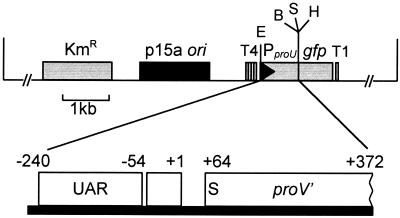
The proU promoter (PproU) from the pOSEX4 vector was cloned as a 612-bp EcoRI-BamHI fragment into the vector pPROBE-KT′ upstream from the gfp gene, creating a PproU-gfp transcriptional fusion; this plasmid was designated pPProGreen. The PproU-gfp fusion was flanked by T1 transcriptional terminators from the E. coli rrnB1 operon to prevent read-through transcription from other promoters on the plasmid (Fig. 1). The PproU-gfp fusion contained two regions of the proU promoter that were necessary for maximum osmoregulated expression, an upstream activating region that allows for maximum inducibility (32) and a downstream silencer that represses basal expression in low-osmolarity media (13) (Fig. 1). The requirement for these elements was confirmed in studies comparing the induction of PproU-gfp fusions containing distinct regions of the proU promoter (data not shown).
FIG. 1.
Map of pPProGreen (15.2 kb). The promoter proU was directionally cloned from the proU expression vector pOSEX4 (19) into the broad-host-range pPROBE-KT′ vector (38). The rrnB transcriptional terminators, with one copy (T1) downstream and four copies (T4) upstream of the fusion, surrounded the PproU-gfp fusion. The proU promoter contains an upstream activating region (UAR) and a silencer (S) located in the proV gene. The transcriptional (+1) and translational (+64) start sites are shown. All of the restriction enzyme sites shown, including HindIII (H), SalI (S), BamHI (B), and EcoRI (E), were unique in pPProGreen.
Two additional plasmids were constructed to confer constitutive gfp expression in control strains. Plasmids containing gfp driven by the promoter of the lactose operon (Plac) or the promoter of the neomycin phosphotransferase gene of Tn5 (PnptII) were constructed as described in Materials and Methods; these plasmids were designated pVLacGreen and pPNptGreen, respectively. Expression of PnptII-gfp resulted in detectable fluorescence in all three strains, i.e., BRT98(pPNptGreen) (143 RFU), DH5α(pPNptGreen) (65 RFU), and B728a(pPNptGreen) (34 RFU). In contrast, Plac-gfp resulted in detectable fluorescence in BRT98(pVLacGreen) (524 RFU) and in DH5α(pVLacGreen) (144 RFU) but not in B728a(pVLacGreen). Plac-gfp expression appeared to be constitutive in both DH5α(pVLacGreen) and BRT98(pVLacGreen) based on the absence of detectable induction by IPTG (isopropyl-β-d-thiogalactopyranoside) (data not shown). One possible reason for this may be because of the titration of the LacI repressor due to the presence of multiple (six to eight) copies of the plasmid (22).
Osmoinduction of PproU-gfp in broth-grown E. coli cells.
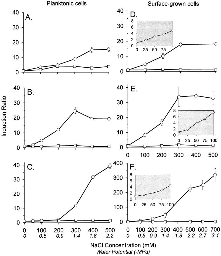
We characterized the effect of NaCl concentration on PproU-gfp expression in DH5α(pPProGreen) in broth-grown cells. Previous studies of PproU expression in S. enterica serovar Typhimurium examined induction by a single NaCl concentration (13, 19, 32) or by multiple concentrations (14, 19), with the latter demonstrating that PproU expression varies with NaCl concentration. We observed a consistent increase in PproU-gfp expression as the NaCl concentration in the growth medium increased from 0 to 400 mM, with a 15-fold increase at 400 mM NaCl (Fig. 2A). The proU promoter in DH5α(pPProGreen) was not induced by NaCl concentrations greater than 400 mM (Fig. 2A), as was observed previously in S. enterica serovar Typhimurium (14). DH5α(pPProGreen) did not grow at salt concentrations of greater than 500 mM NaCl.
FIG. 2.
Induction of PproU-gfp in cells that were grown in 1/2-21C broth (A, B, and C) or on 1/2-21C plates (D, E, and F) containing various concentrations of NaCl. (A and D) E. coli DH5α(pPProGreen) (○) and DH5α(pVLacGreen) (□); (B and E) P. agglomerans BRT98(pPProGreen) (○) and BRT98(pVLacGreen) (□); (C and F) P. syringae B728a(pPProGreen) (○) and B728a(pPNptGreen) (□). The insets show induction over a narrower range of NaCl concentrations. The fluorescence of the cells was measured with a fluorometer. The induction ratio was the mean fluorescence of the cells relative to the mean fluorescence of cells grown with 0 mM NaCl. Values represent the mean ± standard error of the mean (n = 3).
A time course study using fluorometry demonstrated that induction of PproU-gfp in DH5α(pPProGreen) occurred rapidly. In previous studies with E. coli and S. enterica serovar Typhimurium, PproU induction occurred within 10 min following exposure to NaCl (14, 33). We observed a detectable increase in PproU-gfp expression in DH5α(pPProGreen) within 50 min following the addition of NaCl to broth-grown cells (data not shown); the relatively long period probably resulted from the time required for GFP folding (18).
Osmoinduction of PproU-gfp in broth-grown P. agglomerans and P. syringae cells.
BRT98(pPProGreen) and DH5α(pPProGreen) were similar in their basal levels of PproU-gfp transcription in 1/2-21C broth. At NaCl concentrations of 200 mM or greater, PproU-gfp was induced more in BRT98(pPProGreen) than in DH5α(pPProGreen) (Fig. 2A and B). Similar to DH5α(pPProGreen), BRT98(pPProGreen) exhibited a threshold NaCl concentration above which it did not exhibit further increases in fluorescence (Fig. 2B), and it was inhibited in growth by NaCl concentrations of greater than 500 mM.
B728a(pPProGreen) exhibited a significantly lower basal level of PproU-gfp transcription in 1/2-21C broth (0 mM NaCl) than did either DH5α(pPProGreen) or BRT98(pPProGreen) (P < 0.05). For example, in one study, the fluorescence of B728a(pPProGreen) in 1/2-21C broth was 3.3 ± 0.3 RFU, whereas those of DH5α(pPProGreen) and BRT98(pPProGreen) were 33.3 ± 2.2 and 29.3 ± 0.3 RFU, respectively. Although the maximum level of PproU-gfp induction in B728a(pPProGreen), which was achieved with 500 mM NaCl (Fig. 2C), was significantly less than that in DH5α(pPProGreen) or in BRT98(pPProGreen), which were achieved with 400 and 300 mM NaCl, respectively (P < 0.05), the induction ratio for PproU-gfp in B728a(pPProGreen) was larger than those in the other strains at these NaCl concentrations (Fig. 2A to C). Similar to DH5α(pPProGreen) and BRT98(pPProGreen), B728a(pPProGreen) could not grow in 1/2-21C broth with NaCl concentrations of greater than 500 mM.
The fluorescence of the strains DH5α(pVLacGreen), BRT98(pVLacGreen), and B728a(pPNptGreen) was not affected by the NaCl concentration in the medium (Fig. 2A to C). This indicates that the increased fluorescence observed in the PproU-gfp-containing strains was due to PproU-gfp induction and not due to a direct effect of NaCl on the fluorescence of the GFP protein or to an effect of NaCl on some aspect of the cell's physiology that influenced GFP fluorescence.
Osmoinduction of PproU-gfp in surface-grown cells.
We compared the effects of NaCl concentration on PproU expression in planktonic (broth-grown) cells (Fig. 2A to C) versus surface-grown (plate-grown) cells (Fig. 2D to F). The basal level of PproU-gfp expression in cells grown on 1/2-21C plates was similar to that in cells grown in 1/2-21C broth (data not shown). In DH5α(pPProGreen) cells, PproU was induced more by NaCl concentrations of up to 400 mM in cells from plates than in cells from broth (Fig. 2A and D). Similarly, PproU was induced more in BRT98(pPProGreen) and in B728a(pPProGreen) in cells from plates than in cells from broth at comparable NaCl concentrations. In contrast to the two enteric species, BRT98(pPProGreen) and DH5α(pPProGreen), the strain B728a(pPProGreen) exhibited a dramatic increase in fluorescence as the NaCl concentration was increased from 300 to 700 mM in the plates (Fig. 2D to F), and B728a(pPProGreen) grew on plates at NaCl concentrations of greater than 500 mM.
To identify the lower limit of detection by the biosensors, we exposed BRT98(pPProGreen), DH5α(pPProGreen), and B728a(pPProGreen) to NaCl concentrations of between 0 and 100 mM. PproU-gfp expression in all three species detectably increased over the range from 25 to 100 mM NaCl (Fig. 2D to F, insets). In an additional experiment, the minimum NaCl concentration that significantly increased cellular fluorescence in DH5α(pPProGreen) and B728a(pPProGreen) was 10 mM, and that in BRT98(pPProGreen) was 15 mM (P < 0.05) (data not shown).
PproU-gfp induction by a diversity of solutes.
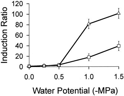
In addition to NaCl, the solutes KCl and Na2SO4 induced PproU-gfp expression in all three bacterial species over a range of water potentials (data not shown). For example, DH5α(pPProGreen) and BRT98(pPProGreen) cells were induced two- to threefold by NaCl, KCl, and Na2SO4 levels conferring a water potential of −1 MPa. PproU-gfp thus responded to high external osmolarity rather than to ion toxicity. The proU promoter similarly responded to high external osmolarity in S. enterica serovar Typhimurium (14). The strains BRT98(pPProGreen), DH5α(pPProGreen), and B728a(pPProGreen) were also grown on plates with the nonpermeating solute PEG 8000 (46), which limits water availability but does so by exerting pressure on the outer membrane rather than on the cytoplasmic membrane (16). PEG 8000 has been used as a dehydrating agent in many microbial and plant studies (see, e.g., references 2, 10, and 16) and is thought to better simulate conditions in low-water-content habitats than do permeating solutes. Although DH5α(pPProGreen) did not grow on 1/2-21C plates with PEG 8000, BRT98(pPProGreen) and B728a(pPProGreen) exhibited increasing PproU-gfp expression with increasing PEG 8000 concentrations (Fig. 3). In fact, the level of induction by PEG 8000 in BRT98(pPProGreen) was similar to the level of induction by NaCl at comparable water potentials (Fig. 2 and 3); for example, BRT98(pPProGreen) was induced 34- and 35-fold by −1.5-MPa conditions imposed by 335 mM NaCl and 33% PEG 8000, respectively (Fig. 2E and 3). In B728a(pPProGreen), PproU-gfp was induced approximately 48- and 100-fold by −1.5-MPa conditions imposed by NaCl and PEG 8000, respectively (Fig. 2F and 3).
FIG. 3.
Induction of PproU-gfp in cells that were grown on 1/2-21C plates containing various levels of PEG 8000. P. agglomerans BRT98(pPProGreen) (□) and P. syringae B728a(pPProGreen) (▵) were grown for 24 h, and the fluorescence intensity of cell suspensions was measured with a fluorometer. The induction ratio was the mean fluorescence of the cells relative to the mean fluorescence of cells grown with 0% PEG 8000. Values represent the mean ± standard error of the mean (n = 3).
Spatial distribution of induced biosensor cells on bean leaf surfaces.
The spatial distribution of P. agglomerans and P. syringae cells exhibiting constitutive gfp expression was evaluated to identify the leaf surface regions that were colonized by these species under both high- and low-moisture conditions. Although individual bacterial cells could often be visualized, the cells were also often in such close proximity to one another that the fluorescence appeared to be confluent. After 24 h of colonization under moist conditions, BRT98(pVLacGreen) cells were visible as individual cells on the planar regions of plant cell surfaces. BRT98(pVLacGreen) and B728a(pPNptGreen) cells were visible in epidermal cell junctions, particularly near veins, at the bases of the hairlike trichomes and glandular trichomes, and in water droplets. Microcolonies of BRT98(pVLacGreen) and B728a(pPNptGreen) were visible on both the abaxial and adaxial leaf surfaces. After 4 and 8 h of drying, i.e., at 4 and 8 h after transferring plants from a moisture tent to the laboratory bench, BRT98(pVLacGreen) and B728a(pPNptGreen) cells were visible as confluent fluorescence in some epidermal cells junctions, in trails between trichomes, and within circular regions that spanned multiple plant cells.
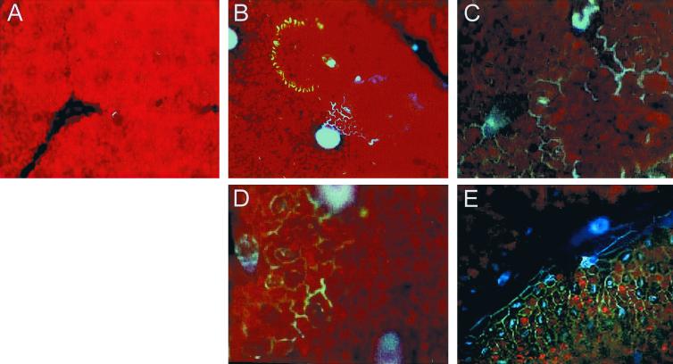
The spatial distribution of induced biosensor cells was evaluated based on the presence of visible fluorescence. After 24 h of colonization under moist conditions, neither BRT98(pPProGreen) nor B728a(pPProGreen) exhibited detectable fluorescence (Fig. 4A). After 4 h of drying, the BRT98(pPProGreen) and B728a(pPProGreen) cells in the epidermal cell junctions exhibited a gradient of increased fluorescence with increasing distance from the vein and sometimes exhibited a gradient of fluorescence in circular regions spanning multiple plant cells, with the most intense fluorescence near the outer edges of the rings (Fig. 4B). Although after 4 h of drying all of the regions that were visibly colonized with BRT98(pVLacGreen) and B728a(pPNptGreen) fostered induced cells of BRT98(pPProGreen) and B728a(pPProGreen), the induced biosensor cells were present in a smaller proportion of these sites than were the cells that constitutively expressed gfp, and fewer microcolonies were visible for B728a(pPProGreen) than for BRT98(pPProGreen) on the leaf surface. In contrast to BRT98(pVLacGreen) and BRT98(pPProGreen), B728a(pPNptGreen) and induced B728a(pPProGreen) cells were uniquely visible in lines radiating out from the glandular trichomes.
FIG. 4.
Spatial distribution of cells exhibiting fluorescence due to PproU-gfp induction on bean leaves (P. vulgaris). (A) P. agglomerans BRT98(pPProGreen) on a bean leaf after 24 h of incubation under moist conditions (no visible bacterial fluorescence); (B) P. agglomerans BRT98(pPProGreen) on a bean leaf 4 h after transfer of the plants from moist conditions to ambient conditions (i.e., after 4 h of drying); (C) P. syringae B728a(pPProGreen) on a bean leaf after 4 h of drying; (D) P. agglomerans BRT98(pPProGreen) on a bean leaf after 8 h of drying; (E) P. syringae B728a(pPProGreen) on a bean leaf after 8 h of drying. Leaves were examined by epifluorescence microscopy. The bacterial cells exhibited green or bluish-green fluorescence, the plant epidermal cells exhibited red autofluorescence, the leaf trichomes exhibited bluish-green (B to D) or blue (E) autofluorescence, and the stomata exhibited blue autofluorescence (E). Magnification, ×100.
Induction of PproU-gfp in BRT98(pPProGreen) on leaves.
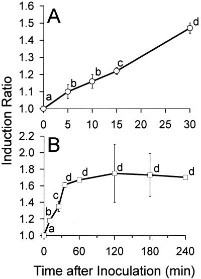
BRT98(pPProGreen) was used to investigate the extent to which water is available to bacteria on leaf surfaces under conditions that favored evaporation of water from the leaf surface. The mean fluorescence of the BRT98(pPProGreen) populations recovered from each of several leaves immediately following inoculation (0 min of drying) was used as a baseline measurement to determine the extent of induction at each subsequent sampling time. After the indicated drying time, the plants were enclosed in premoistened tents to prevent further drying and to allow for GFP maturation to a fluorescent state (49). Among the leaves sampled for each time point in experiment 1, there was relatively little leaf-to-leaf variation in the mean fluorescence of the recovered populations (Fig. 5A). The mean fluorescence of the cells recovered from each leaf consistently increased over the entire 30-min period, although visible water loss from the leaves did not occur until between 10 and 15 min after inoculation. In fact, the increase in the mean fluorescence was significant after only a 5-min drying period (P < 0.05), indicating that a macroscopic evaluation of the presence of water is a poor predictor of water availability at the microscopic level. By 30 min, the mean fluorescence of the population had increased 1.47-fold relative to that at 0 min (Fig. 5A). We generated a curve of the level of PproU-gfp induction in plate-grown BRT98(pPProGreen) cells at NaCl concentrations of 0 to 300 mM, using flow cytometry to measure fluorescence (data not shown). Based on this curve (y = 0.943e0.0089X, in which x = NaCl concentration and y = ratio of the fluorescence at a given NaCl concentration to that at 0 mM), at 30 min after inoculation, the average cell in the population recovered from leaves was exposed to a water potential that was equivalent to that imposed by approximately 55 mM NaCl. The variance of the fluorescence increased as the mean fluorescence increased, and the rate of this increase was similar to that observed with plate-grown cells. The mean forward scatter of the populations consistently decreased over the 30-min period examined, and a regression of the mean forward scatter over time showed that this decrease was significant (slope = −0.1, P = 0.01). Forward scatter reflects cell size (24), and thus this supports previous observations that bacterial cells decrease in size following their arrival on a leaf surface (J.-M. Monier and S. E. Lindow, Abstr. 7th Int. Symp. Microbiol. Aerial Plant Surfaces, abstr. 28, 2000; C. Riffaud and C. E. Morris, Abstr. 7th Int. Symp. Microbiol. Aerial Plant Surfaces, abstr. 35, 2000). This size change, however, was not related to their increased fluorescence, based on the absence of a significant correlation between mean fluorescence and mean forward scatter.
FIG. 5.
Induction of PproU-gfp in P. agglomerans BRT98(pPProGreen) cells recovered from bean leaves at various times following inoculation. (A) Experiment 1, in which the cells were monitored for 30 min (n = 3); (B) experiment 2, in which the cells were monitored for 240 min (n = 2). The induction ratio was the mean fluorescence of the cells relative to the mean fluorescence of the cells at 0 min. Values represent the mean ± standard error of the mean for n samples. Data points indicated by the same letter do not differ by Fisher's least-significant difference test (P < 0.05).
In a replicate experiment, experiment 2, the monitoring period was extended to 4 h, and the control strain BRT98(pVLacGreen) was included. Similar to in experiment 1, there was relatively little leaf-to-leaf variation at any given time point, at least within the first hour (Fig. 5B). The mean fluorescence of the cells recovered from each leaf consistently increased over the first 35 min. Subsequent incubation under ambient conditions did not result in significant increases in the mean fluorescence of the populations. Similar to in experiment 1, the mean fluorescence of the population significantly increased immediately following introduction of the bacterial cells onto the leaf surface (P < 0.05). In contrast to BRT98(pPProGreen), BRT98(pVLacGreen) did not exhibit a significant increase or decrease in fluorescence over the 4-h period examined. For example, at any given time point, the ratio of the mean fluorescence for BRT98(pVLacGreen) to that at 0 min was generally in the range of 0.9 to 1.1. The mean fluorescence of the BRT98(pPProGreen) cells recovered at 35 and 60 min had increased by 1.61- and 1.67-fold relative to that at 0 min (Fig. 5B), which corresponded to an exposure of the average cell in the populations to water potentials equivalent to those imposed by NaCl concentrations of 67 and 71 mM, respectively, at these times. The mean forward scatter of both the BRT98(pPProGreen) and BRT98(pVLacGreen) populations decreased significantly over time (for both strains, slope = −0.02 [P < 0.01]), suggesting that the cells decreased in size. Similar to in experiment 1, this size change was not related to the increased fluorescence, based on the absence of a significant correlation between the mean fluorescence and the mean forward scatter (P < 0.05).
DISCUSSION
We have developed GFP-based biosensors that can be used to quantify bacterial water deprivation. These biosensors allow us to measure the water availability actually sensed by bacteria in a given habitat, rather than simply the general water status of the habitat. Some key features of these biosensors include the following. They respond in a quantitative manner to water deprivation. They are sufficiently sensitive to detect water deprivation levels as low as those imposed by only 10 to 15 mM NaCl. Their detection range is moderately broad, with the upper limit approaching the water deprivation levels that inhibit growth for at least one bacterial species. They contain the PproU-gfp fusion on a multicopy, broad-host-range plasmid, and this effectively amplifies the visible response to water deprivation and facilitates its introduction into a variety of bacterial species. The high level of stability of this plasmid in the species tested, in both the absence and presence of high osmolarity, ensures its maintenance under conditions without antibiotic selection (data not shown). Finally, the absence of a requirement for specific regulatory elements for regulation of the proU promoter suggests that PproU is likely to respond to water deprivation in many bacterial species; this is supported by our finding that PproU-gfp responded to water deprivation in E. coli, P. agglomerans, and P. syringae, as well as in Xanthomonas campestris pv. Vesicatoria (unpublished data).
A key assumption with these biosensors is that cellular fluorescence reflects the activity of the proU promoter. This is supported by the fact that exposure of the biosensor cells to NaCl concentrations of from 0 to 300 mM correlated with both an increase in the amount of GFP protein present, as detected by Western blotting (data not shown), and an increase in cellular fluorescence. More importantly, for the control cells, which were the same strains as the biosensors but which exhibited constitutive gfp expression, exposure to NaCl concentrations of from 0 to 500 mM did not cause detectable changes in cellular fluorescence. This indicates that the cellular fluorescence of the strains was not significantly influenced by water deprivation-induced changes in cell physiology. Such changes include a decreased rate of growth (26), which was observed with B728a at high NaCl concentrations but was not associated with a change in the fluorescence of B728a(pPNptGreen).
This is the first report that the proU promoter from E. coli functions in species other than E. coli and S. enterica serovar Typhimurium. The basal level of PproU expression in P. syringae was lower than that in E. coli, but the levels were similar in P. agglomerans and E. coli. This could be due to the effect of poor promoter recognition or low transcriptional efficiency of an E. coli promoter in P. syringae. The osmoresponsiveness of the proU promoter in P. syringae provides support for the hypothesis that proU osmoregulation does not require specific regulatory proteins, although it is possible that P. syringae provided such proteins.
For all three strains tested, plate-grown cells exhibited higher levels of induction than broth-grown (i.e., planktonic) cells at comparable NaCl concentrations. Surface-grown cells are known to differ physiologically from planktonic cells (42). In fact, this difference has been demonstrated to influence bacterial survival following introduction onto plant leaves (51). In this study, the similarity in the levels of PproU-gfp expression in broth- and plate-grown cells when NaCl was absent indicated that surface-associated and planktonic cells differed not in their basal proU expression but rather in the extent of proU induction. In most terrestrial habitats, bacterial cells are present on surfaces, and thus they are likely to be more similar physiologically to surface-grown than planktonic cells.
As a model system for evaluating the effectiveness of the bacterial biosensors, we examined bacterial exposure to water deprivation during the period of evaporation that followed bacterial introduction onto aerial leaf surfaces. We reasoned that during this period, the visible water film on a leaf disappears, and thus many of the bacteria on the leaf should become limited for water. We focused on water availability specifically following an inoculation event because we could control the presence of water on a leaf during this period and because it seemed highly probable that at least some of the inoculum cells would arrive in sites that would not support the continued presence of water.
The microscopy studies demonstrated that bacteria were exposed to water deprivation on bean leaves under the conditions tested, and they illustrated the spatial distribution of these cells. Only a subset of the colonized sites contained biosensor cells that were exposed to sufficient levels of water deprivation to cause detectable fluorescence. We observed a gradient of decreasing water availability with increasing distance from a vein, demonstrating that the proximity of cells to a vein, where water likely collects, influenced their access to water. The circular regions of fluorescence on the leaves suggest that as the water droplets on a leaf surface evaporated, the bacteria in the receding edge of those droplets became limited for water.
We used flow cytometry to quantify bacterial exposure to water deprivation on leaves; in these studies we examined the fluorescence of each cell in the populations recovered from the leaves. We used BRT98(pPProGreen) because it exhibited a sufficiently high basal level of fluorescence to allow uninduced cells to be distinguished from the indigenous bacteria and particulates that were also recovered from the leaf surface. E. coli DH5α was of less interest because it is not a normal inhabitant of leaf surfaces, and P. syringae B728a(pPProGreen) exhibited a basal level of fluorescence that was too low to effectively separate uninduced cells from the indigenous bacteria. Future studies with this strain, and other strains that similarly exhibit a low basal level of proU expression, will involve introduction of a second fluorescence marker. The consistent level of fluorescence of the control strain BRT98(pVLacGreen) on leaves indicated that the bacteria did not undergo physiological changes that affected their fluorescence, nor were they exposed to environmental conditions that detrimentally affect GFP fluorescence. Such conditions include very low pH (4 to 5.5) and high pH (>12) (7), strong reducing agents (21), and hydrogen peroxide concentrations of greater than 1% (21). The significant increase in the fluorescence of the BRT98(pPProGreen) cells in the 30- to 60-min period following inoculation indicated that many cells were limited for water during this period, as was predicted. The relatively low leaf-to-leaf variability suggests uniformity in the macroenvironment of the leaves, which may have resulted, in part, from a very low leaf density in the pots. The low variability further suggests that biologically based differences in leaf-water relations were relatively small among the leaves.
We predicted that the variability in water availability among microsites on a leaf should be reflected in the variability in fluorescence. We assumed that the variance in the fluorescence values of a population of cells from a leaf is composed of two components, the variance inherent to cells following induction to a given level of water deprivation and the variance that results from the heterogeneity among the microsites in water availability. We estimated the former by examining the variance of cells in a homogenous habitat, i.e., in a laboratory culture. We compared the rates at which the variance increased as the mean fluorescence of the population increased and found that both the cultured cells and the cells from leaves exhibited the same rate of increase over a range of mean fluorescence values. The absence of a detectable increase in the variance in fluorescence of cells from leaves over that of cells in culture indicates that under the conditions tested, the heterogeneity in water availability among the microsites on a leaf, which was detected by microscopy, was not detectable in the flow cytometry analysis of the cells removed from leaves. The two methods of examining the biosensor cells thus each provided unique information. The simplest explanation for a relatively uniform rate of increase in fluorescence across a population is that the decreased water availability following inoculation resulted primarily from the dissolution of solutes that were on the leaf surface preceding inoculation and that the subsequent decreases in water availability resulted from an increase in the concentration of solutes following evaporation.
The level of induction of the average cell in the population indicated that the cells were exposed to a water potential equivalent to that imposed by only 50 to 60 mM NaCl in the 30-min period following arrival on the leaf surface. This is a fairly low level of water deprivation; i.e., it is not sufficiently high to cause cell death or perhaps even to reduce growth. If we assume that bacteria and solutes are uniformly distributed throughout a water layer on a leaf and that solutes remain on a leaf when the water evaporates, then an increase in water deprivation equivalent to that imposed by an increase from 19.2 to 55 mM NaCl, which occurred in this study between 5 and 30 min following inoculation, would correspond to a 65% decrease in the water volume on the leaf in that 25-min period. We have estimated that bean leaves with an area of 50 cm2 hold about 0.5 g of water. If that water was evenly spread across the surface of the leaf, then a 65% decrease in water volume would correspond to a decrease in the water film thickness from 100 to 35 μm.
Macroscale measurements of the water potential of plant leaves are highly variable but are generally much lower than the levels sensed by the biosensor on the leaves in this study. On average, the bacteria on the leaves in this study were exposed to water potentials in the range of −0.05 to −0.42 MPa. In contrast, in published studies, leaf psychrometer measurements of leaf water potential ranged from −0.2 to −2.07 MPa, depending on the species, time of day, and location of the leaf in the canopy (9, 11, 23, 25, 36). The fact that the bacteria in this study were generally exposed to less stressful levels of water deprivation than would be predicted by these leaf psychrometer measurements suggests that the bacteria localized to microsites on leaves that were particularly well hydrated.
In summary, these studies demonstrated that a PproU-gfp fusion-based biosensor was effective at making microscale measurements of water availability to bacteria on bean leaves. The measurements indicated that under the environmental conditions selected, bacteria on the leaves were exposed, on average, to water potentials of as low as −0.42 MPa. This exposure level was unlikely to limit bacterial growth or survival on leaves, based on bacterial growth under water deprivation conditions on plates. Potential uses of these biosensors include evaluating the role of water deprivation in bacterial population dynamics on plants and in other natural habitats and identifying the effect of exopolysaccharides and biofilm formation on bacterial exposure to water deprivation.
Acknowledgments
We thank Kristi Harkins and Donghui Cheng for assistance with the flow cytometer, Philip Dixon for advice on statistics, and Amy Groen, Claudia Lemper, and Jeness Petersen for assistance in constructing plasmids.
This work was supported by grant 9974059 from the National Science Foundation and by Hatch Act and State of Iowa funds.
Footnotes
Paper no. 19692 of the Iowa Agriculture and Home Economics Experiment Station, Ames (project no. 3433).
REFERENCES
- 1.Andersen, J. B., C. Sternberg, L. K. Poulsen, S. P. Bjorn, M. Givskov, and S. Molin. 1998. New unstable variants of green fluorescent protein for studies of transient gene expression in bacteria. Appl. Environ. Microbiol. 64:2240-2246. [DOI] [PMC free article] [PubMed] [Google Scholar]
- 2.Bajii, M., S. Lutts, and J. M. Kinet. 2000. Physiological changes after exposure to and recovery from polyethylene glycol-induced water deficit in roots and leaves of durum wheat (Triticum durum Desf.) cultivars differing in drought resistance. J. Plant Physiol. 157:100-108. [Google Scholar]
- 3.Beattie, G. A., and L. M. Marcell. Comparative dynamics of adherent and non-adherent bacterial populations on maize leaves. Phytopathology, in press. [DOI] [PubMed]
- 4.Belkin, S., D. R. Smulski, A. C. Vollmer, T. K. Van Dyk, and R. A. LaRossa. 1996. Oxidative stress detection with Escherichia coli harboring a katG′::lux fusion. Appl. Environ. Microbiol. 62:2252-2256. [DOI] [PMC free article] [PubMed] [Google Scholar]
- 5.Berg, D. E. 1989. Transposon Tn5, p. 185-210. In D. E. Berg and M. M. Howe (ed.), Mobile DNA. American Society for Microbiology, Washington, D.C.
- 6.Better, M., and D. R. Helinski. 1983. Isolation and characterization of the recA gene of Rhizobium meliloti. J. Bacteriol. 155:311-316. [DOI] [PMC free article] [PubMed] [Google Scholar]
- 7.Bokman, S. H., and W. W. Ward. 1981. Renaturation of Aequorea green fluorescent protein. Biochem. Biophys. Res. Commun. 101:1372-1380. [DOI] [PubMed] [Google Scholar]
- 8.Bringhurst, R. M., Z. G. Cardon, and D. J. Gage. 2001. Galactosides in the rhizosphere: utilization by Sinorhizobium meliloti. Proc. Natl. Acad. Sci. USA 98:4540-4545. [DOI] [PMC free article] [PubMed] [Google Scholar]
- 9.Brown, R. W., and W. T. McDonough. 1977. Thermocouple psychrometer for in situ leaf water potential determinations. Plant Soil 48:5-10. [Google Scholar]
- 10.Busse, M. D., and P. J. Bottomly. 1989. Growth and nodulation responses of Rhizobium meliloti to water stress induced by permeating and nonpermeating solutes. Appl. Environ. Microbiol. 55:2431-2436. [DOI] [PMC free article] [PubMed] [Google Scholar]
- 11.Campbell, C. S., and K. J. McInnes. 1999. Response of in situ psychrometer to cuticle removal by abrasion. Agron. J. 91:859-862. [Google Scholar]
- 12.Cha, H. J., R. Srivastava, V. N. Vakharia, G. Rao, and W. E. Bentley. 1999. Green fluorescent protein as a noninvasive stress probe in resting Escherichia coli cells. Appl. Environ. Microbiol. 65:409-414. [DOI] [PMC free article] [PubMed] [Google Scholar]
- 13.Dattananda, C. S., K. Rajkumari, and J. Gowrishankar. 1991. Multiple mechanisms contribute to osmotic inducibility of proU operon expression in Escherichia coli: demonstration of two osmoresponsive promoters and of a negative regulatory element within the first structural gene. J. Bacteriol. 173:7481-7490. [DOI] [PMC free article] [PubMed] [Google Scholar]
- 14.Dunlap, V. J., and L. N. Csonka. 1985. Osmotic regulation of l-proline transport in Salmonella typhimurium. J. Bacteriol. 163:296-304. [DOI] [PMC free article] [PubMed] [Google Scholar]
- 15.Elasri, M. O., and R. V. Miller. 1999. Study of the response of a biofilm bacterial community to UV radiation. Appl. Environ. Microbiol. 65:2025-2031. [DOI] [PMC free article] [PubMed] [Google Scholar]
- 16.Halverson, L. J., and M. K. Firestone. 2000. Differential effects of permeating and nonpermeating solutes on the fatty acid composition of Pseudomonas putida. Appl. Environ. Microbiol. 66:2414-2421. [DOI] [PMC free article] [PubMed] [Google Scholar]
- 17.Harris, R. F. 1981. Effect of water potential on microbial growth and activity, p. 23-96. In J. F. Parr, W. R. Gardner, and L. F. Elliot (ed.), Water potential relations in soil microbiology. Soil Science Society of America, Madison, Wis.
- 18.Heim, R., A. B. Cubitt, and R. Y. Tsien. 1995. Improved green fluorescence. Nature 373:663-664. [DOI] [PubMed] [Google Scholar]
- 19.Herbst, B., S. Kneip, and E. Bremer. 1994. pOSEX: vectors for osmotically controlled and finely tuned gene expression in Escherichia coli. Gene 151:137-142. [DOI] [PubMed] [Google Scholar]
- 20.Hojberg, O., U. Schnider, H. V. Winteler, J. Sorensen, and D. Haas. 1999. Oxygen-sensing reporter strain of Pseudomonas fluorescens for monitoring the distribution of low-oxygen habitats in soil. Appl. Environ. Microbiol. 65:4085-4093. [DOI] [PMC free article] [PubMed] [Google Scholar]
- 21.Inouye, S., and F. I. Tsuji. 1994. Aequorea green fluorescent protein. Expression of the gene and fluorescence characteristics of the recombinant protein. FEBS Lett. 341:277-280. [DOI] [PubMed] [Google Scholar]
- 22.Itoh, Y., J. M. Watson, D. Haas, and T. Leisinger. 1984. Genetic and molecular characterization of the Pseudomonas plasmid pVS1. Plasmid 11:206-220. [DOI] [PubMed] [Google Scholar]
- 23.Kirkham, M. B. 1986. Water relations of the upper and lower surfaces of maize leaves. Biol. Plant. 28:249-257. [Google Scholar]
- 24.Koch, A. L., B. R. Robertson, and D. K. Button. 1996. Deduction of the cell volume and mass from forward scatter intensity of bacteria analyzed by flow cytometry. J. Microbiol. Methods 27:49-61. [Google Scholar]
- 25.Krenzer, E. G., T. L. Nipp, and R. W. McNew. 1991. Winter wheat mainstem leaf appearance and tiller formation vs. moisture treatment. Agron. J. 83:663-667. [Google Scholar]
- 26.Leveau, J. H., and S. E. Lindow. 2001. Predictive and interpretive simulation of green fluorescent protein expression in reporter bacteria. J. Bacteriol. 183:6752-6762. [DOI] [PMC free article] [PubMed] [Google Scholar]
- 27.Leveau, J. H. J., and S. E. Lindow. 2001. Appetite of an epiphyte: quantitative monitoring of bacterial sugar consumption in the phyllosphere. Proc. Natl. Acad. Sci. USA 98:3446-3453. [DOI] [PMC free article] [PubMed] [Google Scholar]
- 28.Lindow, S. E. 1995. The use of reporter genes in the study of microbial ecology. Mol. Ecol. 4:555-566. [Google Scholar]
- 29.Loper, J. E., and S. E. Lindow. 1994. A biological sensor for iron available to bacteria in their habitats on plant surfaces. Appl. Environ. Microbiol. 60:1934-1941. [DOI] [PMC free article] [PubMed] [Google Scholar]
- 30.Loper, J. E., and S. E. Lindow. 1987. Lack of evidence for in situ fluorescent pigment production by Pseudomonas syringae pv. syringae on bean leaf surfaces. Phytopathology 77:1449-1454. [Google Scholar]
- 31.Lucht, J. M., and E. Bremer. 1994. Adaptation of Escherichia coli to high osmolarity environments: osmoregulation of the high-affinity glycine betaine transport system proU. FEMS Microbiol. Rev. 14:3-20. [DOI] [PubMed] [Google Scholar]
- 32.Lucht, J. M., and E. Bremer. 1991. Characterization of mutations affecting the osmoregulated proU promoter of Escherichia coli and identification of 5′ sequences required for high-level expression. J. Bacteriol. 173:801-809. [DOI] [PMC free article] [PubMed] [Google Scholar]
- 33.Lucht, J. M., P. Dersch, B. Kempf, and E. Bremer. 1994. Interactions of the nucleoid-associated DNA-binding protein H-NS with the regulatory region of the osmotically controlled proU operon of Escherichia coli. J. Biol. Chem. 269:6578-6586. [PubMed] [Google Scholar]
- 34.May, G., E. Faatz, J. M. Lucht, M. Haardt, M. Bolliger, and E. Bremer. 1989. Characterization of the osmoregulated Escherichia coli proU promoter and identification of ProV as a membrane-associated protein. Mol. Microbiol. 3:1521-1531. [DOI] [PubMed] [Google Scholar]
- 35.Mazodier, P., P. Cossart, E. Giraud, and F. Gasser. 1985. Completion of the nucleotide sequence of the central region of Tn5 confirms the presence of three resistance genes. Nucleic Acids Res. 13:195-205. [DOI] [PMC free article] [PubMed] [Google Scholar]
- 36.McCully, M. E. 1999. Root xylem embolisms and refilling. Relation to water potential of soil, roots, and leaves, and osmotic potentials of root xylem sap. Plant Physiol. 119:1001-1008. [DOI] [PMC free article] [PubMed] [Google Scholar]
- 37.Miller, J. H. 1972. Experiments in molecular genetics. Cold Spring Harbor Laboratory, Cold Spring Harbor, N.Y.
- 38.Miller, W. G., J. H. J. Leveau, and S. E. Lindow. 2000. Improved gfp and inaZ broad-host-range promoter-probe vectors. Mol. Plant-Microb. Interact. 13:1243-1250. [DOI] [PubMed] [Google Scholar]
- 39.Miller, W. G., and S. E. Lindow. 1997. An improved GFP cloning cassette designed for prokaryotic transcriptional fusions. Gene 191:149-153. [DOI] [PubMed] [Google Scholar]
- 40.O'Brien, R. D., and S. E. Lindow. 1989. Effects of plant species and environmental conditions on epiphytic population sizes of Pseudomonas syringae and other bacteria. Phytopathology 79:619-627. [Google Scholar]
- 41.Prasher, D. C., V. E. Eckenrode, W. W. Ward, F. G. Prendergast, and M. J. Cormier. 1992. Primary structure of the Aequorea victoria green-fluorescent protein. Gene 111:229-233. [DOI] [PubMed] [Google Scholar]
- 42.Prigent-Combaret, C., O. Vidal, C. Dorel, and P. Lejeune. 1999. Abiotic surface sensing and biofilm-dependent regulation of gene expression in Escherichia coli. J. Bacteriol. 181:5993-6002. [DOI] [PMC free article] [PubMed] [Google Scholar]
- 43.Ptitsyn, L. R., G. Horneck, O. Komova, S. Kozubek, E. A. Krasavin, M. Bonev, and P. Rettberg. 1997. A biosensor for environmental genotoxin screening based on an SOS lux assay in recombinant Escherichia coli cells. Appl. Environ. Microbiol. 63:4377-4384. [DOI] [PMC free article] [PubMed] [Google Scholar]
- 44.Selifonova, O., R. Burlage, and T. Barkay. 1993. Bioluminescent sensors for detection of bioavailable Hg(II) in the environment. Appl. Environ. Microbiol. 59:3083-3090. [DOI] [PMC free article] [PubMed] [Google Scholar]
- 45.Smibert, R. M., and N. R. Kreig. 1994. Phenotypic characterization, p. 607-654. In P. Gerhardt, R. G. E. Murray, W. A. Wood, and N. R. Kreig (ed.), Methods for general and molecular bacteriology. American Society for Microbiology, Washington, D.C.
- 46.Steuter, A. A., A. Mozafar, and J. R. Goodin. 1981. Water potential of aqueous polyethylene glycol. Plant Physiol. 67:64-67. [DOI] [PMC free article] [PubMed] [Google Scholar]
- 47.Stiner, L., and L. J. Halverson. 2002. Development and characterization of a green fluorescent protein-based bacterial biosensor for bioavailable toluene and related compounds. Appl. Environ. Microbiol. 68:1962-1971. [DOI] [PMC free article] [PubMed] [Google Scholar]
- 48.Tauriainen, S., M. Karp, W. Chang, and M. Virta. 1997. Recombinant luminescent bacteria for measuring bioavailable arsenite and antimonite. Appl. Environ. Microbiol. 63:4456-4461. [DOI] [PMC free article] [PubMed] [Google Scholar]
- 49.Tsein, R. Y. 1998. The green fluorescent protein. Annu. Rev. Biochem. 67:509-544. [DOI] [PubMed] [Google Scholar]
- 50.van der Bij, A. J., L. A. de Weger, W. T. Tucker, and B. J. J. Lugtenberg. 1996. Plasmid stability in Pseudomonas fluorescens in the rhizosphere. Appl. Environ. Microbiol. 62:1076-1080. [DOI] [PMC free article] [PubMed] [Google Scholar]
- 51.Wilson, M., and S. E. Lindow. 1993. Effect of phenotypic plasticity on epiphytic survival and colonization by Pseudomonas syringae. Appl. Environ. Microbiol. 59:410-416. [DOI] [PMC free article] [PubMed] [Google Scholar]


