

ABSTRACT
Introduction
Varicocele is a correctable cause of male infertility with management options including catheter embolisation of the testicular vein, done by Interventional Radiologists. Varicoceles can present in childhood and adolescence with scrotal pain or discomfort, or may be subclinical. It involves dilatation of the pampiniform plexus related to congestion and retrograde flow in the testicular vein. If left untreated, the pain and discomfort may impact the quality of life of the paediatric patient, and in the long term, infertility may ensue. However, limited data are available on the long‐term clinical outcomes and complications of Varicocele embolisation in paediatric patients.
Method
Retrospective cohort study of 14 years of endovascular Varicocele embolisation at a single‐operator tertiary paediatric centre.
Results
Eighty‐four patients with a median age of 14 years (range 8–17 years) were identified. The varicoceles were predominantly of grade 1 or 2. 99% of patients had unilateral left‐sided varicoceles. One patient had bilateral varicoceles. This study demonstrated that the technical success rate of endovascular embolisation is 94% and the clinical success rate is 98%. 84% reported feeling well in the immediate postprocedural period. 86% of cases reported excellent long‐term progress. A small proportion (10%) experienced long‐term discomfort or pain. There were no cases of relapse.
Conclusion
Endovascular embolisation is a successful technique for the management of Varicocele in paediatric patients, with good immediate and long‐term clinical outcomes. It has low recurrence rates.
Keywords: adolescent, child, embolisation, interventional radiology, varicocele
1. Introduction
There is limited data, particularly long‐term outcomes, on the clinical efficacy of endovascular embolisation of varicocele in the paediatric population. Varicocele is a common treatable cause of male infertility. Though it may manifest at any age in males, it often presents in childhood or young adulthood [1]. The prevalence of varicocele in the age range of 2–19 years old ranges between 0.79%–14.1% [2]. It may be clinically asymptomatic, but in some patients, varicocele can cause pain and discomfort [3]. In the long term, it may impact the patient's quality of life and result in infertility [4].
1.1. Background
Varicocele is the abnormal dilatation of the pampiniform plexus, a network of venous sinuses that drains the testes and epididymides. Scrotal varicoceles can result in testicular atrophy, including in the paediatric population [2, 5]. The management of varicocele is either by endovascular embolisation or surgery. Microdissection is one of the techniques used by paediatric surgeons for the management of varicocele [6]. Endovascular embolisation is a minimally invasive treatment method performed by Interventional Radiologists to treat varicoceles. In the past two decades, embolisation has predominantly been the mainstay of treatment. Endovascular embolisation has success rates of above 90% and recurrence rates of 0%–4% [7, 8]. Surgery has slightly higher recurrence rates of 11% [9]. The benefits of the endovascular approach compared to surgical treatment include a less invasive procedure, reduced recovery time, and comparable costs [10]. The potential risks associated with endovascular embolisation include an incomplete result, damage to adjacent structures (particularly the testis, vascular structures), inability to reverse pre‐existing injury to the testis, contrast anaphylaxis, and radiation exposure. Potential complaints post‐embolisation include testicular pain, testicular haematoma, and vascular access site pain and haematoma [11].
1.2. Aims of This Study
The aims of this study are to evaluate the technical success rate and immediate and long‐term clinical outcomes of endovascular paediatric varicocele management at our institution.
2. Methods
2.1. Study Design
This is a retrospective cohort study done at a single‐operator paediatric institution. The approval for this study was provided by the local institutional review board. This study follows the STROBE checklist.
2.2. Setting and Data Collection
The study covered the period from March 2010 to May 2024. Patients were identified by clinical records and procedure reports. Data collected include patient demographics (age), clinical presentation, testicular size (volume) on the ipsilateral side and contralateral side on ultrasound, and varicocele diameter from pre‐treatment grey‐scale and colour Doppler ultrasound. Varicoceles are graded based on size on ultrasound: grade 1 (3–4 mm), grade 2 (4–5 mm), grade 3 (> 5 mm) [12]. Follow‐up data collected by telephone call with the patients/patients' parents include: immediate (within two days) postoperative clinical issues, long‐term complications/clinical issues (up to eight years follow‐up), post‐embolisation testicular volume (both ipsilateral and contralateral testes) on ultrasound, and recurrence on the basis of imaging. Post‐treatment ultrasounds are done only in cases where it is clinically indicated or for other unrelated clinical reasons. The short‐term clinical outcome is measured by the successful embolisation of the varicocele. Long‐term clinical outcome is measured by any complications/clinical issues and recurrence on the basis of imaging.
2.3. Inclusion and Exclusion Criteria
Inclusion criteria: Patients under 18 years of age with varicocele who have undergone endovascular treatment at the local institution.
Exclusion criteria: Nil.
2.4. Technique and Treatment Approach
Embolisation has been the procedure of choice at our institution. In children, the procedure is usually performed under general anaesthesia. In the earlier patients of this cohort, vascular access was initially gained via the right common femoral vein and changed to the right internal jugular vein (a more direct approach) and, most recently, via the basilic vein, largely due to patient preference. A 4‐French system is generally used to access the left testicular vein. Depending on the size of the varicocele, two or three coils are usually deployed. Embolisation is performed with detachable/retractable coils. The range of coils used included Boston Scientific Interlock coils (Boston Scientific Corporation, Marlborough, MA, USA) and Cook Retracta Detachable Embolisation Coil (Cook Medical LLC, Bloomington, IN, USA). Currently, the coils used are Penumbra Ruby Detachable coils (Penumbra Inc., Alameda, CA, USA). In addition to coils, sodium tetradecyl sulfate (Fibrovein) 3% foam is administered to primarily occlude collateral vessels, which sometimes were not visible on venography. Post‐coil venography is done to confirm complete closure of the vein. An example case is provided in Figure 1. During foam injection, retrograde flow towards the testis is occluded with compression using an abdominal compression device (F. Spoon Co., Dover, MA, USA) (Figure 2). Radiation dose is kept as low as reasonably achievable, with pulsed fluoroscopy and venography and arterial runs at 1 frame every 2 s, with 4 to 5 runs every procedure. Patients may experience testicular pain, testicular haematoma, and vascular access site pain and haematoma post‐embolisation [11].
FIGURE 1.

A 15‐year‐old male underwent left varicocele embolisation with access via the right basilic vein. (a) Selective catheterisation of the left testicular vein was performed using a ProGreat 2.7 French catheter. (b) Venography of the left testicular vein demonstrated several small vessels supplying the left‐sided varicocele. (c) The vessels supplying the varicocele were sclerosed using Fibrovein 3% (2 mL) foam, with coiling of the main supply vessel with three Penumbra Ruby coils (2 × 12 × 60 mm standard, 1 × 10 × 40 mm soft coil).
FIGURE 2.
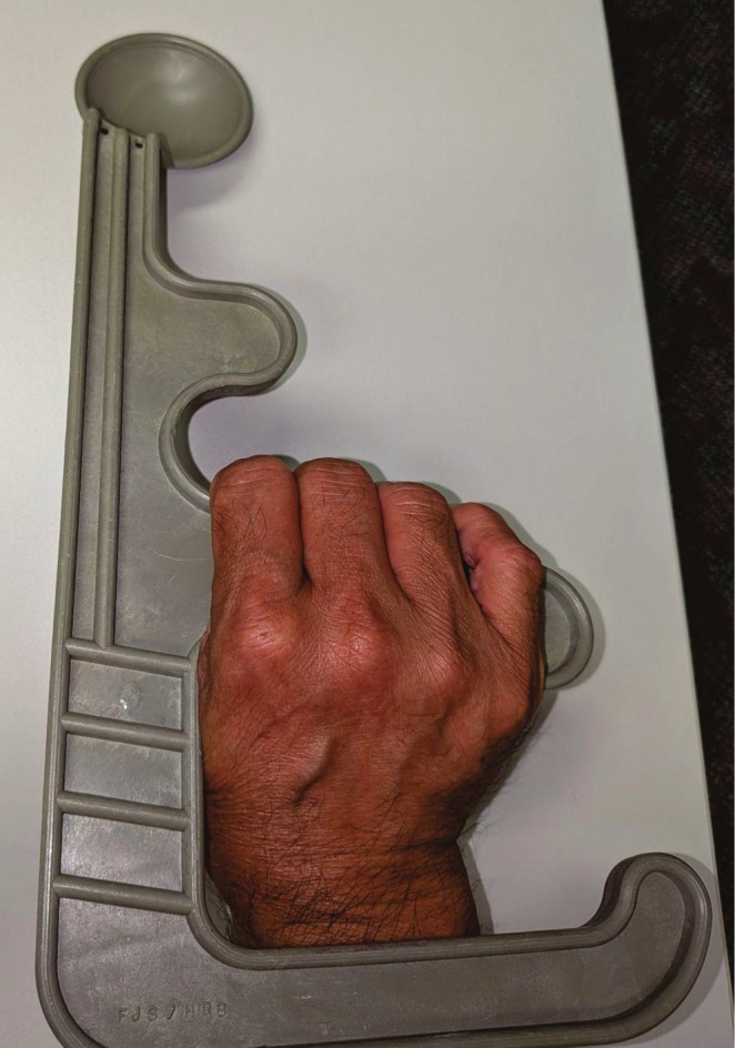
Photograph of the abdominal compression device (F. Spoon Co., Dover, MA, USA) used by this study's Interventional Radiologist during the endovascular varicocele embolisation procedure.
Patients are discharged within two hours of the procedure and are followed up by a telephone call two days later and then by the Paediatric Surgeon. Post‐embolisation ultrasound is generally not needed. In some of the cases where there are patient concerns relating to the procedure or postprocedural complications, ultrasound may be done postoperatively for confirmation of resolution, detection of recurrence, if any, and to measure testicular volume.
3. Results
Over an approximate 14‐year period, 87 cases (84 patients) were identified, with two patients excluded as they were above the age inclusion criteria (ages 24 years old and 38 years old) (Table 1). The median age at the time of procedure was 14 years. The age range was 8 to 17 years old. 83 out of 84 patients had unilateral left‐sided varicoceles with predominantly either grade 1 or 2 varicoceles (84%) [13]. One patient had bilateral varicoceles, with only the left‐sided varicocele treated and a small right‐sided varicocele, which was not treated.
TABLE 1.
Summary statistics of patient demographics.
| Number of cases (number of patients) | 87 (84) |
| Age (years old), median (range) | 14 (8–17) |
| Varicocele on the left side | 100% |
| Unilateral left‐sided varicoceles | 99% |
| Varicocele diameter (mm), median (range) | 4 (1.5–7.3) |
| Varicocele grade (n = 53) | |
| Grade 1 | 25 (47%) |
| Grade 2 | 25 (47%) |
| Grade 3 | 3 (6%) |
| Pre‐existing left testicular atrophy on imaging | 19/44 (43%) |
Information on the varicocele diameter was available in 53 cases. The range of varicocele diameter as measured on ultrasound images was between 1.5 mm and 7.3 mm, with a median of 4 mm.
Varicocele grade distribution was as follows: grade 1 (47%), grade 2 (47%), grade 3 (6%). Prior to varicocele embolisation, 43% of cases had testicular atrophy on the side of the varicocele (defined as at least 20% size difference between the left and right testes) (Table 1).
Of the 72 cases where vascular access site was documented, 31 (43%) were via the right internal jugular vein, 9 (13%) via the right common femoral vein, 26 (36%) via the right basilic vein, 5 (7%) via the right brachial vein, and 1 (1%) via the left internal jugular vein.
Sixty‐six cases had the number of coils used for embolisation documented. The number of cases that used no coils was 6, one coil was 14, two coils 22, three coils 12, four coils 9, six coils 1, and nine coils 1. The cases in which no coils were deployed were either related to adequate embolisation using Fibrovein (i.e., no need for coils), the testicular veins being too small for coils, or the testicular vein could not be catheterised.
Technical success was achieved in 94% of cases. Five cases were unsuccessful. Three of these cases had significant vasospasm, which were re‐attempted, with subsequent successful second attempts and included in the total count of cases. Another two cases were unsuccessful as the testicular vein could not be located on venography: single renal vein with expected position of left testicular vein cannulated, but on venogram runs only the left lumbar plexus was opacified (one case), and the left testicular vein could to be catheterised (one case). Therefore, 82 out of 84 patients had successful embolisation, resulting overall clinical success rate of 98%.
Of the 58 cases with documented immediate postoperative outcome, 49 (84%) had no immediate postoperative issues, eight patients (14%) experienced pain (mostly mild, with one case admitted overnight for monitoring, no severe outcome), and one patient (2%) experienced fatigue. The long‐term clinical outcomes are documented in Table 2, with 86% of cases demonstrating no symptoms on long‐term follow‐up. There were no cases of relapse.
TABLE 2.
Postprocedural immediate and long‐term clinical outcomes.
| Immediate postoperative outcome (n = 58) | |
| Well | 49 (84%) |
| Pain | 8 (14%) |
| Fatigue | 1 (2%) |
| Long‐term progress (n = 71) | |
| No symptoms | 61 (86%) |
| Relapse | 0 (0%) |
| Discomfort/ache/pain | 7 (10%) |
| N/A—recent procedure | 3 (4%) |
Of the 18 cases with smaller measured pre‐treatment ipsilateral testicular volume (defined by at least a 20% difference between testicular size on both sides), five cases demonstrated apparent resolution of the 20% or greater difference in testicular size.
Of the 15 patients who underwent ultrasound both prior to and following varicocele treatment, three patients had new measurements of ipsilateral smaller testes post‐treatment. Five patients did not have smaller measured testes post‐treatment.
Twenty patients underwent post‐procedure ultrasound, with seven patients demonstrating testicular atrophy on the varicocele side.
The majority of cases reported excellent immediate and long‐term progress (i.e., feeling well with no discomfort, ache, or relapse) (84% and 86% respectively). One patient reported pain in the left lateral abdomen and later pain in the scrotum on the day of the procedure. This patient was admitted overnight for monitoring, mainly because of patient/parental anxiety. The patient had no severe outcome and was discharged the following day. There were no cases of relapse. A small proportion (10%) experienced long‐term discomfort or pain.
4. Discussion
This study fills some of the knowledge gaps in existing literature where there is a paucity of data on the endovascular management of paediatric varicoceles, particularly long‐term outcomes. The results of this study over 14 years demonstrated that endovascular embolisation of varicocele in the paediatric population (median age 14 years) is considered a technically successful procedure. The technical success rate of 94% in this study is in line with the literature in both adults and paediatric populations [7, 8, 9]. A South Korean study, which included both adult and paediatric patients, reported a technical success rate of 93%, noting that this study assessed embolisation for varicoceles that recurred or persisted following surgical management [7]. A paediatric study in the U.K. had a technical success rate of 90% (36/40 patients) for embolisation, with four of their procedures abandoned due to an inaccessible vein [8]. Their overall clinical success rate is 94%, compared to 98% in our study [8]. This retrospective U.K. study reported no immediate or long‐term procedural complications in their cohort over a 10‐year period, though our study had a few minor immediate and long‐term postprocedural complications [8]. Our study had no relapse, compared to the U.K. study, which had only 1 patient with relapse [8].
Procedural costs were not evaluated in this study. A cost‐outcome study in the Australian adult setting demonstrated that the cost of varicocele embolisation (median cost AUD$2208.10) is considered relatively low [10]. The cost‐outcome study in adults also found lower per‐pregnancy cost than surgical management in international studies (USD$3429 vs. USD$5610), noting the Australian study examined costs to the health system (patient cost = nil), whereas the North American study assessed costs to the patient [10, 13].
One patient reported pain postprocedurally. There was some extravasation of contrast and possibly some of the sclerosing foam during the procedure. The embolisation coils were surgically removed; however, the patient still reported pain post‐coil removal. The pain lasted for at least four years. On further investigation, the pain was deemed unrelated to and predated the procedure; the patient was diagnosed with chronic pain syndrome. It is the study author's personal observation that there appear to be higher rates of vasospasm and subsequent sclerosant extravasation into the retroperitoneal tissues in children compared to adults.
It is not common clinical practice for all patients to undergo imaging postoperatively. Though patients are followed up two days post‐procedurally with a telephone call and subsequently with the Paediatric Surgeon, if there are new symptoms or concerns at a later stage, it is reliant on the patients to seek further follow‐up to investigate for any recurrence. A small group of patients may undergo imaging postprocedurally for unrelated reasons or to assess for recurrence based on clinical need. Three out of the 15 patients who had an ultrasound both before and after Varicocele treatment had measured smaller testes following embolisation. Though this represents a relatively small number, it may be significant and could be further assessed with a larger cohort. It is noted that this represents a skewed, small subgroup of patients who underwent post‐treatment ultrasound. However, there were no further follow‐up ultrasounds for these cases to confirm whether this observation is due to measurement error or represents truly measured smaller testes. If patients developed true testicular atrophy post‐embolisation, it is expected that these patients would have returned for further review.
Five out of 18 patients with pre‐embolisation left testicular atrophy had apparent volume recovery on a follow‐up scan. The apparent resolution of the 20% or greater difference in testicular volume found in these five cases may be due to measurement error or possible regrowth/recovery of the involved testicular parenchyma.
This study strengthens the notion that the endovascular approach is preferable due to the excellent immediate and long‐term clinical outcomes, including reduced morbidity and length of stay, shorter recovery time, high technical success rates, and low/negligible recurrence rate. Another UK study had a cohort of patients over a period of 10 years [8]. To the best of our knowledge, our study is one of the largest cohorts of paediatric patients over one of the longest follow‐up periods in the literature.
A limitation of this study is that the pre‐embolisation ultrasounds are done at different timepoints relative to the procedure date. For example, one patient may have had their ultrasound three months prior to the procedure, whilst another patient had their ultrasound two years prior to Varicocele treatment. This variability is often due to some patients initially preferring to “watch and wait”, and only undergo management with embolisation if symptoms persist.
Furthermore, the ultrasounds are done by different sonographers, who may have inherent measurement bias, potentially impacting the measured Varicocele size. It is standard practice to measure varicoceles on the Valsalva manoeuvre. Some sonographers have measured the varicocele on the Valsalva manoeuvre, whereas in other cases, it is not documented how the measurement was done.
The retrospective nature of this research relies on pre‐existing available documentation. Some of the medical records, for example, immediate postoperative outcomes, are incomplete for the purpose of data collection, with 25 data points incomplete for this variable in this study. Future studies in the form of a prospectively maintained database may result in fewer incomplete data points.
5. Conclusion
Endovascular embolisation is an excellent management option for the treatment of varicoceles in the paediatric population with high technical and clinical success rates, good immediate and long‐term clinical outcomes, and low recurrence rates.
Ethics Statement
Ethics approval was obtained from the local institution's ethics board.
Conflicts of Interest
The authors declare no conflicts of interest.
Acknowledgements
The authors have nothing to report. Open access publishing facilitated by University of New South Wales, as part of the Wiley ‐ University of New South Wales agreement via the Council of Australian University Librarians.
Funding: The authors received no specific funding for this work.
Data Availability Statement
Research data are not shared.
References
- 1. Masson P. and Brannigan R. E., “The Varicocele,” Urologic Clinics of North America 41, no. 1 (2014): 129–144. [DOI] [PubMed] [Google Scholar]
- 2. Akbay E., Çayan S., Doruk E., Duce M. N., and Bozlu M., “The Prevalence of Varicocele and Varicocele‐Related Testicular Atrophy in Turkish Children and Adolescents,” BJU International 86, no. 4 (2000): 490–493. [DOI] [PubMed] [Google Scholar]
- 3. Paick S. and Choi W. S., “Varicocele and Testicular Pain: A Review,” World Journal of Men's Health 37, no. 1 (2019): 4–11. [DOI] [PMC free article] [PubMed] [Google Scholar]
- 4. Jensen C. F. S., Østergren P., Dupree J. M., Ohl D. A., Sønksen J., and Fode M., “Varicocele and Male Infertility,” Nature Reviews. Urology 14, no. 9 (2017): 523–533. [DOI] [PubMed] [Google Scholar]
- 5. Pinto K. J., Lawrence Kroovand R., and Jarow J. P., “Varicocele Related Testicular Atrophy and Its Predictive Effect Upon Fertility,” Journal of Urology 152 (1994): 788–790. [DOI] [PubMed] [Google Scholar]
- 6. Marmar J. L., DeBenedictis T. J., and Praiss D., “The Management of Varicoceles by Microdissection of the Spermatic Cord at the External Inguinal Ring,” Fertility and Sterility 43, no. 4 (1985): 583–588. [DOI] [PubMed] [Google Scholar]
- 7. Kim J., Shin J. H., Yoon H. K., et al., “Persistent or Recurrent Varicocoele After Failed Varicocoelectomy: Outcome in Patients Treated Using Percutaneous Transcatheter Embolization,” Clinical Radiology 67, no. 4 (2011): 359–365. [DOI] [PubMed] [Google Scholar]
- 8. Wong S., Vigneswaran G., Maclean D., et al., “10‐Year Experience of Paediatric Varicocele Embolization in a Tertiary Centre With Long‐Term Follow‐Up,” Journal of Pediatric Urology 18, no. 2 (2022): 113–116. [DOI] [PubMed] [Google Scholar]
- 9. Cimador M., Pensabene M., Sergio M., Caruso A. M., and Grazia E. D., “Focus on Paediatric and Adolescent Varicocoele: A Single Institution Experience,” Andrology 35, no. 5 (2012): 700–705. [DOI] [PubMed] [Google Scholar]
- 10. Clements W., Chenoweth A., Morphett L., et al., “A Cost Outcome Study of Varicocoele Embolisation and Future Pregnancy in an Australian Public Hospital Setting,” Journal of Medical Imaging and Radiation Oncology 68, no. 3 (2024): 282–288. [DOI] [PubMed] [Google Scholar]
- 11. Funaki B., Lorenz J., and Thuong Van H., Teaching Atlas of Vascular and Non‐Vascular Interventional Radiology (Thieme, 2008). [Google Scholar]
- 12. Patil V., Shetty S. M. C., and Das S. K., “Redefining the Criteria for Grading Varicoceles Based on Reflux Times,” Ultrasound Quarterly 32, no. 1 (2016): 82–85. [DOI] [PubMed] [Google Scholar]
- 13. Kovac J. R., Fantus J., Lipshultz L. I., Fischer M. A., and Klinghoffer Z., “Cost‐Effectiveness Analysis Reveals Microsurgical Varicocoele Repair Is Superior to Percutaneous Embolization in the Treatment of Male Infertility,” Canadian Urological Association Journal 8 (2014): E619–E625. [DOI] [PMC free article] [PubMed] [Google Scholar]
Associated Data
This section collects any data citations, data availability statements, or supplementary materials included in this article.
Data Availability Statement
Research data are not shared.