



Abstract
Background
Early-onset colorectal cancer (EO-CRC, ≤50 years of age) exhibits unique clinical and biological characteristics when compared with average-onset CRC (AO-CRC), but its overall molecular profile is still not well studied.
Materials and methods
We retrospectively analysed 1209 patients with metastatic CRC profiled using FoundationOne® CDx, a clinically validated next-generation sequencing assay targeting 324 cancer-related genes. Patients were classified as EO-CRC (n = 298) or AO-CRC (n = 911). Genomic alterations, including amplifications, deletions, and point mutations, were compared between the groups. Overall survival (OS) was assessed through 1 : 1 propensity-score-matched cohorts adjusted for key clinical and molecular covariates.
Results
Patients with EO-CRC showed a unique genomic profile marked by a higher incidence of MYC, RAD21, GNAS, and MAPK1 amplifications. They also experienced CDKN2B loss and recurrent mutations, including APC∗, NRAS Q61L, PIK3CA, and TP53 G266V. These variations were statistically significant, indicating different oncogenic pathways. When comparing matched analyses, patients with EO-CRC had notably poorer OS than those with AO-CRC: 35 months versus 41 months in the overall matched group (P = 0.0326), 35 months compared with 44 months among Eastern Cooperative Oncology Group performance status 0 patients (P = 0.0026), and 27 months versus 44 months in the RAS/BRAF-mutated subgroup (P = 0.0024).
Conclusions
Patients with EO-CRC show a distinctive and biologically aggressive molecular profile, marked by significant changes in genes associated with cell proliferation and responses to environmental stress. These observations support the classification of EO-CRC as a potentially distinct clinical entity and suggest that personalised treatment strategies tailored to age-related molecular profiles warrant further investigation.
Key words: metastatic colorectal cancer, early-onset colorectal cancer, next-generation sequencing, molecular profiling, precision oncology
Highlights
-
•
Largest NGS study (1209 mCRC) defines EO-CRC molecular landscape.
-
•
EO-CRC enriched for MYC, RAD21, GNAS amplifications and APC R876∗ mutation.
-
•
EO-CRC shows worse OS even in matched RAS/BRAF-mutated or ECOG 0 cohorts.
-
•
Distinct genomic fingerprint suggests environmental stress-driven tumourigenesis.
-
•
Findings support age-specific precision oncology and trial stratification.
Introduction
Colorectal cancer (CRC) is one of the leading causes of cancer-related morbidity and mortality worldwide, ranked as the third most common malignancy and the second leading cause of cancer death globally. In recent decades, the broad adoption of screening programmes and improvements in treatment have contributed to a decrease in CRC incidence and mortality rates among individuals aged ≥50 years.1 Conversely, the occurrence of early-onset CRC (EO-CRC)—which refers to CRC diagnosed at or before age 50 years—has been steadily increasing, especially in high-income countries.2
This concerning trend is especially evident in rectal cancer, with projections indicating that by 2030, as many as 25% of rectal cancers and 10%-12% of colon cancers will be diagnosed in patients <50 years of age. EO-CRC is increasingly acknowledged as a significant clinical and public health issue. Patients in this age group are usually diagnosed outside organised screening programmes, leading to diagnostic delays and more advanced disease at the time of presentation.3
Multiple hypotheses have been proposed to explain the rising incidence of EO-CRC, including increasing rates of obesity, sedentary lifestyle, Westernised diet (rich in red and processed meat, low in fibre), early-life antibiotic exposure, and shifts in gut microbiota composition. These factors may act through epigenetic, inflammatory, or metabolic pathways to promote early carcinogenesis, although the precise mechanisms remain to be elucidated.4, 5, 6
Clinically, EO-CRC has distinct characteristics compared with average-onset CRC (AO-CRC). Younger patients tend to have left-sided or rectal tumours, poorly differentiated histology, and synchronous metastatic disease more often. On a molecular level, EO-CRC displays unique genetic and biological traits. There is a more significant occurrence of RAS mutations (notably KRAS) and a specific pattern of TP53 and APC alterations, and BRAF mutations or mismatch repair-deficient (dMMR)/microsatellite instability-high (MSI-H) are less common phenotypes. Although EO-CRC patients are usually younger and demonstrate better performance status, they paradoxically exhibit worse progression-free survival and overall survival (OS) rates, even within the same molecular subgroups, suggesting possible intrinsic biological aggressiveness.7
Although the clinical and epidemiological aspects of EO-CRC have been increasingly described, molecular profiling data in this population remain limited and fragmented, typically derived from small cohorts or focused on a narrow set of biomarkers. To address this gap, we conducted an in-depth genomic characterisation of EO-CRC tumours using the FoundationOne® CDx assay, a clinically validated next-generation sequencing (NGS) platform that covers >300 cancer-related genes, MSI, and tumour mutational burden.
This analysis aims to identify genomic alterations that are enriched in EO-CRC and explore their potential implications for precision medicine and the biological understanding of this distinct entity.
Materials and methods
Study design and patient selection
We conducted a retrospective observational study involving 1209 patients with histologically confirmed metastatic colorectal cancer (mCRC) from four Italian institutions: the Medical Oncology Unit at the University Hospital of Cagliari, the Medical Oncology Department at Fondazione IRCCS Istituto Nazionale dei Tumori, the Oncology Unit at the University Hospital of Pisa, and Oncology Unit 1 at the Veneto Institute of Oncology IOV—IRCCS. All patients underwent comprehensive genomic profiling as part of standard clinical practice. Patients were categorised based on age at diagnosis into two groups: early-onset CRC (EO-CRC, ≤50 years, n = 298) and average-onset (AO-CRC, >50 years, n = 911). No randomisation was carried out as this was a retrospective observational study.
Next-generation sequencing analysis
Tumour samples were profiled using FoundationOne® CDx (Foundation Medicine, Inc., Cambridge, MA), a clinically validated NGS assay that analyses 324 cancer-related genes in DNA extracted from formalin-fixed paraffin-embedded tumour tissue. The panel detects base substitutions, insertions/deletions, gene copy number alterations (amplifications/deletions), and select gene rearrangements. Genomic alterations were interpreted based on their known or predicted oncogenic significance.
Genomic data comparison
We evaluated the overall mutation frequency per gene (the presence or absence of any alteration) and the distribution of specific variants (e.g. PIK3CA E545K, NRAS Q61L, APC R876∗, etc.). The EO-CRC and AO-CRC groups were compared using Fisher’s exact test for categorical variables. Odds ratios (ORs) with 95% confidence intervals and corresponding P values were calculated for each gene and variant. A P value <0.05 was considered statistically significant.
Heatmap visualisation
Genomic alteration data were visualised using heatmaps to compare mutational patterns between EO-CRC and AO-CRC. Each gene’s mutation frequency and alteration type [e.g. single nucleotide variant, amplification, deletion] were plotted using Microsoft Excel (RRID:SCR_016137) and PowerPoint (RRID:SCR_008728) with conditional formatting and manual annotations. Genes were grouped by functional class or pathway where appropriate.
Survival analysis
OS was operationally defined as the interval from the date of metastatic diagnosis to the date of mortality or the most recent follow-up assessment. To mitigate the influence of clinical confounders, we carried out 1 : 1 propensity score matching (PSM) using logistic regression analysis that incorporated the following covariates: the presence of peritoneal metastases, Eastern Cooperative Oncology Group performance status (ECOG-PS), initial treatment regimen, metastasectomy status, and the presence of RAS/BRAF mutations.
Survival analysis was conducted for the overall matched population, the subset of patients who had ECOG-PS 0, and the RAS/BRAF-mutated and RAS/BRAF wild-type subgroups. Kaplan–Meier curves were generated and compared using the log-rank test, while hazard ratios (HRs) were estimated with Cox proportional hazards models. Survival curves were compared using the log-rank test, assuming proportional hazards and noninformative censoring. All tests were two-sided, and a P value <0.05 was considered statistically significant. All survival analyses were carried out using MedCalc Statistical Software (RRID:SCR_015044) (version 23.2.1). This study was carried out in accordance with the study protocol, the ethical principles stated in the Declaration of Helsinki, as well as those indicated in the International Conference on Harmonization (ICH) Note for Guidance on Good Clinical Practice (GCP; ICH E6, 1995), and all applicable regulatory requirements. The study protocol was approved internally and followed standard procedures as per institutional guidelines.
Statistical software
Statistical analyses were carried out using R software (RRID:SCR_001905) (version 4.5.0) and MedCalc. Visualisations were created with Excel, PowerPoint, and base R plotting tools.
Results
Patient characteristics
A total of 1209 patients with metastatic colorectal cancer (mCRC) were included in the analysis, of whom 298 (24.6%) were classified as EO-CRC (≤50 years of age), and 911 (75.4%) as AO-CRC. Sex distribution differed significantly between EO-CRC and AO-CRC cases. Among patients with EO-CRC, 141 (47.3%) were male and 157 (52.7%) were female, whereas in the AO-CRC group, 532 (58.4%) were male and 379 (41.6%) were female. This difference was statistically significant (χ2 = 10.73, P = 0.001), with an OR of 0.64, indicating a relatively lower likelihood of being male among EO-CRC patients than their AO-CRC counterparts.
No significant difference in tumour site distribution was observed between EO-CRC and AO-CRC cases. In the EO-CRC group, primary tumour location was right-sided colon in 101 cases (33.8%), left-sided colon in 141 cases (47.1%), and rectum in 56 cases (18.7%). Corresponding data in the AO-CRC group were 301 (33.0%), 433 (47.4%), and 177 (19.4%). The distribution was not statistically different (χ2 = 0.10, P = 0.952).
Molecular profile
No significant differences were observed in the distribution of KRAS, NRAS, and BRAF mutations or in the wild-type status between patients with EO-CRC and those with AO-CRC. In the EO-CRC group, 131 cases (43.3%) exhibited KRAS mutations, 14 (4.6%) had NRAS mutations, 31 (10.2%) had BRAF mutations, and 125 (41.3%) were wild-type for all three genes. In the AO-CRC group, the figures were 379 (41.6%), 41 (4.5%), 109 (12.0%), and 392 (43.0%), respectively. The overall distribution between the groups did not differ significantly (χ2 = 0.85, P = 0.837) (Figure 1).
Figure 1.

Heatmap illustrating the mutation frequencies of selected genes in early-onset colorectal cancer (EO-CRC) and average-onset CRC (AO-CRC) patients.
Differences in gene amplifications and deletions
EO-CRC was associated with a significantly higher frequency of various gene amplifications and deletions compared with AO-CRC: MYC amplification (OR 0.60, P = 0.0277), RAD21 amplification (OR 0.47, P = 0.0149), GNAS amplification (OR 9.2, P = 0.04), MAPK1 amplification (OR 9.2, P = 0.04), and CDKN2B loss (OR 5.1, P = 0.02) (Figure 1).
Patients with EO-CRC exhibited a distinct pattern of specific mutations: APC R876∗ (OR 11.56, P = 0.0009), NRAS Q61L (OR 7.7, P = 0.01), PIK3CA E545K (OR 1.78, P = 0.046), and TP53 G266V (OR 9.2, P = 0.04). These alterations were significantly more frequent in EO-CRC than in AO-CRC and may represent early oncogenic drivers in this population (Figure 2).
Figure 2.

Selected gene alterations enriched in early-onset colorectal cancer (EO-CRC) compared with average-onset CRC (AO-CRC). Bar plots show the proportion and absolute number of mutated versus nonmutated cases in each group for: (A) MYC amplification (EO: 34/298, AO: 65/911), (B) RAD21 amplification (EO: 19/298, AO: 28/911), (C) PIK3CA E545K mutation (EO: 23/298, AO: 40/911), (D) APC R876∗ mutation (EO: 20/298, AO: 28/911), (E) CDKN2B loss (EO: 5/298, AO: 3/911), (F) NRAS Q61L mutation (EO: 5/298, AO: 2/911), (G) GNAS amplification (EO: 12/298, AO: 52/911), (H) TP53 G266V mutation (EO: 3/298, AO: 1/911), and (I) MAPK1 amplification (EO: 3/298, AO: 1/911).
We generated a commutation matrix for key driver genes (APC, TP53, KRAS, and NRAS) in EO-CRC tumours (Supplementary Figure S1, available at https://doi.org/10.1016/j.esmoop.2025.105756). Co-occurrence was common, particularly between APC and TP53 (n = 185), and somewhat less so with KRAS. NRAS mutations were infrequent and mutually exclusive of KRAS mutations. These findings suggest that multiple early driver mutations can co-exist in EO-CRC tumours, potentially contributing to their aggressive phenotype.
To put the gene-level results into context, we grouped recurrent alterations by signalling pathway. As shown in Supplementary Figure S2 (available at https://doi.org/10.1016/j.esmoop.2025.105756), EO-CRC tumours showed enrichment of alterations in the WNT (e.g. APC), MAPK (e.g. KRAS, NRAS, MAPK1), PI3K-AKT (e.g. PIK3CA), and TP53 pathways. This pathway-level view supports the idea that EO-CRC is driven by a convergence of oncogenic signals involving cell proliferation, survival, and genome instability.
Propensity-score-matched survival analysis
To assess the clinical relevance of the molecular differences, OS was compared in PSM cohorts. In the overall matched population, patients with EO-CRC exhibited a lower median OS than those with AO-CRC: 35 months versus 41 months (HR 0.8, P = 0.0326) (Figure 3). Among ECOG-PS 0 patients, median OS was significantly lower in EO-CRC than in AO-CRC: 35 months versus 44 months (HR 0.7, P = 0.0026) (Figure 4).
Figure 3.

Kaplan–Meier overall survival curves comparing early-onset colorectal cancer (EO-CRC) and average-onset CRC (AO-CRC) in the propensity-score-matched (PSM) cohort. EO-CRC patients exhibited significantly shorter overall survival compared with AO-CRC patients, despite matched clinical and molecular characteristics.
Figure 4.

Overall survival in early-onset colorectal cancer (EO-CRC) versus average-onset CRC (AO-CRC)among patients with Eastern Cooperative Oncology Group performance status (ECOG-PS) 0. Kaplan–Meier curves show significantly worse survival for EO-CRC patients compared with AO-CRC patients, despite equivalent baseline functional status.
In both the overall matched population and ECOG-PS 0 population, the RAS/BRAF-mutated subgroup exhibited the worst median OS in EO-CRC patients compared with AO-CRC: 27.0 months versus 44.0 months (HR 0.6, P = 0.0024) and 30.0 months versus 44.0 months (HR 0.7, P = 0.0343), respectively (Supplementary Figures S3 and S4, available at https://doi.org/10.1016/j.esmoop.2025.105756).
There was no significant difference in the RAS/BRAF wild-type subgroup (n = 257): 40.0 months versus 41.0 months (HR 0.8, P = 0.3212) and 41.0 months versus 47.0 months (HR 0.7, P = 0.091), respectively (Supplementary Figures S5 and S6, available at https://doi.org/10.1016/j.esmoop.2025.105756).
Discussion
In this study, we conducted a comprehensive molecular comparison between EO-CRC and AO-CRC in a large cohort of patients with metastatic disease, profiled using an extensive NGS panel. EO-CRC is characterised by distinct genetic and biological features, including a higher prevalence of RAS mutations—particularly KRAS—a specific spectrum of TP53 and APC alterations, and a lower frequency of BRAF mutations and dMMR/MSI-H phenotypes.7 Although no significant differences were observed in the frequency or distribution of KRAS, NRAS, or BRAF mutations between patients with EO-CRC and those with AO-CRC, our analysis identified a distinct genomic profile in EO-CRC, marked by a higher prevalence of specific gene amplifications—particularly MYC and RAD21—and recurrent point mutations such as APC R876∗, NRAS Q61L, PIK3CA E545K, and TP53 G266V. These alterations reached statistical significance and may reflect alternative oncogenic pathways in EO-CRC. This underscores the need for more medical oncologists to address these unique challenges in the management of EO-CRC.
This is one of the largest real-world genomic datasets comparing EO-CRC and AO-CRC using a broad, clinically validated panel. Existing literature on the molecular features of EO-CRC remains limited.8,9 A recent study explored gene alterations in EO-CRC, identifying differences in WNT and MAPK pathway involvement; their analysis, however, was based on a smaller gene panel and lacked survival data. Our study extends these findings by integrating both genomic and clinical outcomes, including a PSM survival analysis that demonstrated a poorer prognosis in EO-CRC, particularly in patients with RAS/BRAF-mutated tumours and ECOG-PS 0.
Our findings partially align with those of Li et al., who described enrichment of APC, TP53, KRAS, and CTNNB1 mutations in EO-CRC. We confirmed the enrichment of APC and TP53, whereas KRAS mutations showed no significant difference between EO-CRC and AO-CRC in our metastatic-only cohort. Differences in disease stage or geographical context may contribute to this discrepancy. Interestingly, MYC and RAD21 amplifications, which were enriched in our EO-CRC group, have not been consistently reported in prior large-scale cohorts and may reflect specific environmental or regional factors. Further validation in external populations is warranted.10
Grouping mutations by pathway revealed an enrichment of MAPK and WNT pathway alterations in EO-CRC, consistent with prior studies and supporting the hypothesis of distinct biological trajectories in early-onset disease. These patterns may inform future efforts in pathway-specific therapeutic targeting or biomarker development.
Several limitations of this study should be acknowledged. Firstly, its retrospective nature and reliance on data from routine clinical testing may introduce selection bias. Secondly, although the FoundationOne® CDx panel includes >300 cancer-related genes, its DNA-based design inherently limits the ability to explore transcriptomic, epigenomic, proteomic, or microbiome-related alterations. These biologic layers—which are not assessed in this study—may be essential to elucidate the underlying causes of EO-CRC fully. Additionally, while several alterations appeared enriched in EO-CRC, the number of patients harbouring individual variants such as APC R876∗, NRAS Q61L, or TP53 G266V was limited. Therefore, these results should be interpreted with caution and warrant validation in larger, independent cohorts.
A significant limitation of our study is the absence of germline profiling. Since FoundationOne® CDx is a tumour-only assay, we cannot distinguish between somatic and germline mutations, particularly in genes such as APC and MMR-related genes. Therefore, the possibility of undetected hereditary syndromes (e.g. familial adenomatous polyposis or Lynch syndrome) in the EO-CRC cohort cannot be excluded.
Although our analysis was limited to somatic alterations detectable by DNA sequencing, recent studies have shown that EO-CRC exhibits a distinct epigenetic landscape. Notably, younger patients may display unique DNA methylation signatures, including alterations in CpG island methylator phenotype (CIMP) status, WNT pathway regulation, and other promoter methylation events. These epigenetic features may reflect or mediate the influence of environmental exposures, such as diet or microbiome, and contribute to early tumourigenesis. Future studies incorporating multiomics, including methylation and transcriptomic data, are needed to elucidate these mechanisms fully.11,12
Finally, lifestyle, dietary, and microbiota-related exposures—potentially relevant in EO-CRC—were not assessed.
Despite these limitations, our findings offer novel insights into the molecular profile of EO-CRC and present several hypotheses for further investigation.
MYC overexpression has been implicated in various malignancies, including CRC, where it acts as a global transcriptional amplifier, promoting rapid cellular proliferation.13, 14, 15 Similarly, RAD21 dysregulation has been associated with malignant progression, chemotherapy resistance, and poor prognosis in CRC.16 Both genes are involved in maintaining genomic stability and modulating cellular responses to environmental stress.
Amplifications of MYC and RAD21 observed in EO-CRC may therefore reflect adaptive processes to environmental pressures, including stress-induced signalling and DNA damage responses. Although this remains speculative, such alterations raise the hypothesis that early-life environmental exposures might contribute to the dysregulation of these key genes in EO-CRC.
Understanding the environmental drivers of EO-CRC is crucial for developing effective prevention strategies. Modifiable lifestyle factors such as obesity, physical inactivity, and an unhealthy diet have been linked to an increased risk of EO-CRC.17 Addressing these factors through public health initiatives that promote healthy eating, regular physical activity, and weight management could reduce the incidence of EO-CRC. Furthermore, early screening and awareness programmes tailored to younger populations may facilitate earlier detection and improve outcomes.18
From a therapeutic perspective, the distinct molecular alterations seen in EO-CRC may provide opportunities for targeted therapies. For instance, MYC-driven tumours have demonstrated sensitivity to inhibiting BET bromodomain-containing proteins, suggesting a possible therapeutic target.19 Similarly, RAD21 overexpression has been linked to resistance against certain chemotherapies, indicating that targeting RAD21 could enhance treatment efficacy.16 Further research into these targeted approaches is needed to improve outcomes for EO-CRC patients.
In conclusion, this study reinforces the concept that EO-CRC represents a biologically and clinically distinct entity, with implications for precision oncology, early detection, and public health policy. Prospective studies incorporating multiomics approaches and environmental data will be crucial to deepen our understanding of this emerging cancer subtype.
Acknowledgments
Funding
This work was supported by the European Union—NextGenerationEU through the Italian Ministry of University and Research [grant number: PNRR—M4C2-I1.3 Project PE_00000019 “HEAL ITALIA”] to AP CUP F53C22000750006 University of Cagliari.
The views and opinions expressed are those of the authors only and do not necessarily reflect those of the European Union or the European Commission. Neither the European Union nor the European Commission can be held responsible for them.
Disclosure
The authors have declared no conflicts of interest.
Data Sharing
Datasets generated during and/or analysed during the current study are available from the corresponding author on reasonable request.
Supplementary data
Supplementary Figure 1.

Supplementary Figure 2.
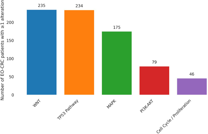
Supplementary Figure 3.

Supplementary Figure 4.

Supplementary Figure 5.

Supplementary Figure 6.

References
- 1.Siegel R.L., Miller K.D., Wagle N.S., Jemal A. Cancer statistics, 2023. CA Cancer J Clin. 2023;73(1):17–48. doi: 10.3322/caac.21763. [DOI] [PubMed] [Google Scholar]
- 2.Bailey C.E., Hu C.Y., You Y.N., et al. Increasing disparities in the age-related incidences of colon and rectal cancers in the United States, 1975-2010. JAMA Surg. 2015;150(1):17–22. doi: 10.1001/jamasurg.2014.1756. [DOI] [PMC free article] [PubMed] [Google Scholar]
- 3.Araghi M., Soerjomataram I., Bardot A., et al. Changes in colorectal cancer incidence in seven high-income countries: a population-based study. Lancet Gastroenterol Hepatol. 2019;4(7):511–518. doi: 10.1016/S2468-1253(19)30147-5. [DOI] [PMC free article] [PubMed] [Google Scholar]
- 4.Hua H., Jiang Q., Sun P., Xu X. Risk factors for early-onset colorectal cancer: systematic review and meta-analysis. Front Oncol. 2023;13 doi: 10.3389/fonc.2023.1132306. [DOI] [PMC free article] [PubMed] [Google Scholar]
- 5.McDowell R., Perrott S., Murchie P., Cardwell C., Hughes C., Samuel L. Oral antibiotic use and early-onset colorectal cancer: findings from a case-control study using a national clinical database. Br J Cancer. 2022;126(6):957–967. doi: 10.1038/s41416-021-01665-7. [DOI] [PMC free article] [PubMed] [Google Scholar]
- 6.Atchade A.M., Williams J.L., Mermelstein L., Nemesure B. Unraveling the complexities of early-onset colorectal cancer: a perspective on dietary and microbial influences. Front Public Health. 2024;12 doi: 10.3389/fpubh.2024.1370108. [DOI] [PMC free article] [PubMed] [Google Scholar]
- 7.Pretta A., Ziranu P., Perissinotto E., et al. Early onset metastatic colorectal cancer patients as a distinctive clinical and molecular phenomenon. Br J Cancer. 2024;132(2):188–194. doi: 10.1038/s41416-024-02902-5. [DOI] [PMC free article] [PubMed] [Google Scholar]
- 8.van Riggelen J., Yetil A., Felsher D.W. MYC as a regulator of ribosome biogenesis and protein synthesis. Nat Rev Cancer. 2010;10(4):301–309. doi: 10.1038/nrc2819. [DOI] [PubMed] [Google Scholar]
- 9.Lin C.Y., Lovén J., Rahl P.B., et al. Transcriptional amplification in tumor cells with elevated c-Myc. Cell. 2012;151(1):56–67. doi: 10.1016/j.cell.2012.08.026. [DOI] [PMC free article] [PubMed] [Google Scholar]
- 10.Li J., Pan Y., Guo F., et al. Patterns in genomic mutations among patients with early-onset colorectal cancer: an international, multicohort, observational study. Lancet Oncol. 2025;26(8):1055–1066. doi: 10.1016/S1470-2045(25)00239-6. [DOI] [PubMed] [Google Scholar]
- 11.Willauer A.N., Liu Y., Pereira A.A.L., et al. Clinical and molecular characterization of early-onset colorectal cancer. Cancer. 2019;125(12):2002–2010. doi: 10.1002/cncr.31994. [DOI] [PMC free article] [PubMed] [Google Scholar]
- 12.Joo J.E., Clendenning M., Wong E.M., et al. DNA methylation signatures and the contribution of age-associated methylomic drift to carcinogenesis in early-onset colorectal cancer. Cancers (Basel) 2021;13(11):2589. doi: 10.3390/cancers13112589. [DOI] [PMC free article] [PubMed] [Google Scholar]
- 13.Lin C.Y., Lovén J., Rahl P.B., et al. Transcriptional amplification in tumor cells with elevated c-Myc. Cell. 2012;151(1):56–67. doi: 10.1016/j.cell.2012.08.026. [DOI] [PMC free article] [PubMed] [Google Scholar]
- 14.Sabò A., Kress T.R., Pelizzola M., et al. Selective transcriptional regulation by Myc in cellular growth control and lymphomagenesis. Nature. 2014;511(7510):488–492. doi: 10.1038/nature13537. [DOI] [PMC free article] [PubMed] [Google Scholar]
- 15.Walz S., Lorenzin F., Morton J., et al. Activation and repression by oncogenic MYC shape tumour-specific gene expression profiles. Nature. 2014;511(7510):483–487. doi: 10.1038/nature13473. [DOI] [PMC free article] [PubMed] [Google Scholar]
- 16.Deb S., Xu H., Tuynman J., et al. RAD21 cohesin overexpression is a prognostic and predictive marker exacerbating poor prognosis in KRAS mutant colorectal carcinomas. Br J Cancer. 2014;110(6):1606–1613. doi: 10.1038/bjc.2014.31. [DOI] [PMC free article] [PubMed] [Google Scholar]
- 17.Adigun A.O., Adebile T.M., Okoye C., et al. Causes and prevention of early-onset colorectal cancer. Cureus. 2023;15(9) doi: 10.7759/cureus.45095. [DOI] [PMC free article] [PubMed] [Google Scholar]
- 18.Otegbeye E.E., Colditz G.A., Cao Y. Prevention of early-onset colorectal cancer: not one size fits all. JNCI Cancer Spectr. 2021;5(3):kab030. doi: 10.1093/jncics/pkab030. [DOI] [PMC free article] [PubMed] [Google Scholar]
- 19.Mertz J.A., Conery A.R., Bryant B.M., et al. Targeting MYC dependence in cancer by inhibiting BET bromodomains. Proc Natl Acad Sci U S A. 2011;108(40):16669–16674. doi: 10.1073/pnas.1108190108. [DOI] [PMC free article] [PubMed] [Google Scholar]
Associated Data
This section collects any data citations, data availability statements, or supplementary materials included in this article.