

Abstract
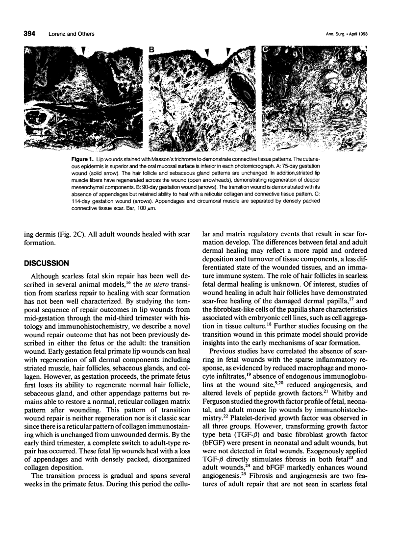
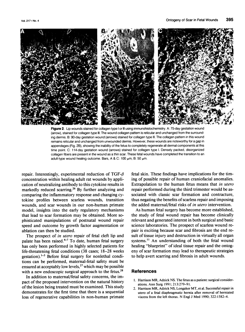
OBJECTIVE: This study determined how scar formation develops in a non-human primate model of fetal skin repair. SUMMARY BACKGROUND DATA: A transition from healing scarlessly to healing with scar formation characterizes skin repair in rat and sheep fetuses. New knowledge of the regulatory processes occurring in the fetal wound at the initial stages of scar formation may provide insights into the early mechanisms of scar formation. METHODS: Full-thickness wounds were made in fetal rhesus monkey lips from 75 through 114 days gestation (n = 6, term = 165 days). Wounds were harvested at 14 days postwounding and processed for histology (hematoxylin & eosin, Masson's trichrome) as well as immunohistochemistry (human type I or type III collagen). RESULTS: Wounds healed with complete restoration of normal tissue architecture in the 75-day gestation fetus. However in the 85-100 day gestation fetuses, wounds healed with an absence of hair follicles and sebaceous glands, but the dermal collagen pattern remained reticular and similar to that in unwounded dermis. At 107 days, a thin scar was present in the wound, thereby demonstrating a transition to scar formation between 100 and 107 days gestation (early 3rd trimester) in the non-human primate. CONCLUSIONS: In the non-human primate fetus, a transition from scarless repair to adult-type repair with scar formation occurs in the early third trimester. These data provide insight into the transition process; the ontogeny of scar formation is characterized initially by wounds healing without the presence of epidermal appendages but with a normal reticular dermal collagen pattern, which we term the "transition wound."
Full text
PDF
Images in this article
Selected References
These references are in PubMed. This may not be the complete list of references from this article.
- Adzick N. S., Longaker M. T. Animal models for the study of fetal tissue repair. J Surg Res. 1991 Sep;51(3):216–222. doi: 10.1016/0022-4804(91)90097-6. [DOI] [PubMed] [Google Scholar]
- Adzick N. S., Longaker M. T. Scarless fetal healing. Therapeutic implications. Ann Surg. 1992 Jan;215(1):3–7. doi: 10.1097/00000658-199201000-00004. [DOI] [PMC free article] [PubMed] [Google Scholar]
- Boon L., Manicourt D., Marbaix E., Vandenabeele M., Vanwijck R. A comparative analysis of healing of surgical cleft lip corrected in utero and neonates. Plast Reconstr Surg. 1992 Jan;89(1):11–20. [PubMed] [Google Scholar]
- Dado D. V., Kernahan D. A., Gianopoulos J. G. Intrauterine repair of cleft lip: what's involved. Plast Reconstr Surg. 1990 Mar;85(3):461–467. [PubMed] [Google Scholar]
- Demarchez M., Sengel P., Prunieras M. Wound healing of human skin transplanted onto the nude mouse. I. An immunohistological study of the reepithelialization process. Dev Biol. 1986 Jan;113(1):90–96. doi: 10.1016/0012-1606(86)90110-7. [DOI] [PubMed] [Google Scholar]
- Démarchez M., Hartmann D. J., Herbage D., Ville G., Pruniéras M. Wound healing of human skin transplanted onto the nude mouse. II. An immunohistological and ultrastructural study of the epidermal basement membrane zone reconstruction and connective tissue reorganization. Dev Biol. 1987 May;121(1):119–129. doi: 10.1016/0012-1606(87)90145-x. [DOI] [PubMed] [Google Scholar]
- Estes J. M., Whitby D. J., Lorenz H. P., Longaker M. T., Szabo Z., Adzick N. S., Harrison M. R. Endoscopic creation and repair of fetal cleft lip. Plast Reconstr Surg. 1992 Nov;90(5):743–749. [PubMed] [Google Scholar]
- Folkman J., Klagsbrun M. Angiogenic factors. Science. 1987 Jan 23;235(4787):442–447. doi: 10.1126/science.2432664. [DOI] [PubMed] [Google Scholar]
- Hallock G. G., Rice D. C., McClure H. M. In utero lip repair in the rhesus monkey: an update. Plast Reconstr Surg. 1987 Dec;80(6):855–858. doi: 10.1097/00006534-198712000-00021. [DOI] [PubMed] [Google Scholar]
- Harrison M. R., Adzick N. S., Jennings R. W., Duncan B. W., Rosen M. A., Filly R. A., Goldberg J. D., deLorimier A. A., Golbus M. S. Antenatal intervention for congenital cystic adenomatoid malformation. Lancet. 1990 Oct 20;336(8721):965–967. doi: 10.1016/0140-6736(90)92420-m. [DOI] [PubMed] [Google Scholar]
- Harrison M. R., Adzick N. S., Longaker M. T., Goldberg J. D., Rosen M. A., Filly R. A., Evans M. I., Golbus M. S. Successful repair in utero of a fetal diaphragmatic hernia after removal of herniated viscera from the left thorax. N Engl J Med. 1990 May 31;322(22):1582–1584. doi: 10.1056/NEJM199005313222207. [DOI] [PubMed] [Google Scholar]
- Ihara S., Motobayashi Y., Nagao E., Kistler A. Ontogenetic transition of wound healing pattern in rat skin occurring at the fetal stage. Development. 1990 Nov;110(3):671–680. doi: 10.1242/dev.110.3.671. [DOI] [PubMed] [Google Scholar]
- Jahoda C. A., Oliver R. F. Histological studies of the effects of wounding vibrissa follicles in the hooded rat. J Embryol Exp Morphol. 1984 Oct;83:95–108. [PubMed] [Google Scholar]
- Jahoda C. A., Oliver R. F. Vibrissa dermal papilla cell aggregative behaviour in vivo and in vitro. J Embryol Exp Morphol. 1984 Feb;79:211–224. [PubMed] [Google Scholar]
- Krummel T. M., Michna B. A., Thomas B. L., Sporn M. B., Nelson J. M., Salzberg A. M., Cohen I. K., Diegelmann R. F. Transforming growth factor beta (TGF-beta) induces fibrosis in a fetal wound model. J Pediatr Surg. 1988 Jul;23(7):647–652. doi: 10.1016/s0022-3468(88)80638-9. [DOI] [PubMed] [Google Scholar]
- Longaker M. T., Whitby D. J., Adzick N. S., Crombleholme T. M., Langer J. C., Duncan B. W., Bradley S. M., Stern R., Ferguson M. W., Harrison M. R. Studies in fetal wound healing, VI. Second and early third trimester fetal wounds demonstrate rapid collagen deposition without scar formation. J Pediatr Surg. 1990 Jan;25(1):63–69. doi: 10.1016/s0022-3468(05)80165-4. [DOI] [PubMed] [Google Scholar]
- Longaker M. T., Whitby D. J., Adzick N. S., Kaban L. B., Harrison M. R. Fetal surgery for cleft lip: a plea for caution. Plast Reconstr Surg. 1991 Dec;88(6):1087–1092. doi: 10.1097/00006534-199112000-00023. [DOI] [PubMed] [Google Scholar]
- Lorenz H. P., Longaker M. T., Perkocha L. A., Jennings R. W., Harrison M. R., Adzick N. S. Scarless wound repair: a human fetal skin model. Development. 1992 Jan;114(1):253–259. doi: 10.1242/dev.114.1.253. [DOI] [PubMed] [Google Scholar]
- Roberts A. B., Sporn M. B., Assoian R. K., Smith J. M., Roche N. S., Wakefield L. M., Heine U. I., Liotta L. A., Falanga V., Kehrl J. H. Transforming growth factor type beta: rapid induction of fibrosis and angiogenesis in vivo and stimulation of collagen formation in vitro. Proc Natl Acad Sci U S A. 1986 Jun;83(12):4167–4171. doi: 10.1073/pnas.83.12.4167. [DOI] [PMC free article] [PubMed] [Google Scholar]
- Shah M., Foreman D. M., Ferguson M. W. Control of scarring in adult wounds by neutralising antibody to transforming growth factor beta. Lancet. 1992 Jan 25;339(8787):213–214. doi: 10.1016/0140-6736(92)90009-r. [DOI] [PubMed] [Google Scholar]
- Smith L. T., Holbrook K. A., Madri J. A. Collagen types I, III, and V in human embryonic and fetal skin. Am J Anat. 1986 Apr;175(4):507–521. doi: 10.1002/aja.1001750409. [DOI] [PubMed] [Google Scholar]
- Sullivan W. G. In utero cleft lip repair in the mouse without an incision. Plast Reconstr Surg. 1989 Nov;84(5):723–732. [PubMed] [Google Scholar]
- Whitby D. J., Ferguson M. W. Immunohistochemical localization of growth factors in fetal wound healing. Dev Biol. 1991 Sep;147(1):207–215. doi: 10.1016/s0012-1606(05)80018-1. [DOI] [PubMed] [Google Scholar]
- Whitby D. J., Ferguson M. W. The extracellular matrix of lip wounds in fetal, neonatal and adult mice. Development. 1991 Jun;112(2):651–668. doi: 10.1242/dev.112.2.651. [DOI] [PubMed] [Google Scholar]