Abstract
Variola virus, the most virulent member of the genus Orthopoxvirus, specifically infects humans and has no other animal reservoir. Variola causes the contagious disease smallpox, which has a 30–40% mortality rate. Conversely, the prototype orthopoxvirus, vaccinia, causes no disease in immunocompetent humans and was used in the global eradication of smallpox, which ended in 1977. However, the threat of smallpox persists because clandestine stockpiles of variola still exist. Although variola and vaccinia share remarkable DNA homology, the strict human tropism of variola suggests that its proteins are better suited than those of vaccinia to overcome the human immune response. Here, we demonstrate the functional advantage of a variola complement regulatory protein over that of its vaccinia homologue. Because authentic variola proteins are not available for study, we molecularly engineered and characterized the smallpox inhibitor of complement enzymes (SPICE), a homologue of a vaccinia virulence factor, vaccinia virus complement control protein (VCP). SPICE is nearly 100-fold more potent than VCP at inactivating human C3b and 6-fold more potent at inactivating C4b. SPICE is also more human complement-specific than is VCP. By inactivating complement components, SPICE serves to inhibit the formation of the C3/C5 convertases necessary for complement-mediated viral clearance. SPICE provides the first evidence that variola proteins are particularly adept at overcoming human immunity, and the decreased function of VCP suggests one reason why the vaccinia virus vaccine was associated with relatively low mortality. Disabling SPICE may be therapeutically useful if smallpox reemerges.
Public health concerns regarding the possible reemergence of variola virus have led to renewed interest in the pathogenesis of smallpox and in the development of therapies and safer vaccines (1–3). At a fundamental level, the strict human tropism of variola virus has never been explained (4, 5). Except for DNA similarity studies between proteins of variola and other orthopoxvirus family members (6–8), minimal data exist on authentic variola proteins (5). In vivo models involving orthopoxviruses are limited primarily to vaccinia, cowpox, and ectromelia viruses, which do not cause disease in immunocompetent humans (2, 9–11). To test the hypothesis that variola is particularly suited to infect humans, in part because of an ability to evade complement-dependent inactivation of the virus, we characterized the orthopoxvirus complement regulatory proteins (CRPs) of variola and vaccinia and compared their effectiveness in overcoming human complement activation.
The severity of orthopoxvirus infections depends on multiple factors including both the host immune response and viral mechanisms that function to undermine it (6, 10). The innate immune response includes the complement system, which acts to destroy microbial pathogens, including viruses and virally infected cells (12). Investigation of the role of host complement in orthopoxvirus pathogenesis revealed that mice deficient in complement component C5 (required for the completion of the complement pathway) had an uncontrolled inflammatory response with marked ulceration and tissue damage when infected with cowpox in the footpads (11). Conversely, complement-sufficient mice had weaker and rapidly resolving responses to cowpox injections, without tissue destruction. The differences between the two groups of mice were only observed with the first exposure to the virus, and no lesions were seen when the mice were rechallenged with cowpox after complete resolution of the initial lesions, which suggested that the adaptive immune responses were sufficient to neutralize the virus on reinfection and that complement was critical in the initial immune response.
Viruses encode proteins specifically to overcome host complement (12). Our studies concern CRPs encoded in the terminal regions of orthopoxviruses, where proteins important in immune modulation and virulence are encoded (4, 7, 13, 14). Some virally encoded CRPs are similar in structure and function to mammalian CRPs (15). Under normal conditions, mammalian CRPs preserve the integrity of bystander host cells in the face of complement activation. Virally encoded CRPs, however, presumably deflect complement destruction of infected host cells to allow for more effective viral spread (15, 16). Because nucleated mammalian cells are able to “vesiculate” portions of membrane damaged by complement, it is the rate of complement activation that determines the fate of the cell (16). Conversely, the efficiency of viral CRPs may govern whether an infected cell is destroyed or remains a site of viral replication.
CRPs differ with respect to ligand specificity and the mechanism of convertase inactivation (17). CRPs may accelerate the normal decay of the convertases or function as cofactors for the serine protease, factor I, to cleave enzymatically the α′-chains of C3b and C4b into smaller, inactive fragments (17). The extent of α′-chain inactivation differs for each cofactor and partly depends on cofactor concentration and ionic strength. Structurally, CRPs are composed of 4–56 homologous motifs, termed short consensus repeats (SCRs) (17). Each SCR is approximately 60 aa long with four invariant cysteines that are disulfide-linked. Human complement receptor type 1 (CR1; CD35), for example, has 30 SCRs and represents the most potent mammalian CRP, with both decay-accelerating and cofactor activity (18–20).
Vaccinia virus complement control protein (VCP) is secreted by vaccinia-infected cells (21, 22) and functions primarily as a cofactor for the serine protease factor I, rather than as a decay accelerator (23–25). Deletion of VCP does not affect the growth of vaccinia in vitro (21, 23). However, VCP expression in vivo enhances the virulence of the virus in rabbits and guinea pigs and causes larger lesions when injected intradermally (26). Vaccinia's natural host is unknown, but it has been extensively studied in laboratory animals.
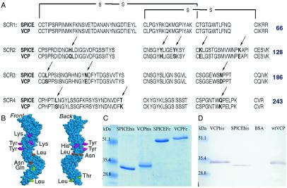
DNA comparison studies reveal that the genomes of all variola strains encode a CRP consisting of four SCRs (14), that we named smallpox inhibitor of complement enzymes (SPICE). Variola, strain Bangladesh (5, 8) encodes for SPICE in the D15L terminal region. SPICE differs from the VCP amino acid sequence by 4.6% (Fig. 1A). The different variola strains sequenced to date, including India-1967, Somalia-1977, Congo-1970, and Garcia-1966, each encode a SPICE protein sequence that differs by 0.4% from that of the Bangladesh strain (14). The recently described crystal structure of VCP (27) provides a platform on which to describe the amino acid similarities and differences between VCP and SPICE. VCP's structure reveals an extended molecule with discrete SCRs. The 11-aa differences between VCP and SPICE are dispersed throughout SCRs 2, 3, and 4. Assuming the VCP platform is applicable for SPICE, no clustering of the amino acid differences occurs (Fig. 1B).
Figure 1.
Sequences of SPICE and VCP; identification of rSPICE, rVCP, wild-type VCP. (A) Aligned amino acid sequences of SPICE and VCP, with arrows identifying residue differences. Conserved cysteines that dictate SCR folding are schematically linked, and the last amino acid of each SCR is numbered. (B) Front and back schematic views of SPICE mapped onto the structure of VCP, visualized by RASMOL V.2.7.2.1. The amino acid differences are pink in SCR2, orange in SCR3, and green in SCR4. (C) Coomassie-stained SDS/PAGE of SPICEhis, VCPhis, SPICEFc, and VCPFc. (D) Western blot of VCPhis, SPICEhis, and wild-type VCP. Proteins were identified by using polyclonal anti-VCP/SPICE antiserum. Wild-type VCP and VCPhis migrate identically at approximately 35 kDa.
In the absence of wild-type variola proteins, we describe the molecular engineering, expression, and characterization of SPICE. Our studies demonstrate that, despite the remarkable sequence identity between SPICE and VCP, SPICE holds a functional advantage, human complement specificity, and a cofactor activity more like that of the human CRPs than of VCP. These data provide insight into the pathogenesis and species specificity of variola.
Methods
Molecular Engineering, Expression, and Purification of Recombinant SPICE.
SPICE was generated by mutating the VCP amino acid sequence into that of SPICE. Thirteen nucleotide mutations resulted in 11 aa substitutions (Fig. 1A). Twenty-two primers (Life Technologies, Grand Island, NY) were designed with the following point mutations: T was substituted for A at position 339 of the VCP DNA sequence (22), T for C400, A for C416, A for G430, A for G466, T for C500, G for C501, A for G538, T for G540, A for G640, T for C686, C for A749, and C for A814. The DNA sequence was confirmed by a 377 Stretch sequencer (Applied Biosystems). SPICE was substituted for VCP in three plasmids [pRelVCP1234, pApHygVCPFc, and pVCPFc (28)] previously used to express VCPFc in 293T cells. Recombinant baculoviruses were created by subcloning SPICEFc and VCPFc EcoRI/XbaI fragments into pFastBacHTa (Bac-to-Bac system, Life Technologies) to create pFastBacHTaSPICEFc and pFastBacHTaVCPFc and recombinant baculovirus strains containing SPICEFc and VCPFc.
A baculovirus construct containing a placental alkaline phosphatase leader sequence (29) and six histidines at the carboxyl terminus was created by using a PCR-based approach. Oligonucleotides 99AMR1 (5′-GCACCCGGGAGTTCTATCATGCTGTACTATTCCG-3′) and HISSTOP (5′-GCTCTAGACTCGAGCTAGTGGTGGTGGTGGTGGTGGCGTACACATTTTGGAAGTTC-3′) were used. The SPICE and VCP PspA1/XhoI fragments were ligated into pBluescript II KS phagemid (Stratagene) and subsequently subcloned into pFastBac at XbaI/XhoI sites to generate pFBSPICEhis and pFBVCPhis. Concurrently, the alkaline phosphatase leader from pPbac (Stratagene) was cleaved by using NheI/PspAI and ligated to generate pFBapSPICEhis and pFBapVCPhis, which were used to generate baculovirus containing SPICEhis and VCPhis.
Recombinant proteins were produced in 293T cells (28) or in Spodoptera frugiperda (Sf9 cells) (Life Technologies) and affinity purified from culture supernatant with CL-4B protein A beads (Amersham Pharmacia) or Ni-NTA Superflow (Qiagen, Valencia, CA). Recombinant proteins were dialyzed against PBS (Life Technologies). SPICEhis and VCPhis were characterized by electrophoresis on an SDS/12% polyacrylamide gel and by mass spectrometry (M-Scan, West Chester, PA).
Production of Anti-VCP and Anti-SPICE Antiserum to Identify rVCP, rSPICE, and Wild-Type VCP.
Anti-VCP antiserum was produced by standard methods (30). Four- to six-week-old BALB/c mice (The Jackson Laboratory) were immunized i.p. with 35 μg of rVCP in complete Freund's adjuvant and later boosted with 35 μg of protein in incomplete Freund's adjuvant. Medium from BS-C-1 cells (CCL-26, American Type Culture Collection), previously infected with 10 plaque-forming units/cell of vaccinia strain Western Reserve (generously provided by J. Boyer, University of Pennsylvania), was concentrated 100-fold, fractionated by SDS/PAGE, and transferred to nitrocellulose membrane. Wild-type VCP was identified by Western blot analysis with anti-VCP antiserum (1:1,000 dilution).
C3b or C4b Degradation in the Presence of SPICE and Factor I.
Cofactor activity was measured by incubating C3b or C4b with each cofactor at 37°C. Samples were removed at 0, 4, and 24 h, as indicated. Nine micrograms of C3b or C4b (Advanced Research Technologies, San Diego) were incubated with 3 μg of factor I (Advanced Research Technologies) and 6 μg of VCPhis, SPICEhis, factor H (Advanced Research Technologies) or soluble CR1 (sCR1) (provided by Novartis Pharmaceutical, Hanover, NJ) in a total volume of 33 μl at 37°C, as described (25). Ten microliters were removed at 0, 4, and 24 h, mixed with sample buffer containing 2-mercaptoethanol (Bio-Rad), boiled 2 min, and subjected to electrophoresis on a SDS-9% polyacrylamide gel. C3b and C4b cleavage products were visualized by staining the gel with Coomassie blue. The C3bα′ cleavage fragments migrate as follows: 68 kDa, 46 kDa (iC3b1), 43 kDa (iC3b2), and <35 kDa (C3dg/C3c). C4bα′ cleavage fragments migrate as follows: 64 kDa (iC4b), 46 kDa (C4d), and 25 kDa (C4c).
Rate of Cofactor Activity of SPICEhis, VCPhis, and sCR1.
Kinetic experiments were performed by incubating C3b or C4b with factor I (13 μM) and 0.65 pM SPICEhis, VCPhis, or sCR1 in a total volume of 66 μl at 37°C. Samples (10 μl) were removed at the times indicated and subjected to electrophoresis. Coomassie-stained gels were scanned for densitometric analysis with a transmissive spectrophotometer (model 355A, Molecular Dynamics) and analyzed by using imagequant 3.2 software (Molecular Dynamics). Reported values represent the optical density (OD) relative to the original sample of the C3bα′ or C4bα′ chains. Experiments were performed five times.
Inhibition of Complement-Mediated Hemolysis of Sheep and Rabbit Erythrocytes.
Complement-mediated hemolysis was evaluated by determining the highest dilution of human, dog, guinea pig, and baboon (data not shown) sera that resulted in 100% hemolysis of antibody-sensitized sheep erythrocytes (EA) or unsensitized rabbit erythrocytes (Er) (1 × 10⁸ per ml GVB++) (Advanced Research Technologies) (31) at 37°C. Thereafter, the sera were serially diluted in U-bottom polypropylene 96-well plates (Corning) until no hemolysis was detected. Reactions were stopped after 1 h with PBS (180 μl per well). Maximum hemolysis was obtained by the addition of water instead of PBS. Supernatant (100 μl) was read at 405 nm. Serum heated to 56°C for 30 min established background hemolysis.
To determine the influence of various CRPs on complement-mediated hemolysis of EA or Er, 25 μl of serially diluted serum was added, followed by 25 μl of 0.1–0.25 mg/ml CRP or PBS, and 50 μl of erythrocytes (1 × 10⁸ per ml in GVB++). Reactions were incubated at 37°C and stopped after 1 h with the addition of PBS as described above. Reported values represent percent of maximum hemolysis for CRP-treated wells relative to PBS-treated wells at identical serum dilutions. All experiments were performed between three and ten times, and reproducible results were obtained in all cases.
Results
Molecular Engineering, Expression, and Purification of Recombinant SPICE.
Neither variola virus, native variola proteins, nor variola genomic DNA is available to the general scientific community. Therefore, we generated the SPICE coding sequence from that of VCP by site-directed mutagenesis of 13 VCP nucleotides, which resulted in 11 aa substitutions (Fig. 1A), consistent with the predicted amino acid sequence of SPICE. To facilitate purification of recombinant SPICE and VCP proteins, we designed two constructs for each. One construct encoded for a carboxyl-terminal histidine tag (his), while the other encoded for a carboxyl-terminal mouse IgG2a Fc domain (Fc).
Recombinant SPICE (rSPICE) and VCP (rVCP) proteins were produced in mammalian and insect cells and affinity purified. SPICEhis, VCPhis, SPICEFc, and VCPFc migrated as single bands under reducing conditions on 12% SDS/PAGE at 33, 35, 53, and 55 kDa, respectively (Fig. 1C). Under nonreducing conditions, the Fc-fusion proteins migrated at approximately 110 kDa, representing dimers due to disulfide bonds between two Fc domains (32). The recombinant proteins produced in mammalian and insect cells functioned identically and migrated similarly on SDS/PAGE (data not shown).
The 28,936- and 28,818-Da expected molecular weights of SPICEhis and VCPhis, respectively, were confirmed by mass spectrometry, although the migration on SDS/PAGE was slightly slower than expected. Wild-type VCP, secreted into the media of vaccinia-infected cells, and recombinant VCP were identified on Western blot (Fig. 1D). Both wild-type VCP and VCPhis migrated at approximately 35 kDa, consistent with the published mass of wild-type VCP (23).
Factor I Cofactor Activity of SPICE.
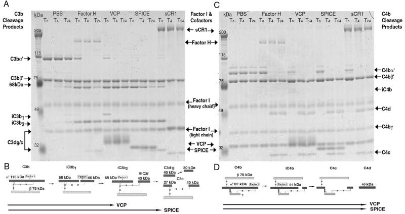
The molecular engineering and expression of SPICE confirmed that variola produces a compact, functionally active cofactor for factor I. C3b degradation experiments performed as described (25, 33) compared the cofactor activity of SPICE, VCP, soluble human CR1 (sCR1) (33), and factor H (Fig. 2A) at normal ionic strength. Soluble CR1 is a recombinant, soluble protein consisting of the 30 SCRs of the membrane-bound CR1 but lacking its transmembrane and cytoplasmic domains (33). The cofactor activity of SPICE was robust, with C3b cleavage to iC3b2 occurring immediately, followed by cleavage to C3c/C3dg (sampled at 4 and 24 h). In the presence of VCP, C3b was degraded at a slower rate, first to iC3b1 and then to iC3b2. However, VCP did not participate in further degradation of iC3b2, even after 24 h. Therefore, qualitatively, SPICE functions more like factor H, which serves as a cofactor for factor I in the degradation of iC3b2 to C3c/C3dg after longer incubations (34) (Fig. 2B). Although not as potent as sCR1, SPICE can result in a similar degradation pattern (33). For C4b, we performed identical experiments, replacing C4b for C3b (Fig. 2C). SPICEhis and VCPhis seem to function identically and result in the degradation of C4b to C4c/C4d (Fig. 3D). Factor H also serves as a cofactor for factor I degradation of C4b in the fluid phase (35).
Figure 2.
C3b and C4b degradation in the presence of SPICEhis and VCPhis. (A) Appearance of C3b cleavage products in the presence of factor I and either factor H, VCPhis, SPICEhis, sCR1, or PBS at 0, 4, and 24 h. (B) Schematic of C3b degradation in the presence of factor I (45) and cofactors, VCP or SPICE. (C) Appearance of C4b cleavage products in the presence of factor I and either factor H, VCPhis, SPICEhis, sCR1, or PBS at 0, 4, and 24 h. (D) Schematic of C4b degradation (46) in the presence of factor I and cofactors, VCP or SPICE.
Figure 3.
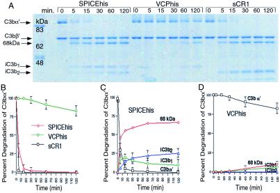
Time course of C3b degradation with SPICEhis, VCPhis, and sCR1. (A) Coomassie-stained SDS/PAGE of C3b degradation products from reactions containing C3b, factor I, and either SPICEhis, VCPhis, or sCR1. Reaction samples were removed at 0, 5, 15, 30, 60, and 120 min. (B) Rate of degradation of C3bα′. Gels were scanned for densitometric analysis by using transmissive spectrophotometer, and the OD of the C3bα′ band at each time point was compared with the OD of the original sample of C3bα′. The percent degradation is plotted against time. Experiments were performed five times and error bars represent SD. (C) Rate of degradation of C3bα′ chain and the generation of other C3b cleavage products in the presence of SPICEhis are plotted as a percent of the original C3bα′ chain. Analysis was performed as described in B. (D) Rate of degradation of C3b and generation of C3b cleavage products in the presence of VCPhis are plotted as a percentage of the original C3bα′ chain. Analysis was performed as described in B.
Kinetic experiments were performed to compare the rate of cofactor activity at limiting concentrations of recombinant cofactors (Fig. 3A). By using 100-fold lower concentrations (0.68 pM) of recombinant cofactors than those used in the previous experiment (Fig. 2A), differences in activity could be established. In the presence of SPICEhis, 50% of the C3bα′ chain was degraded in ≈3 min (Fig. 3 A and B), and entirely degraded by 15 min. Soluble CR1 led to nearly complete degradation of the C3bα′ chain in 7 min. However, only 20% of the C3bα′ chain was degraded after 120 min with VCP. Therefore, SPICEhis functions as a cofactor for factor I in the degradation of C3b with nearly 100-fold greater efficiency than VCPhis. Soluble CR1 functions at least twice as fast as SPICEhis. All experiments were performed at least five times, and reproducible results were obtained in all cases. Furthermore, the degradation patterns showed that with SPICEhis, the C3bα′ chain was immediately cleaved to iC3b1, followed by cleavage to iC3b2 (Fig. 3C), whereas with VCPhis, no such degradation to iC3b2 (Fig. 3D) was observed, consistent with Sahu et al. (25).
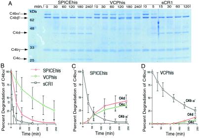
Similarly, kinetic experiments were performed by using C4b (Fig. 4A). At limiting concentrations of recombinant proteins (0.68 pM), 50% of the C4bα′ chain was degraded in 5 min with sCR1 (Fig. 4B), 20 min with SPICEhis, and 120 min with VCPhis. Therefore, SPICEhis functions approximately 6-fold faster than VCPhis but slower than sCR1. Minimal improvement in SPICEhis and VCPhis cofactor activity for C3b and C4b was observed at half-ionic strength solution (data not shown).
Figure 4.
Time course of C4b degradation with SPICEhis, VCPhis, and sCR1. (A) Coomassie-stained SDS/PAGE of C4b degradation products from reactions containing C4b, factor I, and either SPICEhis, VCPhis, or sCR1. Reaction samples were removed at 0, 30, 60, 120, 180, and 240 min for SPICEhis and VCPhis and 0, 5, 15, 30, 60, and 120 min for sCR1. (B) Rate of degradation of C4bα′. The gels were scanned for densitometric analysis by using transmissive spectrophotometer and the OD of the C4bα′ band at each time point was compared with the OD of the original sample of C4bα′. The percent degradation is plotted against time. Experiments were performed five times and error bars represent SD. (C) Rate of degradation of C4bα′ chain and generation of C4b cleavage products in the presence of SPICEhis are plotted as a percent of the original C4bα′ chain. Analysis was performed as described in B. (D) Rate of degradation of C4b and generation of C4b cleavage products in the presence of VCPhis are plotted as a percentage of the original C4bα′ chain. Analysis was performed as described in B.
Species Preferences of SPICE.
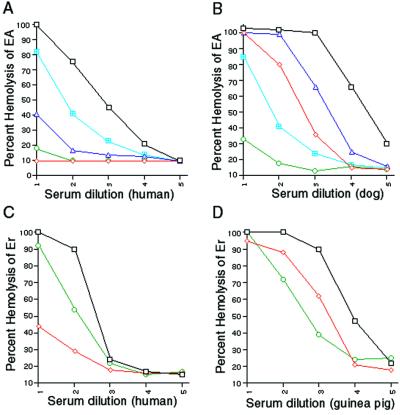
Because mammalian CRPs exhibit homologous restriction, that is, they function best against complement from phylogenetically related species (36), we asked whether variola might exhibit “host complement restriction,” particularly because of its strict host range. We compared the ability of rSPICE and rVCP to inhibit complement from human, dog, guinea pig (Fig. 5 A–D), and baboon (data not shown) in hemolytic assays. EA are lysed in the presence of an increasing amount of complement because of activation of the classical pathway, whereas Er are sensitive to the alternative pathway. Analysis of the data shows that both rVCP and rSPICE inhibited both complement pathways. However, rSPICE preferentially inhibited human and baboon complement, whereas rVCP preferentially inhibited dog and guinea pig complement. The Fc constructs behaved less efficiently than the His-tagged constructs, possibly because of the dimerization of the Fc domain, which could influence function. The hemolysis studies suggest that the preference of SPICE for human complement parallels the preference of variola for a human host.
Figure 5.
Inhibition of complement from different species by viral CRPs. (A) Influence of viral CRPs on the classical pathway-mediated hemolysis of EA in the presence of human complement. EA were incubated with human serum in the presence of no inhibitor (black squares), or equimolar amounts of SPICEhis (red diamonds) and VCPhis (green circles) or SPICEFc (blue triangles) and VCPFc (aqua quartered squares) for 1 h at 37°C. Reported values represent the amount of hemolysis relative to maximum hemolysis, as determined by the addition of water to EA. Results are representative of ten separate experiments. (B) Influence of viral CRPs on the classical pathway-mediated hemolysis of EA in the presence of dog complement. Experiments were performed as described above except that dog serum was used as the source of complement. Results are representative of six separate experiments. (C) Influence of viral CRPs on the alternative pathway-mediated hemolysis of Er in the presence of human complement. Results are representative of four separate experiments. (D) Influence of viral CRPs on the alternative pathway-mediated hemolysis of Er in the presence of guinea pig complement. Results are representative of three separate experiments.
Discussion
Although the virulence of orthopoxviruses represents the cumulative effects of all their proteins, no studies have compared the function of any vaccinia and variola homologues. We compared the soluble viral complement regulatory proteins, SPICE and VCP, and demonstrated that SPICE is a more potent inhibitor of human complement and that this finding could not have been predicted by DNA analysis alone. The efficiency of SPICE was nearly 100-fold higher than that of VCP. Although sCR1 functioned better than SPICE, its better function may be explained by the three ligand-binding sites of CR1 (37–40). Because SPICE efficiently prevents the stability of human C3b and C4b, it essentially inactivates the convertases and, therefore, both the classical and alternative pathways of complement. Consequently, the production of the inflammatory mediators C3a, C4a, and C5a is also decreased. Therefore, the purpose of SPICE may be to create a microenvironment around variola-infected cells to protect them from complement-mediated attack while they serve as a site of viral replication.
SPICE and VCP demonstrate a preference for complement from different species in hemolytic experiments. The fact that SPICE functions better against human complement quantitatively and qualitatively demonstrates that variola exhibits human preference at the protein level, a concept previously suggested, but not shown. The fact that VCP functions relatively poorly at inactivating human C3b and C4b suggests one reason for its relative safety and effectiveness as a smallpox vaccine.
The only other poxvirus that specifically causes human disease is molluscum contagiosum virus, which results in benign skin nodules that spontaneously regress. Genome sequence analysis demonstrates that despite the marked sequence similarities among the molluscum contagiosum virus, vaccinia, and variola genomes, molluscum contagiosum virus lacks homologous sequences to known vaccinia proteins that modulate the host immune response (41). Included in the list of approximately 20 absent proteins is a VCP or SPICE homologue. It is possible that the absence of a CRP may contribute to the benign disease pattern of molluscum contagiosum virus.
The minimal amino acid differences between VCP and SPICE suggest that a few residues are critical to the function of CRPs. Several regions that are identical between VCP and SPICE correlate with regions previously shown to be important in the mammalian CRP, membrane cofactor protein (MCP; CD46). MCP functions as a cofactor for factor I to inactivate both C3b and C4b (27, 42). Region 191–196 of MCP is important in C3b binding and correlates with regions 184–189 of VCP and SPICE (Fig. 1A). In addition, sequence comparisons of VCP and MCP suggest that Phe-201 and Arg-203 in VCP, both present in SPICE, are important in C3b interactions.
The enhanced function of SPICE suggests that one or more of the 11 aa differences between SPICE and VCP may be critical in determining the functional advantage of SPICE. A Lys-120 for Glu-120 substitution in SCR2 correlates with an identical substitution made in SCR2 of CR1 (STEPPIC to STKPPIC) that resulted in a gain of C3b-binding and cofactor activity (38, 40). Moreover, Liszewski et al. demonstrated that the sequence NEGYYLIGEE (regions 94–103) of MCP represented both a C4b-binding/cofactor activity site and a C3b cofactor activity site (42). A nearly identical sequence is in regions 94–103 of SPICE and VCP, although greater sequence identity is in SPICE, which contains the two sequential Tyr (positions 97 and 98) vs. VCP's one Tyr at position 97. Because an Ala for Tyr-98 substitution in MCP abrogated C4b cofactor activity (42), the Tyr-98 in SPICE may be important in C4b cofactor activity. Furthermore, Murthy et al. proposed that Asn-94, Ser-95, His-98, Glu-102, and Ser-103 in VCP are surface-exposed, and therefore are structurally important in intermolecular interactions (27), but even greater sequence identity is seen in this region between SPICE and MCP. The value of the different amino acid residues between VCP and SPICE will be the focus of future mutagenesis studies. The previously characterized crystal structure of SCR1 and 2 of MCP could not be compared because this portion of MCP contains the measles-binding site, not the complement ligand-binding site (43).
In future studies, it will be important to investigate the CRPs of other orthopoxviruses such as cowpox. However, the cowpox CRP has relatively large deletions in SCR1 and twice the number of amino acid differences than between VCP and SPICE (44), which may limit the conclusions about the relevance of specific amino acids in CRP function.
The global eradication of smallpox was a major medical triumph. However, because variola virus still exists, its potential use in a bioterrorist attack could result in worldwide smallpox outbreaks, because most of the population is not immunized against smallpox. In fact, only 20% of individuals previously vaccinated are still protected because of waning immunity (3). The threat of smallpox must serve as an impetus for the development of effective therapies to cure this disease. In addition, a safer vaccine is needed to immunize the population. When previously used, the vaccine currently available was associated with considerable complications, particularly in those who were immunosuppressed (3). A better understanding of variola pathogenesis is necessary to achieve the goals of developing both a therapy for smallpox and a safer vaccine.
Ethical and public health concerns preclude in vivo work on variola, and the World Health Organization prohibits DNA recombination studies between variola and other orthopoxvirus genomes. Therefore, the study of variola presents multiple obstacles. Although in vivo studies of orthopoxviruses that lack human tropism can suggest virulence factors important in variola, the relatively benign nature of many orthopoxviruses in humans may underestimate the importance of any homologous proteins in variola. Only the integration of in vitro studies of variola proteins with in vivo studies of other orthopoxviruses can suggest a blueprint for therapeutic intervention in smallpox. Because smallpox was eradicated before techniques for the manipulation of DNA were widely available, very few molecular studies of this virus are available.
In this study, we demonstrate an approach to molecularly engineer variola proteins, which are necessary to understand the pathogenesis of smallpox. We provide in vitro evidence that SPICE is a highly specific and potent inhibitor of human complement, partially explaining the species specificity and the pathogenesis of variola. Assuming that complement is as critical to orthopoxvirus containment in humans as it is in rodents, SPICE seems to be a virulence factor for variola virus. Thus, the disabling of SPICE could represent an approach to the treatment of smallpox. In addition, the disabling of VCP could represent an approach to developing a safer smallpox vaccine.
Acknowledgments
We are grateful to Drs. Steven Eck, Joseph Ahearn, Dennis Hourcade, and Jonni Moore for invaluable discussions and to Jeremy Reid for reviewing the manuscript. Thanks to Dr. Jean Boyer for providing media from vaccinia-infected cells, and to Drs. Richard Harrison, Tomasz Sablinski, and Manfred Schulz of Novartis Pharmaceutical for sCR1 (TP-10). This work was supported in part by National Institutes of Health Grant HLB31331.
Abbreviations
- C3b
proteolytically cleaved form of C3
- C3bα′
α-chain of C3b
- iC3b1
inactivated form of C3b resulting from factor I cleavage of the α-chain
- iC3b2
inactivated form of C3b resulting from a second factor I cleavage of the α-chain
- C4b
proteolytically cleaved form of C4
- C4bα′
α-chain of C4b
- CR1
complement receptor type 1 (CD35)
- CRP
complement regulatory protein
- EA
antibody-sensitized sheep erythrocytes
- Er
unsensitized rabbit erythrocytes
- MCP
membrane cofactor protein (CD46)
- SCR
short consensus repeat
- sCR1
soluble CR1
- SPICE
smallpox inhibitor of complement enzymes
- VCP
vaccinia virus complement control protein
- Fc
carboxyl-terminal mouse IgG2a Fc domain
Footnotes
See commentary on page 8461.
References
- 1.Fenner F. Rev Infect Dis. 1982;4:916–930. doi: 10.1093/clinids/4.5.916. [DOI] [PubMed] [Google Scholar]
- 2.Moss B. Proc Natl Acad Sci USA. 1996;93:11341–11348. doi: 10.1073/pnas.93.21.11341. [DOI] [PMC free article] [PubMed] [Google Scholar]
- 3.Henderson D A. Science. 1999;283:1279–1282. doi: 10.1126/science.283.5406.1279. [DOI] [PubMed] [Google Scholar]
- 4.Buller R M, Palumbo G J. Microbiol Rev. 1991;55:80–122. doi: 10.1128/mr.55.1.80-122.1991. [DOI] [PMC free article] [PubMed] [Google Scholar]
- 5.Massung R F, Esposito J J, Liu L I, Qi J, Utterback T R, Knight J C, Aubin L, Yuran T E, Parsons J M, Loparev V N, et al. Nature (London) 1993;366:748–751. doi: 10.1038/366748a0. [DOI] [PubMed] [Google Scholar]
- 6.Aguado B, Selmes I P, Smith G L. J Gen Virol. 1992;73:2887–2902. doi: 10.1099/0022-1317-73-11-2887. [DOI] [PubMed] [Google Scholar]
- 7.Shchelkunov S N, Resenchuk S M, Totmenin A V, Blinov V M, Marennikova S S, Sandakhchiev L S. FEBS Lett. 1993;327:321–324. doi: 10.1016/0014-5793(93)81013-p. [DOI] [PubMed] [Google Scholar]
- 8.Massung R F, Liu L I, Qi J, Knight J C, Yuran T E, Kerlavage A R, Parsons J M, Venter J C, Esposito J J. Virology. 1994;201:215–240. doi: 10.1006/viro.1994.1288. [DOI] [PubMed] [Google Scholar]
- 9.Smith G L, Symons J A, Khanna A, Vanderplasschen A, Alcami A. Immunol Rev. 1997;159:137–154. doi: 10.1111/j.1600-065x.1997.tb01012.x. [DOI] [PubMed] [Google Scholar]
- 10.Alcami A, Smith G L. Immunol Today. 1995;16:474–478. doi: 10.1016/0167-5699(95)80030-1. [DOI] [PubMed] [Google Scholar]
- 11.Miller C G, Justus D E, Jayaraman S, Kotwal G J. Cell Immunol. 1995;162:326–332. doi: 10.1006/cimm.1995.1086. [DOI] [PubMed] [Google Scholar]
- 12.Cooper N R. In: The Human Complement System in Health and Disease. Volanakis J E, Frank M M, editors. New York: Dekker; 1998. pp. 393–407. [Google Scholar]
- 13.Smith G L. J Gen Virol. 1993;74:1725–1740. doi: 10.1099/0022-1317-74-9-1725. [DOI] [PubMed] [Google Scholar]
- 14.Massung R F, Loparev V N, Knight J C, Totmenin A V, Chizhikov V E, Parsons J M, Safronov P F, Gutorov V V, Shchelkunov S N, Esposito J J. Virology. 1996;221:291–300. doi: 10.1006/viro.1996.0378. [DOI] [PubMed] [Google Scholar]
- 15.Shchelkunov S N, Blinov V M, Sandakhchiev L S. FEBS Lett. 1993;319:80–83. doi: 10.1016/0014-5793(93)80041-r. [DOI] [PubMed] [Google Scholar]
- 16.Lachmann P J, Davies A. Immunol Rev. 1997;159:69–77. doi: 10.1111/j.1600-065x.1997.tb01007.x. [DOI] [PubMed] [Google Scholar]
- 17.Liszewski M K, Atkinson J P. In: The Human Complement System in Health and Disease. Volanakis J E, Frank M M, editors. New York: Dekker; 1998. pp. 149–166. [Google Scholar]
- 18.Fearon D T. Proc Natl Acad Sci USA. 1979;76:5867–5871. doi: 10.1073/pnas.76.11.5867. [DOI] [PMC free article] [PubMed] [Google Scholar]
- 19.Iida K, Nussenzweig V. J Exp Med. 1981;153:1138–1150. doi: 10.1084/jem.153.5.1138. [DOI] [PMC free article] [PubMed] [Google Scholar]
- 20.Medof M E, Iida K, Mold C, Nussenzweig V. J Exp Med. 1982;156:1739–1754. doi: 10.1084/jem.156.6.1739. [DOI] [PMC free article] [PubMed] [Google Scholar]
- 21.Moss B, Winters E, Cooper J A. J Virol. 1981;40:387–395. doi: 10.1128/jvi.40.2.387-395.1981. [DOI] [PMC free article] [PubMed] [Google Scholar]
- 22.Kotwal G J, Moss B. Nature (London) 1988;335:176–178. doi: 10.1038/335176a0. [DOI] [PubMed] [Google Scholar]
- 23.Kotwal G J, Isaacs S N, McKenzie R, Frank M M, Moss B. Science. 1990;250:827–830. doi: 10.1126/science.2237434. [DOI] [PubMed] [Google Scholar]
- 24.McKenzie R, Kotwal G J, Moss B, Hammer C H, Frank M M. J Infect Dis. 1992;166:1245–1250. doi: 10.1093/infdis/166.6.1245. [DOI] [PubMed] [Google Scholar]
- 25.Sahu A, Isaacs S N, Soulika A M, Lambris J D. J Immunol. 1998;160:5596–5604. [PubMed] [Google Scholar]
- 26.Isaacs S N, Kotwal G J, Moss B. Proc Natl Acad Sci USA. 1992;89:628–632. doi: 10.1073/pnas.89.2.628. [DOI] [PMC free article] [PubMed] [Google Scholar]
- 27.Murthy K H, Smith S A, Ganesh V K, Judge K W, Mullin N, Barlow P N, Ogata C M, Kotwal G J. Cell. 2001;104:301–311. doi: 10.1016/s0092-8674(01)00214-8. [DOI] [PubMed] [Google Scholar]
- 28.Rosengard A M, Alonso L C, Korb L C, Baldwin W M, III, Sanfilippo F, Turka L A, Ahearn J M. Mol Immunol. 1999;36:685–697. doi: 10.1016/s0161-5890(99)00081-4. [DOI] [PubMed] [Google Scholar]
- 29.Mroczkowski B S, Huvar A, Lernhardt W, Misono K, Nielson K, Scott B. J Biol Chem. 1994;269:13522–13528. [PubMed] [Google Scholar]
- 30.Yokoyama W M. In: Current Protocols in Immunology. Coligan J, Kruisbeek A, Margulies D, Shevach E, Strober W, editors. Vol. 1. New York: Wiley; 2001. pp. 2.5–2.6. [Google Scholar]
- 31.Kitamura H. In: The Complement System. Rother K T, Till G O, Hänsch G M, editors. Berlin: Springer; 1998. [Google Scholar]
- 32.De Preval C, Pink J R, Milstein C. Nature (London) 1970;228:930–932. doi: 10.1038/228930a0. [DOI] [PubMed] [Google Scholar]
- 33.Weisman H F, Bartow T, Leppo M K, Marsh H C, Jr, Carson G R, Concino M F, Boyle M P, Roux K H, Weisfeldt M L, Fearon D T. Science. 1990;249:146–151. doi: 10.1126/science.2371562. [DOI] [PubMed] [Google Scholar]
- 34.Ross G D, Lambris J D, Cain J A, Newman S L. J Immunol. 1982;129:2051–2060. [PubMed] [Google Scholar]
- 35.Pangburn M K, Schreiber R D, Muller-Eberhard H J. J Exp Med. 1977;146:257–270. doi: 10.1084/jem.146.1.257. [DOI] [PMC free article] [PubMed] [Google Scholar]
- 36.Dalmasso A P. In: The Complement System. Rother K, Till G O, Hänsch G M, editors. Vol. 1. Berlin: Springer; 1997. pp. 471–486. [Google Scholar]
- 37.Klickstein L B, Bartow T J, Miletic V, Rabson L D, Smith J A, Fearon D T. J Exp Med. 1988;168:1699–1717. doi: 10.1084/jem.168.5.1699. [DOI] [PMC free article] [PubMed] [Google Scholar]
- 38.Krych M, Hourcade D, Atkinson J P. Proc Natl Acad Sci USA. 1991;88:4353–4357. doi: 10.1073/pnas.88.10.4353. [DOI] [PMC free article] [PubMed] [Google Scholar]
- 39.Kalli K R, Hsu P H, Bartow T J, Ahearn J M, Matsumoto A K, Klickstein L B, Fearon D T. J Exp Med. 1991;174:1451–1460. doi: 10.1084/jem.174.6.1451. [DOI] [PMC free article] [PubMed] [Google Scholar]
- 40.Krych M, Clemenza L, Howdeshell D, Hauhart R, Hourcade D, Atkinson J P. J Biol Chem. 1994;269:13273–13278. [PubMed] [Google Scholar]
- 41.Senkevich T G, Bugert J J, Sisler J R, Koonin E V, Darai G, Moss B. Science. 1996;273:813–816. doi: 10.1126/science.273.5276.813. [DOI] [PubMed] [Google Scholar]
- 42.Liszewski M K, Leung M, Cui W, Subramanian V B, Parkinson J, Barlow P N, Manchester M, Atkinson J P. J Biol Chem. 2000;275:37692–37701. doi: 10.1074/jbc.M004650200. [DOI] [PubMed] [Google Scholar]
- 43.Casasnovas J M, Larvie M, Stehle T. EMBO J. 1999;18:2911–2922. doi: 10.1093/emboj/18.11.2911. [DOI] [PMC free article] [PubMed] [Google Scholar]
- 44.Miller C G, Shchelkunov S M, Kotwal G J. Virology. 1997;229:126–133. doi: 10.1006/viro.1996.8396. [DOI] [PubMed] [Google Scholar]
- 45.Lambris J D, Sahu A, Wetsel R A. In: The Human Complement System in Health and Disease. Volanakis J E, Frank M M, editors. New York: Dekker; 1998. pp. 83–118. [Google Scholar]
- 46.Press E M, Gagnon J. Biochem J. 1981;199:351–357. doi: 10.1042/bj1990351. [DOI] [PMC free article] [PubMed] [Google Scholar]


