



SUMMARY
The development of a Zika virus (ZIKV) vaccine is complicated by the high homology between ZIKV and dengue virus (DENV) envelope (E) proteins, resulting in immunological cross-reactivity that can exacerbate disease through antibody-dependent enhancement (ADE). Here, we screen 121 anti-DENV monoclonal antibodies (mAbs) for cross-reactivity with ZIKV E proteins. We identify 70 cross-reactive mAbs, 66 of which have epitopes that included at least one of seven E protein residues conserved among DENV1–DENV4 and ZIKV (R73, E79, W101, L107, F108, K110, and W212), establishing these residues as the key determinants of DENV-ZIKV cross-reactivity. Using these data, we engineer a ZIKV E protein variant with 10 mutations (“ZIKVm10”) that reduces cross-reactivity with DENV mAbs in vitro and minimizes the induction of anti-DENV antibodies in immunized mice. Passive serum transfer from ZIKVm10-immunized mice confers near-complete protection against lethal ZIKV challenge and reduced ADE for DENV infection, providing a pathway for improved ZIKV vaccine design.
In brief
Using the epitope maps of 121 DENV antibodies, Grinyo-Escuer et al. identify seven key residues driving DENV-ZIKV cross-reactivity. They engineer a ZIKV E protein variant, “ZIKVm10,” that minimizes DENV cross-reactivity, reduces ADE risk, and protects against lethal ZIKV challenge in mice, offering a pathway for safer, more targeted ZIKV vaccines.
Graphical Abstract

INTRODUCTION
In 2015, a Zika virus (ZIKV) infection epidemic in the Americas resulted in an estimated 500,000–1,500,000 human cases.1,2 ZIKV infection can be accompanied by severe clinical manifestations, including the development of Guillain-Barré syndrome and congenital brain defects in developing fetuses.3 Vaccine development for ZIKV is complicated by the high degree of sequence and structural homology to dengue virus (DENV), as cross-reactive antibodies can cause antibody-dependent enhancement (ADE) of infection and exacerbated disease.4–7 Because these viruses share geographical distribution, a thorough understanding of their shared humoral immunity is needed to assist in the design of a safe and effective ZIKV vaccine.
ZIKV is an enveloped virus of the Flaviviridae virus family, which includes other human pathogenic flaviviruses, such as DENV, yellow fever, and West Nile viruses. The single-stranded, positive-sense flavivirus RNA genome encodes seven non-structural proteins and three structural proteins: capsid, premembrane (prM), and envelope (E). The E glycoprotein is a type II fusion protein that enables host cell entry and is the primary target of neutralizing antibodies. The E protein monomer contains three structural domains—domain I (DI), DII, and DIII—with a fusion loop at the tip of DII. During virus maturation within a host cell, the E proteins undergo conformational changes, causing transition from an immature prM/E trimer to a prM/E heterodimer, followed by prM cleavage and, ultimately, dissociation of the pr peptide after the virus leaves the cell (reviewed in Heinz and Allison, Lindenbach and Rice, and Perera and Kuhn8–10).
The ZIKV E protein shares high homology with those of DENV and other flaviviruses, particularly in the fusion loop, which is absolutely conserved across many flaviviruses.11,12 The structural similarity between DENV and ZIKV E proteins raises the probability that antibodies developed against one virus can recognize but not neutralize the other virus. In the context of ADE, weak or non-neutralizing antibody binding can exacerbate infection by increasing virus uptake via cells expressing Fc-gamma receptors. ADE is proposed to occur among the four antigenically distinct serotypes of DENV (DENV1–DENV4), where prior infection by one serotype can lead to a more severe disease course in individuals re-infected with another serotype.13,14 ADE has been observed in animal models15,16 and in humans.5,17 This type of enhancement may also explain the characteristics of dengue disease seen in some recipients of Dengvaxia, a tetravalent dengue vaccine.4 For ZIKV, a prior ZIKV infection increased the risk of subsequent symptomatic DENV2 infection and severe disease in human pediatric cohorts6 and in a mouse model of DENV infection.18 Similarly, DENV cross-reactive human antibodies can enhance ZIKV infection through ADE19–21 as well as in mice administered Dengvaxia.22 Overall, these data suggest that the course of ZIKV and DENV infection can be more severe in people who have previously been exposed to the other virus due to the presence of cross-reactive antibodies. As a result, cross-reactivity is a significant concern for administering a ZIKV vaccine in areas where DENV is co-circulating,23 and a ZIKV vaccine would ideally be designed to elicit minimal cross-reactivity with DENV. Previous approaches to this problem have focused on engineering the conserved fusion loop region24–26 but have not systematically identified the energetically critical residues that drive cross-reactive binding.
Previously, we generated a comprehensive library of point mutations (largely alanine substitutions) covering all prM/E residues across DENV1–DENV4, used those libraries to epitope map 171 DENV monoclonal antibodies (mAbs), and performed functional studies to identify key residues responsible for expression, infectivity, and budding.27 Here, we performed binding studies with DENV1–DENV4 and ZIKV for a large panel of these mAbs and integrated the results with our prior epitope mapping data to identify 7 E protein residues, conserved among DENV1–DENV4 and ZIKV, that are key determinants of DENV-ZIKV cross-reactivity for the mAbs identified in our screen. We then used this information to engineer a ZIKV immunogen (“ZIKVm10”) that minimized DENV-ZIKV antibody cross-reactivity. Sera from mice immunized with ZIKVm10 subviral particles (SVPs) showed greatly reduced binding to DENV prM/E (relative to sera from mice immunized with wild-type [WT] ZIKV SVPs), reduced ADE for dengue infection, and, upon transfer to AG129 mice, provided near-complete protection against lethal ZIKV challenge. These results suggest the feasibility of designing a ZIKV prM/E immunogen with a modified structure that confers protection against ZIKV without eliciting cross-reactive antibodies against DENV.
RESULTS
Epitope mapping of 171 DENV mAbs
To identify mAb binding sites on prM/E, we previously epitope-mapped 171 anti-DENV mAbs (primarily human) using mutation libraries of prM/E. This “shotgun mutagenesis” mapping method identifies individual E protein residues whose side chains make the highest energetic contributions to the antibody-epitope interaction.28,29 DENV mAbs were obtained from several laboratories, each using their own isolation strategy, with the majority derived from human survivors of DENV infection.30–35 The epitopes we identified showed that these mAbs recognized all regions of the DENV envelope ectodomain: prM, DI, DII, and DIII of E protein (Figure 1A). The DII fusion loop was the most common antigenic site bound by this collection of mAbs, with 81 of the 171 (47%) mAbs binding in this region. The next most common antigenic sites were E protein DII (non-fusion loop, 36 mAbs, 21%) and prM (38, 22%), while DIII (11, 6%) and DI (5, 3%) were represented at lower levels. Epitope mapping identified 73 distinct E protein residues broadly distributed across the E ectodomain that were required for binding this panel of mAbs (Figures 1B and 1C). The 38 anti-prM mAbs targeted 9 residues on the prM protein (Figure 1B).
Figure 1. Epitope mapping of 171 mAbs on DENV E glycoprotein.
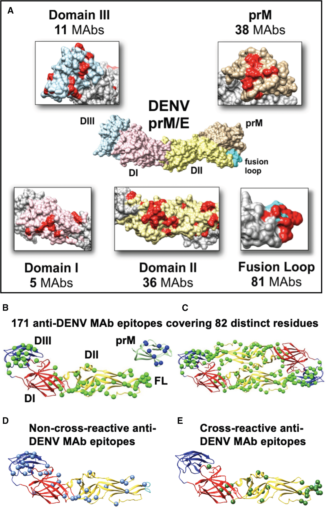
The DENV E glycoprotein consists of five major antigenic regions: the prM protein; DI, DII, and DIII; and the fusion loop of the E protein.
(A) Insets show epitope residues (red) identified by shotgun mutagenesis, mapped onto the DENV structure highlighted in each region. The epitopes shown are a summary of mAbs described and mapped previously.30–36 82 residues of DENV prM/E protein are recognized by these mAbs.
(B and C) 73 distinct residues on E protein form the epitopes of the 133 mapped DENV anti-E mAbs, shown on a DENV2 E monomer (B) and homodimer (C) (PDB: 1OAN).
(B–E) Epitope residues are shown as spheres. DI is colored red, DII yellow, and DIII blue. Also shown are the 9 distinct residues shown in dark blue on prM (PDB: 3C5X) that form the epitopes of the 38 mapped DENV anti-prM mAbs.
(D) Epitope residues for anti-DENV mAbs that do not cross-react with ZIKV are shown as blue spheres mapped onto a DENV2 monomer (PDB: 1OAN).
(E) Epitope residues for anti-DENV mAbs that are cross-reactive with ZIKV are shown as green spheres.
Identification of sites on ZIKV E proteins that cross-react with anti-DENV mAbs
We next analyzed the ZIKV cross-reactivity of 121 of the mapped anti-DENV mAbs (chosen based on their availability at the time of this study). These 121 mAbs represented a broad cross-section of the mapped anti-DENV mAbs, binding to prM; E protein DI, DII, and DIII; and the fusion loop.
Cross-reactivity of the anti-DENV mAbs with ZIKV prM/E was determined by performing a flow cytometry analysis of mAb binding to human cells expressing prM/E from DENV1–DENV4 or ZIKV (ZIKV binding is shown in Figure 2, and all data are shown in Tables S1–S5; Figure S1). These screens identified 70 anti-DENV mAbs that also bound to ZIKV prM/E (signal:background of 3 or higher). Generally, ZIKV cross-reactive mAbs were reactive with all four DENV serotypes, consistent with broad flavivirus cross-reactivity. DENV3 and DENV4 were the most frequently recognized DENV serotypes, with 100% of cross-reactive mAbs recognizing both ZIKV and DENV4 and 99% binding DENV3. DENV1 was the least cross-reactive, with 64% (45/70) of ZIKV binding mAbs also binding DENV1.
Figure 2. Identification of anti-DENV mAbs that cross-react with ZIKV prM/E.

mAb cross-reactivity against HEK-293T cells expressing ZIKV prM/E was determined for anti-DENV mAbs targeting the fusion loop, DI, DII, DIII, fusion loop/DII, bc loop, and prM. Reactivity is reported as fluorescence signal over background. Data represent the mean and range of two replicate measurements. The x axis indicates individual mAbs ordered by ZIKV reactivity values.
Of the 70 DENV mAbs identified as cross-reactive, 46 targeted the E protein fusion loop (94% of fusion loop mAbs were cross-reactive, 46 of 49 mAbs), 5 targeted the fusion loop/DII (42% cross-reactive, 5 of 12 mAbs), and 17 targeted non-fusion loop epitopes within DII (53% cross-reactive, 17 of 32 mAbs). Additionally, 1 of 2 mAbs targeting DI and 1 of 2 mAbs targeting the DII bc loop were cross-reactive with ZIKV. None of the anti-DENV mAbs binding prM (17 tested) or E protein DIII (7 tested) bound ZIKV above background levels.
To identify the residues in ZIKV E protein that are recognized by cross-reactive DENV mAbs, we analyzed the residues comprising the epitopes identified for these mAbs and visualized them on the structure of the DENV2 E protein monomer (Figures 1D and 1E; residue numbering uses DENV2 for Tables S1–S5, and protein alignment is provided in Figure S2). A total of 30 cross-reactive epitope residues were identified and were further analyzed by domain (Table 1). The residue most frequently associated with cross-reactive DENV mAbs was E protein fusion loop residue W101, present in 89% (41 of 46) of cross-reactive fusion loop mAb epitopes. Fusion loop residues L107 (37%, 17 of 46) and F108 (39%, 18 of 46) were also frequently represented in the epitopes of cross-reactive mAbs. Cross-reactivity in other regions of DII was primarily attributed to residue W212, present in 88% of DII epitopes that did not include the fusion loop (15 of 17 cross-reactive DII mAbs). The importance of residues W101 and W212 is highlighted by the fact that at least one of them is involved in binding 81% (57 of 70) of all cross-reactive mAbs.
Table 1.
Summary of anti-DENV mAbs cross-reacting with ZIKV
| Domain | No. of DENV mAbs tested | No. ZIKV cross-reactive mAbs | Cross-reactive mAb epitope residues in DENV E protein (no. of mAbs) |
|---|---|---|---|
|
| |||
| prM | 17 | 0 | none |
| DI | 2 | 1 | V151 (1), R188 (1), G281 (1), H282 (1), K284 (1) |
| DII | 32 | 17 | K128 (1), E133 (1), H209 (3), W212 (15), D215 (5), P217 (4), P222 (1), A245 (1), Q256 (1), A267 (1) |
| bc loop | 2 | 1 | R73 (1), G78 (1), E79 (1) |
| Fusion loop | 49 | 46 | R99 (3), G100 (5), W101 (41), G102 (2), G104 (5), G106 (7), L107 (17), F108 (18), G111 (7) |
| DII/fusion loop | 12 | 5 | R73 (2), G78 (4), E79 (1), R99 (1), G100 (2), W101 (1), G104 (3), G106 (2), L107 (2), K110 (4), G111 (2), V113 (1), A245(1), T262 (1) |
| DIII | 7 | 0 | none |
Summary of the 121 tested anti-DENV mAbs that cross-react with ZlKV (70 mAbs) and the 30 distinct DENV E protein residues to which they bind (note that some residues are listed twice in the table because of overlapping regions). Residue numbering follows DENV2 (sequence alignment is provided in Figure S2). For each domain, the number of mAbs that bound to each residue is shown in parentheses. Residues found to make major contributions to DENV-ZIKV cross-reactivity are underlined, based on the representation of that residue by the cross-reactive mAb epitopes within each domain. The 7 underlined residues were chosen to cover the broadest epitope space with the most conserved residues. Other residues were not included despite being critical for multiple mAbs, as they were often redundant with already selected residues.
One epitope was identified in the DII bc loop, adjacent to the fusion loop, which is recognized by the highly cross-reactive mAb 1C19,32 and is comprised of critical residues R73, G78, and E79. Also behind the fusion loop, residues V113 and A245 were components of some cross-reactive DII/fusion loop epitopes. Two other mAbs recognized a loop adjacent to DI, with one of these mAbs cross-reactive with ZIKV.
Overall, our data show that 94% (66 of 70) of cross-reactive mAbs recognize one or more of seven critical E protein residues: R73, E79, W101, L107, F108, K110, and W212, with 57 of these 70 mAbs (81%) binding to either W101 or W212. 25 of the 30 E protein residues in the cross-reactive DENV mAb epitopes are identical among ZIKV and all four DENV serotypes, including W101 and W212. Of the exceptions, 4 residues (V113, K128, P222, and T262) are conserved between ZIKV and at least one DENV serotype, and only 1 residue (V151) is not present in ZIKV at all (substituted by isoleucine), although it is conserved among all DENV serotypes (an alignment of ZIKV and DENV1–DENV4 E proteins is provided in Figure S2).
Some cross-reactive mAbs were identified previously as neutralizing (* inTables S1–S5), broadly neutralizing for DENV1–DENV4 (**), or cross-neutralizing for ZIKV (***, mAb 1C1937). The broadly neutralizing mAbs have epitope residues located at or near the fusion loop, while cross-neutralizing mAb 1C19 binds DII behind the fusion loop (Figure 3A). While fusion loop mAbs recognize a short stretch of residues in DENV, their binding typically shows conformational dependence on disulfide bonds.30–35 Similarly, all fusion loop and other cross-reactive mAbs that we mapped also recognize conformational epitopes (Figure 3B), with binding dependent on Cys residues that form disulfide bonds. Nearly all epitope residues showed high conservation between ZIKV and all 4 DENV serotypes (Figures 3C and S2). The epitope residues that were bound by the most mAbs were those in the fusion loop (Figure 3D).
Figure 3. Summary of cross-reactive epitopes.

Cross-reactive epitopes of DENV mAbs were characterized based on (A) neutralization status; mAb critical residues are shown as ZIKV cross-neutralizing mAbs (XNAbs), broadly neutralizing mAbs (bNAbs) for DENV1–DENV4, and DENV-neutralizing mAbs (NAbs).
(B–D) Conformational binding, determined from epitope mapping data or prior denaturing western blot experiments (B),30 conserved (or non-conserved) residue identity across all DENV serotypes and ZIKV (C), and the number of mAbs that bind to each epitope residue (D).
Cross-reactive fusion loop mAbs that neutralize DENV do not neutralize ZIKV
To further characterize anti-DENV mAb reactivity to ZIKV, we tested ZIKV neutralization using a selection of cross-reactive anti-DENV mAbs. Many of the mAbs tested here neutralized DENV infectivity, as described in the publications that originally characterized these mAbs (Tables S1–S5). We investigated the neutralizing abilities of four anti-DENV mAbs that were previously shown to neutralize DENV and that are cross-reactive with ZIKV (mAbs 1N5, 4E8, and 1C1932 and 4G238). These mAbs target the fusion loop (mAbs 1N5, 4E8, and 4G2) and the DII bc loop located behind the fusion loop (mAb 1C19). We compared the ability of these mAbs to neutralize ZIKV reporter virus particles (RVPs).37 Neutralization assays confirmed that the fusion loop mAbs neutralized all DENV serotypes (Figure S3A), yet none neutralized ZIKV (Figure S3B). mAb 1C19 (binding to the bc loop) was previously characterized as possessing potent neutralizing activity for DENV32 and also neutralizes ZIKV.37 In contrast to the fusion loop mAbs, mAb 1C19 effectively neutralized ZIKV RVPs with a 50% inhibitory concentration (IC50) value of 0.19 μg/mL, comparable to the IC50 values obtained with DENV1 (0.32 μg/mL) and DENV3 (0.96 μg/mL). These results show that, while fusion loop mAbs are highly cross-reactive, they do not necessarily cross-neutralize, consistent with previous studies.19 The inability of fusion loop DENV mAbs to neutralize ZIKV, despite strong binding, suggests that they could facilitate ADE. This potential for ADE highlights the fusion loop residues as critical targets for mutation in ZIKV vaccine development, where modifying these epitopes could reduce the generation of non-neutralizing, cross-reactive antibodies and lower the risk of ADE.
ENGINEERING A ZIKV IMMUNOGEN TO ELIMINATE CROSS-REACTIVITY WITH DENV mAbs
With the goal of reducing DENV-ZIKV antibody cross-reactivity, we next sought to engineer ZIKV prM/E using data from our analyses of mAb cross-reactivity, neutralization, and epitope mapping. We identified seven E protein residues that together were critical for the binding of nearly all (94%) cross-reactive DENV mAbs: DII bc loop R73 and E79; fusion loop W101, L107, and F108; fusion loop/DII K110; and DII W217 (the ZIKV E protein residue corresponding to DENV W212) (Figure 4A). With the aim of generating a ZIKV prM/E immunogen that would induce minimal cross-reactivity to DENV envelope, we individually mutated each of these 7 residues on ZIKV prM/E to 16 different amino acids to identify suitable alternate residues at each position.
Figure 4. Engineering of ZIKV E protein reduces DENV mAb cross-reactivity.

(A) Key residues on ZIKV E protein that determine cross-reactivity with anti-DENV mAbs were mutated. Fusion loop residues W101, L107, and F108; fusion loop/DII residue K110; bc loop residues R73 and E79; and DII residue W217 (W212 on DENV) were each mutated to 16 different amino acids (shown on ZIKV E: PDB: 5IRE). Mutation to Cys and Pro was avoided due to probable structural perturbations, and mutation to Ala was unnecessary here due to prior Ala-scan mutational studies. Residue numbering aligns with ZIKV.
(B) SVP production for each mutant was measured by ELISA. Data points are the average of 3 replicates and are shown as a percentage of non-mutated WT ZIKV SVP production.
(C) Cross-reactivity with anti-DENV mAbs that target the specific mutated residue on ZIKV E was evaluated by flow cytometry. Results are shown as a percentage of binding to the WT and are an average of 2 replicates.
(D) Cells expressing the engineered ZIKV prM/E construct (ZIKVm10) showed high reactivity with conformational anti-ZIKV mAbs (ZIKV-195, ZIKV-394, and ZIKV-161) binding to different epitopes of E protein and low reactivity with anti-DENV mAbs targeting DII fusion loop (3F21) and the DII bc loop (1C19) but high activity with mAb 1N18 targeting DII at non-engineered ZIKV residue W217. Binding was evaluated by flow cytometry; results are shown as a percentage of binding to the WT and show the average and standard deviation of 4 replicates.
(E) SVPs were produced, incorporating WT or ZIKVm10 prM/E, with expression confirmed by ELISA, using titration with a conformational anti-DII mAb ZIKV-117 (after capture by conformational anti-DIII mAb ZV-56). Each data point is the average and range of 2 replicates.
We tested the ability of each prM/E variant to be incorporated into ZIKV subviral particles (SVPs), generated by expression of each variant ZIKV prM/E construct without capsid. To confirm SVP production of each variant, SVP secretion was determined by ELISA (Figure 4B). Most clones displayed some SVP secretion capability; however, ZIKV residue W217 was found to be essential for secretion, as no mutations of this residue allowed SVP production. Next, to determine whether mutations could reduce cross-reactivity, we used flow cytometry analysis to evaluate cross-reactivity of DENV mAbs that target the specific residues that we had mutated (Figure 4C). Several mutations resulted in reduced cross-reactivity. For example, variant R73K demonstrated a high level of SVP production (150% of WT) while reducing reactivity with DENV mAbs 1C19 (40%) and 2.10H-30.10C (0%). Similarly, variant F108R retained SVP secretion at levels close to those of the WT (95%) but demonstrated reduced reactivity with DENV mAbs DENV 1C5 (14%) and DENV114 (0%). Some mutations, including W217E and W217V, greatly reduced DENV mAb binding, but no mutations at this residue supported SVP secretion.
Based on these data, we selected 5 mutations for further testing: R73K, E79L, W101K, F108R, and K110L, chosen because they reduced DENV mAb cross-reactivity in vitro while preserving (or increasing) SVP secretion. We also selected 5 additional mutations, identified from prior functional screens,27 that increased stability (E55A), neutralization (V153A), and particle production (G14A, V257A, V303A). We engineered a variant ZIKV prM/E combining all 10 of these mutations, termed ZIKVm10. We confirmed high reactivity of ZIKVm10 with four anti-ZIKV mAbs targeting different regions of the E protein (Figure 4D). As designed, the construct demonstrated low reactivity with anti-DENV mAbs targeting the fusion loop and bc loop but high reactivity with cross-reactive DENV mAb 1N18, which targets unmutated residue W217 (W212 in DENV E). To test the engineered prM/E as an immunogen, WT and ZIKVm10 SVPs were produced, with expression confirmed by ELISA (Figure 4E).
ZIKVm10 protects against ZIKV challenge and reduces DENV cross-reactivity and ADE
To test ZIKVm10 (containing mutations G14A, E55A, R73K, E79L, W101K, F108R, K110L, V153A, V257A, and V303A) for the ability to induce an immune response specific to ZIKV, we immunized CD-1 mice with WT or ZIKVm10 SVPs using three rounds of injections. Serum samples were collected prior to each immunization and at the end of the immunization protocol. Sera from both WT and the ZIKVm10 immunogen bound to WT ZIKV prM/E (Figure 5A). Sera from WT immunizations showed significant cross-reactivity to DENV prM/E on days 20, 39, and 55 (p < 0.05 vs. serum from a naive mouse) (Figure 5B). In contrast, sera from mice immunized with ZIKVm10 SVPs showed no significant cross-reactivity to DENV prM/E on days 20 and 55 (p > 0.05 vs. naive mouse serum) and only a slight increase in binding on day 39. Endpoint serum neutralization experiments demonstrated significant ZIKV neutralization by sera from both the WT ZIKV and ZIKVm10 immunizations (Table S6). Sera from mice immunized with ZIKVm10 SVPs showed 50% neutralization titer (NT50) values at lower dilutions than sera from mice immunized with WT SVPs on days 20 and 39, but NT50 values were broadly comparable for sera drawn on day 55.
Figure 5. Immunization with ZIKVm10 SVPs reduces DENV cross-reactivity while maintaining protection from live ZIKV.

(A–E) Average reactivity of serum (shown as signal:background) from 15 naive CD-1 mice or mice immunized with WT or ZIKVm10 SVPs, tested by flow cytometry against cells expressing prM/E from (A) ZIKV and (B) DENV2 (*p < 0.05 vs. naive, **p < 0.01, ***p < 0.001; data are represented as mean ± SEM). AG129 mice then received passive immunizations of serum from naive mice or mice immunized with WT SVPs or ZIKVm10 SVPs and were then challenged with live ZIKV. The AG129 mice were monitored for (C) survival, (D) body weight (data represented as mean ± SD), and (E) health score (data are represented as mean ± SD).
(F) Viral load in serum from the AG129 mice was determined 4 days post infection (line at median). Tukey’s multiple comparisons test was used to compare differences between experimental groups for antibody binding. One-way ANOVA and Dunn’s multiple comparison test were used to compare viremia load. Kaplan-Meier survival curves were analyzed using the Mantel-Cox log rank test. Results were considered statistically significant if p < 0.05.
To assess the ability of the immune sera to protect against ZIKV infection, we performed passive serum transfer experiments using the ZIKV-susceptible AG129 mouse model. AG129 mice are deficient in type I and type II interferon receptors, which are important for the innate antiviral response and modulating adaptive immunity.39 Therefore, AG129 mice are highly susceptible to ZIKV infection, and any protection from lethality can be attributed solely to mAbs gained from passive immunization.40 AG129 mice were infused with sera collected from naive mice, from mice immunized with WT ZIKV SVPs, or from mice immunized with ZIKVm10 SVPs. Infused mice were then challenged with live ZIKV and monitored for weight loss and health score for 20 days post infection. As expected, ZIKV challenge in the naive group led to complete lethality by 11 days post challenge (Figure 5C). Infusion with WT or ZIKVm10 serum showed significantly enhanced survival, with 100% survival in mice given the WT prM/E serum and 87.5% survival in mice given the ZIKVm10 prM/E serum, with no statistical difference between these two groups. There was also no significant loss of body weight or increase in health score in either ZIKV immunization group (Figures 5D and 5E). Viral load on day 4 post infection was elevated in the naive group but minimal in the WT and ZIKVm10 immunization groups, with no statistical difference between them (Figure 5F).
To further investigate the effectiveness of ZIKVm10 in eliminating interactions with DENV, we tested sera from individual mice immunized with either WT or ZIKVm10 SVPs for their ability to cause ADE. We mixed serial dilutions of sera with ZIKV or DENV RVPs and tested their infectivity of K562 cells, which naturally express the Fc-gamma receptor, to provide an entry pathway for virions bound by antibody.37,41 ZIKVm10 sera required significantly higher concentrations (lower log reciprocal values, p = 0.02) to reach maximum ADE compared to the WT (Figure 6A), whereas WT ZIKV sera mediated ADE of DENV at more dilute concentrations, reflecting better binding to DENV (Figure 6B). In several ZIKVm10-immunized mice, no or only very low ADE of DENV was detectable (Figure 6C). Both WT ZIKV- and ZIKVm10-immunized mice showed enhancement of ZIKV infectivity at similar levels, as expected. Collectively, these data demonstrate that our engineered immunogen ZIKVm10 can produce similar protection from ZIKV challenge while significantly decreasing DENV cross-reactivity and DENV ADE.
Figure 6. Sera from mice immunized with ZIKVm10 SVPs show reduced ADE of DENV infectivity.

(A) The serum dilution at which maximum ADE was observed (maximum of the fitted curve) is plotted for all samples that demonstrate ADE (WT ZIKV, n = 6; ZIKVm10, n = 8). WT ZIKV sera facilitated ADE at more dilute concentrations (higher log reciprocal values), while ZIKVm10 sera required significantly higher concentrations for maximum ADE (*p < 0.05 by Student’s t test, line at mean ± SD).
(B and C) ADE-mediated infectivity of DENV2 and ZIKV reporter virus particles (RVPs) in K562 cells (which express the Fc-gamma receptor) after incubation with serum from individual mice immunized with WT ZIKV SVPs (B) or ZIKVm10 SVPs (C). All infectivity values for ZIKV RVPs are shown on the left y axis, while DENV RVPs are shown on the right y axis, with axis ranges set based on the maximum values for each dataset. Data were fit to a Gaussian distribution and are plotted as the mean ± range from duplicate assays.
DISCUSSION
The induction of neutralizing antibodies is a key goal of an efficient vaccine. However, the high homology between ZIKV and DENV E proteins complicates the development of a ZIKV vaccine due to potential exacerbation of DENV disease by cross-reactive antibodies promoting ADE. Identification and elimination of cross-reactive epitopes, as well as the induction of antibodies that bind at ZIKV epitopes not targeted by any known anti-DENV antibodies, is a promising strategy for the development of an effective ZIKV vaccine.24–26 Eliminating cross-reactive epitopes in a vaccine immunogen would minimize ADE by inducing an immune response against only ZIKV E protein epitopes that are not shared by DENV.
In the current study, we screened 121 epitope-mapped anti-DENV mAbs for cross-reactivity with ZIKV to identify antigenically important residues shared between DENV and ZIKV. We identified fusion loop residues W101, L107, F108, and K110; non-fusion loop DII residue W212; and bc loop residues R73 and E79 as the primary determinants of cross-reactivity. These seven residues were determined as critical for the binding of 94% of cross-reactive mAbs identified. By mutating the ZIKV E protein, we identified specific mutations of these residues that reduced DENV mAb cross-reactivity in vitro while maintaining secretion of ZIKV SVPs, highlighting the potential of this approach for vaccine design. Mutations that reduced cross-reactivity were combined with additional mutations (found previously to enhance SVP production) to create an engineered ZIKV variant immunogen (ZIKVm10) that showed decreased binding to cross-reactive DENV mAbs. We investigated the antigenicity of ZIKVm10 in vivo by immunizing mice with SVPs containing either WT prM/E ZIKV protein or ZIKVm10 and then used sera isolated from these mice for passive immunization of AG129 mice (which are highly susceptible to ZIKV infection). After challenge of passively immunized mice with live ZIKV, we did not detect a significant difference between the WT and ZIKVm10 immunization groups in survival, body weight loss, health score, or viral load, indicating effective protection induced by ZIKVm10. At the same time, sera from mice immunized with ZIKVm10 demonstrated low cross-reactivity with DENV and showed decreased in vitro ADE activity with DENV, suggesting that most cross-reactive epitopes in ZIKVm10 were eliminated as intended.
Our study identified a total of 70 anti-DENV mAbs (58% of all mAbs tested) that were cross-reactive with ZIKV. Cross-reactive mAb epitopes localized to the fusion loop, DII, DI, and bc loop of the E protein, and cross-reactivity was not observed for mAbs with epitopes in prM or E protein domain DIII. Previous reports similarly demonstrated DENV-ZIKV cross-reactivity in the fusion loop.19,42 Here, we extend these findings by systematically identifying individual residues throughout prM/E that contribute to cross-reactivity. Major contributors to this cross-reactivity are fusion loop residue W101 and DII residue W212, both conserved across ZIKV and all four DENV serotypes. Between them, these two residues were determined to be critical for binding of 81% of cross-reactive mAbs, with W101 a target residue for the vast majority of mAbs (43 of 49) that bound the fusion loop. W212 was also found to be critical for the majority of cross-reactive mAbs in DII. The lack of cross-reactivity of DENV prM antibodies with ZIKV is consistent with the relatively low conservation between DENV and ZIKV prM, and similar low cross-reactivity was observed in previous western blot analyses.43 Neutralization studies confirmed that DENV-neutralizing mAbs targeting the fusion loop did not neutralize ZIKV, even though these same mAbs can be highly cross-reactive in binding, as seen in previous studies.44 The fusion loop is accessible in immature regions of flavivirus virions; the discrepancy between ZIKV and DENV in neutralization may reflect differences in virus stability and breathing that influence the exposure of fusion loop epitopes.45,46 The non-neutralizing cross-reactivity among antibodies recognizing the fusion loop is an important consideration for vaccine design, since it has the potential to enhance infection through ADE.19,20 Cross-reactivity outside of the fusion loop also has important implications for ADE. Generally, mAbs with epitopes in the fusion loop and DII region can neutralize DENV serotypes but only bind ZIKV weakly and display ADE.32,37,42
In addition to the cross-reactive mAbs described above, a class of broadly neutralizing anti-DENV mAbs bind quaternary envelope dimer epitopes (EDEs) on the E protein of all 4 DENV serotypes and also bind and potently neutralize ZIKV.44,47,48 EDE mAbs were not represented in our original panel of 171 DENV mAbs, but we epitope mapped the EDE mAbs C8 and C10 on ZIKV E protein separately.37 We note that their binding was greatly reduced by several conserved residues identified here for cross-reactive DENV mAbs, including R73, W101, and F108, which were mutated in ZIKVm10.
While data focused on the cross-reactivity between ZIKV and DENV are useful, identification of regions that are not cross-reactive is also important to determine targets for ZIKV-specific antibodies and vaccine candidates. Our data suggest that the prM and DIII regions of prM/E are generally not cross-reactive between DENV and ZIKV and therefore are areas likely to be good targets for ZIKV-specific mAbs. The lack of cross-reactive epitopes in DIII is particularly notable, as other flavivirus vaccine strategies have used DIII as the primary target for immunization,49,50 and our data suggest that DIII from ZIKV would not induce antibodies that cross-react with DENV. Our data are consistent with previous reports of DIII mAbs against ZIKV being strongly neutralizing for ZIKV with limited cross-reactivity to DENV.18,51,52 Anti-prM mAbs generally do not neutralize and are poorly described for ZIKV.
Identifying immunization strategies that induce in vivo protection through an engineered ZIKV immunogen without inducing cross-reactivity to DENV is an active area of research. Previous studies have evaluated engineered vaccine candidates in mice, including dengue and Zika virus-like particles (VLPs),40,53 mRNA,24 and prM/E genes expressed from adenoviruses.26 A major goal of engineering a ZIKV immunogen is to eliminate cross-reactivity in the fusion loop to minimize the generation of non-neutralizing anti-fusion loop mAbs that would result in ADE.24 However, mutations can have unwanted consequences. For example, in one study, ZIKV VLPs with a single amino acid change in the fusion loop (F108A) resulted in a completely immature VLP with no detectable prM processing by furin.54 AG129 mice immunized with the immature F108A VLPs were significantly diminished in their ability to induce neutralizing antibodies and to protect against ZIKV challenge. Similarly, in our screen, multiple mutations at W217 reduced mAb cross-reactivity but failed to support SVP secretion.
Mutating residues to minimize cross-reactivity may also disrupt epitopes targeted by known neutralizing antibodies, such as EDE mAbs. Consequently, lower levels of ZIKV cross-reactive antibodies may be accompanied by a slight reduction in the total pool of antibodies capable of neutralization, which may explain the relatively lower NT50 values elicited by ZIKVm10 compared to the WT on days 20 and 39 (Table S6). Similar observations have been made previously by others,24 with several reasons proposed for lower neutralizing responses induced by mutated E protein, including mutations changing the display of neutralizing epitopes.
Other successful vaccine candidates have taken a similar approach but focused primarily on engineering the ZIKV fusion loop.24,26 These immunogens conferred immunity against ZIKV without enabling ADE for DENV serotypes 1–4 and could also abrogate maternal-neonatal transmission.26 Similarly, immunization with an E dimer subunit engineered to minimize fusion loop exposure resulted in ZIKV-neutralizing antibodies with minimal DENV cross-reactivity.55 Also, immunization with an E protein containing mutations in and near the fusion loop elicited antibodies with neutralizing capacity equal to that induced by WT E protein but induced much less serological cross-reactivity and did not cause ADE in vitro.25 Finally, engineering and directed evolution strategies have been used to obtain a mutated but functional DENV fusion loop that is not recognized by anti-fusion loop mAbs.56
Our study identifies a set of residues from the epitope mapping of 171 anti-DENV mAbs and uses the epitope data to identify residues that determine the cross-reactivity between DENV and ZIKV. Mutations at cross-reactive residues allowed the generation of ZIKV SVPs that no longer react with DENV mAbs. Immunization of mice with ZIKVm10 SVPs incorporating these mutations reduces serum DENV cross-reactivity and protects against ZIKV challenge similar to the WT, suggesting the utility of using information on cross-reactive epitope residues to optimize a ZIKV vaccine.
Limitations of the study
The DENV mAbs studied here were obtained from several laboratories, each using their own isolation strategy, but still represent a limited set of mAbs. Most of the mAbs we tested (70%) bind the fusion loop or prM, and so our cross-reactivity assays are biased toward these classes of antibodies. We also did not observe DENV serotype cross-reactivity with the panel of DIII mAbs we tested; all mAbs we tested that bound DIII recognized only DENV4. This is likely due to the relatively limited samples available in our study, as cross-reactive DIII mAbs have been described elsewhere.57 Our results likely reflect biases in the ways that mAbs were originally identified and screened during their isolation (by isolation method, against what serotype, etc.) and the diversity of epitopes recognized by the mAbs obtained. The testing of more mAbs and more diverse mAbs in the future could potentially identify additional residues involved in DENV-ZIKV cross-reactivity.
RESOURCE AVAILABILITY
Lead contact
Requests for further information and reagents may be directed to the lead author, Benjamin J. Doranz (bdoranz@integralmolecular.com).
Materials availability
Requests for reagents may be directed to the lead contact, Benjamin J. Doranz (bdoranz@integralmolecular.com). Reagents generated in this study will be made available on request, but we may require a payment and/or a completed materials transfer agreement if there is potential for commercial application.
Data and code availability
No original code was generated in this study. All original viral sequences used in this study were previously deposited; GenBank identifiers are provided in the STAR Methods. Any additional information required to reanalyze the data reported in this paper is available from the lead contact upon request.
STAR★METHODS
EXPERIMENTAL MODEL AND STUDY PARTICIPANT DETAILS
Cell lines
The following cell lines were used in this study: K562, HEK-293T and Vero (all from ATCC, Vero cell line was authenticated by ATCC services), QT6 quail cells (kindly provided by Paul Bates, University of Pennsylvania), BHK-DRRZ cells expressing DENV2 replicon,27,37 and BHK cells expressing DC-SIGN (derived from BHK21 clone 15, Center for Vector-borne Diseases, UC Davis). All cell lines were grown at 37°C at 5% CO2 in DMEM complete medium (DMEM with 10% FBS and 1% penicillin-streptomycin, 2 mM L-glutamine, 10 mM HEPES, 1 mM sodium pyruvate and 1% MEM non-essential amino acids) (Corning). All cell lines tested negative for mycoplasma contamination before use.
Mouse studies
All animal experiments were conducted in accordance with IACUC guidelines and regulations and were performed at IBT Bioservices (Rockville, MD); the IBT Bioservices review board approved this study. The immunogenicity study used 45 female Swiss-ICR CD-1 mice aged 6–8 weeks, which may limit the generalizability of the findings to both sexes. 6–8-week-old AG129 mice (IBT Bioservices) deficient in type-I and type-II interferon receptors were used for passive immunization studies (28 male and 28 female). Animals were housed and cared for under conditions that meet the requirements specified in the NRC’s Guide for the Care and Use of Laboratory Animals.
METHOD DETAILS
Flow cytometry detection of MAb or serum binding
Expression constructs for the wild-type prM/E envelope proteins of DENV1 (WestPac), DENV2 (16803), DENV3 (CH53489), DENV4 (TVP360), and ZIKV (SPH2015), or ZIKV prM/E mutants were transfected into HEK-293T cells and allowed to express for 22 h. Cells were washed and fixed in 4% (vol/vol) paraformaldehyde (Electron Microscopy Sciences), permeabilized with 0.1% (wt/vol) saponin (Sigma-Aldrich), and incubated with purified MAbs diluted in PBS, 10% normal goat serum (NGS) (Sigma), and 0.1% saponin, followed by an AlexaFluor 488-conjugated secondary antibody (Jackson ImmunoResearch Laboratories) in 10% NGS/0.1% saponin. Cells were washed 3 times with PBS supplemented with Ca2+ and Mg2+ (PBS++)/0.1% saponin, followed by 2 washes in PBS. Mean cellular fluorescence was detected using a flow cytometer (Intellicyt). For each antibody, the binding data are given as a signal:background value (background fluorescence obtained by incubating MAbs with cells transfected with a negative control plasmid) and data shown are the average of 2 replicates.
To test mouse sera reactivity with ZIKV and DENV2 prM/E, these proteins were expressed in avian QT6 cells and assayed as above, except that sera were pre-adsorbed with fixed QT6 cells before the assays. For this, 500uL of 1:1,650 serum dilution was incubated with 2.5 × 10⁶ fixed QT6 cells in deep-well plates for 1h with shaking at 4°C. Cells were then removed by centrifugation.
To screen sera binding to ZIKV and DENV2 prM/E, QT6 cells were co-transfected for 22h with GFP and ZIKV prM/E, DENV2 prM/E, or pUC, fixed, permeabilized and blocked in 10% NGS, as above. After a 1h incubation, cells were washed at room temp with the 1:1,650 diluted pre-adsorbed sera. Cells were washed before detection with anti-mouse secondary conjugated with AF647 and read on an Intellicyt flow cytometer, as above. Fluorescence was measured after gating for transfected cells (GFP+). Binding by each sample was calculated as a signal:background and data shown is the average of 4 replicates.
Production of ZIKV and DENV reporter virus particles (RVPs)
ZIKV and DENV RVPs were produced as described previously37,41 by co-expression of virus CprM/E structural genes transfected into the stable cell line BHK-DRRZ, which expresses a full-length DENV2 replicon in which the CprM/E genes are replaced by a gene for Renilla luciferase. After 72h, supernatants containing RVPs were harvested, passed through 0.45 μm filters, aliquoted, and stored at −80°C. For all experiments, frozen RVPs were thawed for 3 min in a 37°C water bath and then placed on ice before use.
Production of ZIKV SVPs
ZIKV subviral particles (SVPs) lacking capsid protein were produced by transfection of HEK-293T cells with a ZIKV prM/E expression plasmid. For small-scale production, cells in 96-well plates were transfected with WT or mutant prM/E expression plasmids, and supernatants were harvested 48 h after transfection. Large scale production of ZIKV SVPs for immunization was performed by calcium phosphate transfection of ZIKV prM/E expression plasmid into HEK-293T cells. SVPs were harvested at 48 h. Clarified harvest medium was concentrated via tangential flow filtration (TFF) (70 kDa MWCO) then layered over 20% sucrose and centrifuged for 3 h at 150,000 × g. Pelleted material was resuspended in PBS (pH 7.5) and concentrated through a second sucrose cushion. The resulting pellet was resuspended in PBS (pH 7.5), aliquoted, and frozen at −80°C.
Neutralization assays
Neutralization assays were carried out using Reporter Virus Particles (RVPs), as described previously.37 The sequences from E proteins presented on RVPs were derived from the following strains: DENV1 (WestPac), DENV2 (16681), DENV3 (CH53489), DENV4 (TVP360), and ZIKV (SPH2015). MAbs were diluted to 120 μg/mL in 250 μL Infection Media (DMEM supplemented with 10% FBS adjusted to pH 8 ± 0.05 with NaOH) in a 96 well V bottom plate, and 3-fold dilutions were made thereafter using aerosol barrier tips. Sera were heat inactivated for 30 min at 56°C prior to use. Serum samples were initially diluted 5-fold and then 3-fold dilutions were made thereafter. 90 μL of diluted MAbs were transferred in duplicate to a black 96-well plate pre-coated with Poly-D-Lysine. ZIKV RVPs were thawed and diluted 1:4.5, to a previously determined working concentration, using Infection Media. 90 μL of diluted RVPs were added to each well of the neutralization plate containing MAbs. As a negative control, cells were incubated without ZIKV RVPs. Plates were covered and placed in a 37°C tissue culture incubator. Following a 1 h incubation, 3×10⁴ BHK-DC-SIGN cells were added to each well in 50 μL of Infection Media and plates were incubated for 72 h at 37°C before measurement of luciferase activity. Luciferase activity was detected using the Promega Renilla-Glo Luciferase Assay System (Promega #E2710). Plates were spun for 10 min at 2,000 rpm (581g, Sorvall Legend XTR), supernatant was removed, and cells were lysed using 25 μL of freshly prepared 1X Renilla luciferase assay lysis buffer. Covered plates were incubated at room temperature with shaking at 500 rpm for 30 min. Luciferase activity (relative luminescence units, RLU) was measured by adding 30 μL of luciferase assay buffer (buffer + substrate) per well, gently swirling the plate manually, and then incubating for 5 min at room temperature before reading on an Envision plate reader (PerkinElmer). Data are the average of two replicate measurements and are reported as percent infection by normalizing to wells containing cells infected by RVPs in the absence of antibody or serum (defined as 100% infectivity). 50% neutralization titers (NT50) for sera or inhibitory concentrations (IC50) for MAbs were calculated using GraphPad Prism. NT50 and IC50 values were calculated by fitting the data to the equation for sigmoidal dose-response with variable slope.
Antibody-dependent enhancement (ADE) assays
ZIKV and DENV RVPs were mixed with serial dilutions of sera from mice immunized with WT ZIKV SVPs or ZIKVm10 SVPs in black PDL-coated 96-well plates and incubated for 1 h at 37°C. K562 target cells were then added to each well and incubated for 72 h at 37°C. Culture medium was removed, cells were lysed, and plates were analyzed for luciferase activity as described above. Data were curve-fit using the Gaussian function in GraphPad Prism.
SVP production ELISA
A 384-well white flat-bottom microtiter plate was coated with mouse DIII MAb ZV-5658 (5 μg/mL) in 0.1 M NaHCO3, pH 8.6, and incubated overnight at 4°C. The plate was blocked with 3% (wt/vol) BSA in PBS for 2 h at room temperature. To compare the SVP budding abilities of engineered variant ZIKV prM/E constructs, a 96-well SVP production plate containing frozen SVPs was thawed, and equal volumes (50 μL) of supernatant were transferred to the blocked ELISA plate and incubated overnight at 4°C to allow capture of SVPs. A human anti-DIII MAb, ZIKV-394, or DII MAb ZIKV-11751 in blocking buffer was incubated on the plate for 1 h at room temperature, followed by addition of rabbit anti-human HRP-conjugated secondary antibody (1:5,000) in blocking buffer for 1 h at room temperature. Reactivity was detected using SuperSignal West Femto Chemiluminescent Substrate. Serial dilutions of both capture and detection antibodies were used to optimize detection conditions (Z′ > 0.3). All luminescence values were background-subtracted and normalized to the average luciferase signal generated from wild-type SVPs.
Mouse studies
All animal experiments were conducted in accordance with IACUC guidelines and regulations and were performed at IBT Bioservices (Rockville, MD).
Immunogenicity study in CD-1 mice
Immune sera for passive transfer studies were generated by immunizing 6–8 week old Swiss-ICR CD-1 mice (Charles River) on days 0, 21, and 40 using a dose of 2 μg of ZIKV WT or ZIKVm10 SVPs formulated with SAS adjuvant (Sigma Adjuvant System, Sigma Aldrich). Mice (n = 15/group) were immunized by intramuscular (IM) injection at the tail base. Blood samples were collected on days −3 (pre-immune), 20, 39 and 55 (terminal bleed). Serum was tested for neutralizing activity of ZIKV, for binding to ZIKV and DENV prM/E by flow cytometry, and for ADE with DENV and ZIKV RVPs (as described above). Terminal sera were then pooled for passive immunization studies.
Passive immunization studies in AG129 mice
Groups (n = 8/group) of 6–8-week-old AG129 mice (IBT Bioservices) were passively transferred with 200 μL of pooled day 55 sera by intraperitoneal (i.p.) injection. After one hour, mice were challenged subcutaneously (s.c.) with 104 PFU of ZIKV strain FSS13025. Blood samples were collected by retro-orbital bleed 4 days post challenge for analysis of viremia by plaque titration. Following virus challenge, mouse weight, morbidity, and mortality were monitored daily. A 1-to-5 morbidity scale was adapted from60: 1) Healthy; 2) Displaying mild signs of lethargy, some fur ruffling, and no hunched posture; 3) Fur ruffling, mild signs of lethargy, early signs of hind leg paralysis possible; 4) Fur ruffling, increased lethargy and limited mobility, and signs of paralysis common; and 5) Moribund, minimal mobility consistent with inability to reach food or water, neurological signs evident (paralysis/seizures). Mice were euthanized if weight loss was equal to or greater than 20% of their original weight and/or if they scored 5 on the morbidity scale.
ZIKV plaque reduction neutralization test (PRNT)
Endpoint 50% PRNT titers were performed essentially as previously described.37 Sera were diluted as described above for neutralization assays and then incubated for 1 h at 37°C with approximately 80 PFU of ZIKV (SPH2015). Vero cells were seeded the previous day in 6-well tissue culture plates at a density of 6×10⁵ cells/well. Medium was aspirated and replaced with the antibody/virus mixture, and plaques were allowed to develop under agar overlays. After 72 h, 2 mL of 0.6% agar containing 50 μg/mL neutral red was overlaid and plaques were counted after an additional 72 h.
QUANTIFICATION AND STATISTICAL ANALYSIS
The Student’s t-test was used to compare differences between experimental groups for neutralizing antibody titers and binding. One-way ANOVA and Dunn’s multiple comparison test was used to compare viremia load. Kaplan-Meier survival curves were analyzed using the Mantel-Cox log-rank test. Tukey’s multiple comparisons test was used to compare sera binding to SVPs. Results were considered statistically significant if the p-value was <0.05. All statistical analysis was performed using GraphPad Prism software.
ADDITIONAL RESOURCES
A detailed protocol of infection for the DENV and ZIKV RVPs used here can be found at: https://www.integralmolecular.com/wp-content/uploads/2023/06/A01-Infectivity-of-DENV-ZIKV-RVPs.pdf
Supplementary Material
SUPPLEMENTAL INFORMATION
Supplemental information can be found online at https://doi.org/10.1016/j.celrep.2025.116098.
KEY RESOURCES TABLE.
| REAGENT or RESOURCE | SOURCE | IDENTIFIER |
|---|---|---|
|
| ||
| Antibodies | ||
|
| ||
| ZIKV-195 | Sapparapu et al.51 | N/A |
| ZIKV-394 | This manuscript, Crowe lab | N/A |
| ZIKV-161 | This manuscript, Crowe lab | N/A |
| ZIKV-117 | Sapparapu et al.51 | N/A |
| 4G2 | Henchal et al.38 | N/A |
| Alexa Fluor 488-conjugated goat anti-human secondary antibody | Jackson ImmunoResearch Laboratories | Cat# 109-545-003, RRID:AB_2337831 |
| Anti-mouse secondary conjugated with AF647 | Jackson ImmunoResearch Laboratories | Cat# 115-605-003; RRID:AB_2338902 |
| ZV-56, mouse DIII MAb | Zhao et al.58 | N/A |
| D11C | Costin et al.30 | N/A |
| 2.4A-16.11F | This manuscript, Schieffelin lab | N/A |
| 3.6C-40.8G | This manuscript, Schieffelin lab | N/A |
| 1B19 | Smith et al.32 | N/A |
| 1C17 | This manuscript, Crowe lab | N/A |
| 1C18 | Smith et al.32 | N/A |
| 1D11 | This manuscript, Crowe lab | N/A |
| 1E4 | Smith et al.32 | N/A |
| 1F12.2 | Smith et al.32 | N/A |
| 1G23 | This manuscript, Crowe lab | N/A |
| 1H20 | Smith et al.32 | N/A |
| 1J14 | This manuscript, Crowe lab | N/A |
| 1J21 | This manuscript, Crowe lab | N/A |
| 1K16 | Smith et al.32 | N/A |
| 1K19 | This manuscript, Crowe lab | N/A |
| 1L4 | Smith et al.32 | N/A |
| 1M6.2 | Smith et al.32 | N/A |
| 1M12.2 | Smith et al.32 | N/A |
| 1N5 | Smith et al.32 | N/A |
| 1N8 | Smith et al.32 | N/A |
| 2B12 | This manuscript, Crowe lab | N/A |
| 2B21 | This manuscript, Crowe lab | N/A |
| 2C9 | This manuscript, Crowe lab | N/A |
| 2F4 | This manuscript, Crowe lab | N/A |
| 2H15 | This manuscript, Crowe lab | N/A |
| 2M11 | This manuscript, Crowe lab | N/A |
| 3B4 | Smith et al.32 | N/A |
| 3D18 | Smith et al.32 | N/A |
| 3G5 | Smith et al.32 | N/A |
| 3H4 | Smith et al.32 | N/A |
| 4E8 | Smith et al.32 | N/A |
| 5C7 | This manuscript, Crowe lab | N/A |
| 5C8 | Smith et al.32 | N/A |
| 5K17 | Smith et al.32 | N/A |
| 5N6 | This manuscript, Crowe lab | N/A |
| 2.9G | This manuscript, Schieffelin lab | N/A |
| 1.9E | This manuscript, Schieffelin lab | N/A |
| 17.11E | This manuscript, Schieffelin lab | N/A |
| DV4-E4 | This manuscript, Diamond lab | N/A |
| WNV E18 | Smith et al.31 | N/A |
| WNV E60 | Smith et al.31 | N/A |
| WNV E86 | Smith et al.31 | N/A |
| 1B H1L1 | Xu et al.36 | N/A |
| 3H H1L1 | Xu et al.36 | N/A |
| 5A H6L1 | Xu et al.36 | N/A |
| 5D H6L1 | Xu et al.36 | N/A |
| 6E H1L1 | Xu et al.36 | N/A |
| 8F H1L1 | Xu et al.36 | N/A |
| 1F7 | This manuscript, Crowe lab | N/A |
| 2.11B-18.12D | This manuscript, Schieffelin lab | N/A |
| 5.3A-34.9H | This manuscript, Schieffelin lab | N/A |
| 2J21 | Smith et al.32 | N/A |
| 2.8H | This manuscript, Schieffelin lab | N/A |
| 1.8B | This manuscript, Schieffelin lab | N/A |
| 1D H4L1 | Xu et al.36 | N/A |
| 1.6B-36.9E | This manuscript, Schieffelin lab | N/A |
| 2.10H-30.10C | This manuscript, Schieffelin lab | N/A |
| 5O16 | This manuscript, Crowe lab | N/A |
| 15.3D | This manuscript, Schieffelin lab | N/A |
| 14.5F | This manuscript, Schieffelin lab | N/A |
| 3.5E | This manuscript, Schieffelin lab | N/A |
| 3.1E-61.7F | This manuscript, Schieffelin lab | N/A |
| 4.2C-13.7F | This manuscript, Schieffelin lab | N/A |
| 5.11D-33.11B | This manuscript, Schieffelin lab | N/A |
| 9.4F-8.10E | This manuscript, Schieffelin lab | N/A |
| 1A15 | This manuscript, Crowe lab | N/A |
| 1B13 | This manuscript, Crowe lab | N/A |
| 1D6 | This manuscript, Crowe lab | N/A |
| 1D7 | This manuscript, Crowe lab | N/A |
| 1D8 | This manuscript, Crowe lab | N/A |
| 1K21 | This manuscript, Crowe lab | N/A |
| 1M12 | This manuscript, Crowe lab | N/A |
| 1M21 | This manuscript, Crowe lab | N/A |
| 1N18 | This manuscript, Crowe lab | N/A |
| 1O18 | This manuscript, Crowe lab | N/A |
| 2A10 | This manuscript, Crowe lab | N/A |
| 2D7 | This manuscript, Crowe lab | N/A |
| 2L3 | This manuscript, Crowe lab | N/A |
| 1M19 | This manuscript, Crowe lab | N/A |
| 3G22 | This manuscript, Crowe lab | N/A |
| 3M15 | This manuscript, Crowe lab | N/A |
| 4G16 | This manuscript, Crowe lab | N/A |
| 5E6 | This manuscript, Crowe lab | N/A |
| 5E8 | This manuscript, Crowe lab | N/A |
| 5F2 | This manuscript, Crowe lab | N/A |
| 5I3 | This manuscript, Crowe lab | N/A |
| DENV | This manuscript, Crowe lab | N/A |
| 1C5 | Kotaki et al.59 | N/A |
| 4.4F | This manuscript, Schieffelin lab | N/A |
| 2.7F | This manuscript, Schieffelin lab | N/A |
| 4.7A | This manuscript, Schieffelin lab | N/A |
| 2C H3L2 | Xu et al.36 | N/A |
| 6C H8L1 | Xu et al.36 | N/A |
| 5J7 | Messer et al.35 | N/A |
| 2C5 | This manuscript, Crowe lab | N/A |
| 3H16 | This manuscript, Crowe lab | N/A |
| 7E H1L1 | Xu et al.36 | N/A |
| DV4-E29 | Sukupolvi-Petty et al.31 | N/A |
| DV4-E33 | Sukupolvi-Petty et al.31 | N/A |
| DV4-E40 | Sukupolvi-Petty et al.31 | N/A |
| DV4-E76 | Sukupolvi-Petty et al.31 | N/A |
| DV4-E78 | Sukupolvi-Petty et al.31 | N/A |
| DV4-E87 | Sukupolvi-Petty et al.31 | N/A |
| DV4-E121 | Sukupolvi-Petty et al.31 | N/A |
| 1H21 | This manuscript, Crowe lab | N/A |
| 1C19 | Smith et al.32 | N/A |
| 1B22 | Smith et al.33 | N/A |
| 1E23 | Smith et al.33 | N/A |
| 1I12 | Smith et al.33 | N/A |
| 1K20 | Smith et al.33 | N/A |
| 1O6 | Smith et al.33 | N/A |
| 2B17 | Smith et al.33 | N/A |
| 2G3 | Smith et al.33 | N/A |
| 2H12 | Smith et al.33 | N/A |
| 2H21 | Smith et al.33 | N/A |
| 2J9 | Smith et al.33 | N/A |
| 2K2 | Smith et al.33 | N/A |
| 4F8 | Smith et al.32 | N/A |
| 4G21 | Smith et al.33 | N/A |
| 5E15 | Smith et al.33 | N/A |
| 5G22 | Smith et al.33 | N/A |
| 5M22 | Smith et al.33 | N/A |
| 1L10 | This manuscript, Crowe lab | N/A |
|
| ||
| Bacterial and virus strains | ||
|
| ||
| DENV1 (WestPac) | N/A | GenBank: U88535.1 |
| DENV2 (16681) | N/A | GenBank: OP909734.1 |
| DENV3 (CH53489) | N/A | GenBank: DQ863638.1 |
| DENV4 (TVP360) | N/A | GenBank: KU513442.1 |
| ZIKV (SPH2015) | N/A | GenBank: ALU33341.1 |
| ZIKV (FSS13025) | N/A | GenBank: JN860885.1 |
|
| ||
| Chemicals, peptides, and recombinant proteins | ||
|
| ||
| Paraformaldehyde | Electron microscopy sciences | Cat# 15700 |
| Saponin | Sigma-Aldrich | Cat# SEA0073 |
| DPBS(++) | Cytiva | Cat# SH30264.02 |
| Goat serum | Sigma-Aldrich | Cat# G9023 |
| Fetal bovine serum (FBS) | Sigma-Aldrich | Cat# F9665 |
| DMEM complete medium | Corning | Cat# 10-013-CM |
|
| ||
| Critical commercial assays | ||
|
| ||
| Renilla-Glo Luciferase Assay System | Promega | (Promega #E2710) |
|
| ||
| Experimental models: Cell lines | ||
|
| ||
| K562 | ATCC | CRL-3344, RRID:CVCL_UC14 |
| HEK-293T | ATCC | CRL-3216, RRID:CVCL_0063 |
| Vero | ATCC | CCL-81; RRID: CVCL_0059 |
| QT6 quail cells | Gift from Paul Bates | N/A |
| BHK cells expressing DC-SIGN (derived from BHK21 clone 15) | Center for Vector-borne Diseases, UC Davis | N/A |
|
| ||
| Experimental models: Organisms/strains | ||
|
| ||
| AG129 mice | IBT Bioservices | N/A |
| Swiss-ICR CD-1 mice | Charles River | Strain code: 022 |
|
| ||
| Recombinant DNA | ||
|
| ||
| pZika prM/E, expressing ZIKV prM/E | This manuscript | N/A |
| Whitbeck et al.37; Mattia et al.41 | N/A | |
|
| ||
| Software and algorithms | ||
|
| ||
| Structure Visualization Software | UCSF Chimera | https://www.cgl.ucsf.edu/chimera |
| GraphPad Prism Graphing software | GraphPad | v 9.0.0 https://graphpad.com/ |
| Biorender | Biorender | https://biorender.com/ |
|
| ||
| Other | ||
|
| ||
| Flow cytometer | Sartorius (Intellicyt) | iQue3 |
| SuperSignal West Femto Chemiluinescent substract | ThermoFisher | Cat# 34094 |
| Black 96-well plate pre-coated with Poly-D-Lysine | ThermoFisher | Cat# 152037 |
| Sorvall Legend XTR centrifuge | ThermoFisher | N/A |
| EnVision plate reader | Perkin Elmer | N/A |
| SAS adjuvant (Sigma Adjuvant System) | Sigma Aldrich | Cat# S6322 |
Highlights.
DENV-ZIKV cross-reactivity is primarily driven by seven critical residues
An engineered ZIKV E variant, “ZIKVm10,” is designed to minimize cross-reactivity and ADE
ZIKVm10 reduces DENV cross-reactivity in vitro and reduces ADE of DENV infection
Sera from ZIKVm10-immunized mice confer protection against ZIKV infection
ACKNOWLEDGMENTS
This work was supported by the National Institute of Allergy and Infectious Diseases (NIAID), NIH, contract 75N93019C00073.
Footnotes
DECLARATION OF INTERESTS
J.C.W., K.D., D.M.N., E.D., and B.J.D. are shareholders of Integral Molecular. M.J.A. is a shareholder of IBT Bioservices. M.S.D. is a consultant or member of a scientific advisory board for Inbios, Ocugen, Vir Biotechnology, Topspin Therapeutics, GlaxoSmithKline, Merck, and Moderna. The Diamond laboratory has received unrelated funding support in sponsored research agreements from Emergent BioSolutions, Moderna, Topspin Therapeutics, and Vir Biotechnology. J.E.C. has served as a consultant for Luna Labs USA, Merck Sharp & Dohme Corporation, Emergent BioSolutions, and BTG International Inc., and is a member of the scientific advisory board of Meissa Vaccines, a former member of the scientific advisory board of Gigagen (Grifols), and the founder of IDBiologics. The laboratory of J.E.C. received unrelated sponsored research agreements from AstraZeneca, Takeda Vaccines, and IDBiologics during the conduct of the study. The engineered protein described here is subject to patent 63/827,340 by A.G.-E, K.D., E.D., and B.J.D.
REFERENCES
- 1.Bogoch II, Brady OJ, Kraemer MUG, German M, Creatore MI, Kulkarni MA, Brownstein JS, Mekaru SR, Hay SI, Groot E, et al. (2016). Anticipating the international spread of Zika virus from Brazil. Lancet 387, 335–336. 10.1016/S0140-6736(16)00080-5. [DOI] [PMC free article] [PubMed] [Google Scholar]
- 2.Petersen E, Wilson ME, Touch S, McCloskey B, Mwaba P, Bates M, Dar O, Mattes F, Kidd M, Ippolito G, et al. (2016). Rapid Spread of Zika Virus in The Americas–Implications for Public Health Preparedness for Mass Gatherings at the 2016 Brazil Olympic Games. Int. J. Infect. Dis. 44, 11–15. 10.1016/j.ijid.2016.02.001. [DOI] [PubMed] [Google Scholar]
- 3.Lazear HM, and Diamond MS (2016). Zika Virus: New Clinical Syndromes and Its Emergence in the Western Hemisphere. J. Virol. 90, 4864–4875. 10.1128/JVI.00252-16. [DOI] [PMC free article] [PubMed] [Google Scholar]
- 4.Halstead SB (2017). Dengvaxia sensitizes seronegatives to vaccine enhanced disease regardless of age. Vaccine 35, 6355–6358. 10.1016/j.vaccine.2017.09.089. [DOI] [PubMed] [Google Scholar]
- 5.Katzelnick LC, Gresh L, Halloran ME, Mercado JC, Kuan G, Gordon A, Balmaseda A, and Harris E (2017). Antibody-dependent enhancement of severe dengue disease in humans. Science 358, 929–932. 10.1126/science.aan6836. [DOI] [PMC free article] [PubMed] [Google Scholar]
- 6.Katzelnick LC, Narvaez C, Arguello S, Lopez Mercado B, Collado D, Ampie O, Elizondo D, Miranda T, Bustos Carillo F, Mercado JC, et al. (2020). Zika virus infection enhances future risk of severe dengue disease. Science 369, 1123–1128. 10.1126/science.abb6143. [DOI] [PMC free article] [PubMed] [Google Scholar]
- 7.Wells TJ, Esposito T, Henderson IR, and Labzin LI (2025). Mechanisms of antibody-dependent enhancement of infectious disease. Nat. Rev. Immunol. 25, 6–21. 10.1038/s41577-024-01067-9. [DOI] [PubMed] [Google Scholar]
- 8.Heinz FX, and Allison SL (2003). Flavivirus structure and membrane fusion. Adv. Virus Res. 59, 63–97. 10.1016/s0065-3527(03)59003-0. [DOI] [PubMed] [Google Scholar]
- 9.Lindenbach BD, and Rice CM (2003). Molecular biology of flaviviruses. Adv. Virus Res. 59, 23–61. 10.1016/s0065-3527(03)59002-9. [DOI] [PubMed] [Google Scholar]
- 10.Perera R, and Kuhn RJ (2008). Structural proteomics of dengue virus. Curr. Opin. Microbiol. 11, 369–377. 10.1016/j.mib.2008.06.004. [DOI] [PMC free article] [PubMed] [Google Scholar]
- 11.Lai CY, Tsai WY, Lin SR, Kao CL, Hu HP, King CC, Wu HC, Chang GJ, and Wang WK (2008). Antibodies to envelope glycoprotein of dengue virus during the natural course of infection are predominantly cross-reactive and recognize epitopes containing highly conserved residues at the fusion loop of domain II. J. Virol. 82, 6631–6643. 10.1128/JVI.00316-08. [DOI] [PMC free article] [PubMed] [Google Scholar]
- 12.Seligman SJ (2008). Constancy and diversity in the flavivirus fusion peptide. Virol. J. 5, 27. 10.1186/1743-422X-5-27. [DOI] [PMC free article] [PubMed] [Google Scholar]
- 13.Halstead SB (2014). Dengue Antibody-Dependent Enhancement: Knowns and Unknowns. Microbiol. Spectr. 2. 10.1128/microbiolspec.AID-0022-2014. [DOI] [PubMed] [Google Scholar]
- 14.Screaton G, Mongkolsapaya J, Yacoub S, and Roberts C (2015). New insights into the immunopathology and control of dengue virus infection. Nat. Rev. Immunol. 15, 745–759. 10.1038/nri3916. [DOI] [PubMed] [Google Scholar]
- 15.Ng JKW, Zhang SL, Tan HC, Yan B, Martinez JM, Tan WY, Lam JH, Tan GKX, Ooi EE, and Alonso S (2014). First experimental in vivo model of enhanced dengue disease severity through maternally acquired heterotypic dengue antibodies. PLoS Pathog. 10, e1004031. 10.1371/journal.ppat.1004031. [DOI] [PMC free article] [PubMed] [Google Scholar]
- 16.Pinto AK, Brien JD, Lam CYK, Johnson S, Chiang C, Hiscott J, Sarathy VV, Barrett AD, Shresta S, and Diamond MS (2015). Defining New Therapeutics Using a More Immunocompetent Mouse Model of Antibody-Enhanced Dengue Virus Infection. mBio 6, e01316. 10.1128/mBio.01316-15. [DOI] [PMC free article] [PubMed] [Google Scholar]
- 17.Waggoner JJ, Katzelnick LC, Burger-Calderon R, Gallini J, Moore RH, Kuan G, Balmaseda A, Pinsky BA, and Harris E (2020). Antibody-Dependent Enhancement of Severe Disease Is Mediated by Serum Viral Load in Pediatric Dengue Virus Infections. J. Infect. Dis. 221, 1846–1854. 10.1093/infdis/jiz618. [DOI] [PMC free article] [PubMed] [Google Scholar]
- 18.Stettler K, Beltramello M, Espinosa DA, Graham V, Cassotta A, Bianchi S, Vanzetta F, Minola A, Jaconi S, Mele F, et al. (2016). Specificity, cross-reactivity, and function of antibodies elicited by Zika virus infection. Science 353, 823–826. 10.1126/science.aaf8505. [DOI] [PubMed] [Google Scholar]
- 19.Dejnirattisai W, Supasa P, Wongwiwat W, Rouvinski A, Barba-Spaeth G, Duangchinda T, Sakuntabhai A, Cao-Lormeau VM, Malasit P, Rey FA, et al. (2016). Dengue virus sero-cross-reactivity drives antibody-dependent enhancement of infection with zika virus. Nat. Immunol. 17, 1102–1108. 10.1038/ni.3515. [DOI] [PMC free article] [PubMed] [Google Scholar]
- 20.Paul LM, Carlin ER, Jenkins MM, Tan AL, Barcellona CM, Nicholson CO, Michael SF, and Isern S (2016). Dengue virus antibodies enhance Zika virus infection. Clin. Transl. Immunology 5, e117. 10.1038/cti.2016.72. [DOI] [PMC free article] [PubMed] [Google Scholar]
- 21.Bardina SV, Bunduc P, Tripathi S, Duehr J, Frere JJ, Brown JA, Nachbagauer R, Foster GA, Krysztof D, Tortorella D, et al. (2017). Enhancement of Zika virus pathogenesis by preexisting antiflavivirus immunity. Science 356, 175–180. 10.1126/science.aal4365. [DOI] [PMC free article] [PubMed] [Google Scholar]
- 22.Shukla R, Beesetti H, Brown JA, Ahuja R, Ramasamy V, Shanmugam RK, Poddar A, Batra G, Krammer F, Lim JK, et al. (2020). Dengue and Zika virus infections are enhanced by live attenuated dengue vaccine but not by recombinant DSV4 vaccine candidate in mouse models. EBioMedicine 60, 102991. 10.1016/j.ebiom.2020.102991. [DOI] [PMC free article] [PubMed] [Google Scholar]
- 23.Zimmerman MG, Wrammert J, and Suthar MS (2020). Cross-Reactive Antibodies during Zika Virus Infection: Protection, Pathogenesis, and Placental Seeding. Cell Host Microbe 27, 14–24. 10.1016/j.chom.2019.12.003. [DOI] [PMC free article] [PubMed] [Google Scholar]
- 24.Richner JM, Himansu S, Dowd KA, Butler SL, Salazar V, Fox JM, Julander JG, Tang WW, Shresta S, Pierson TC, et al. (2017). Modified mRNA Vaccines Protect against Zika Virus Infection. Cell 168, 1114–1125.e10. 10.1016/j.cell.2017.02.017. [DOI] [PMC free article] [PubMed] [Google Scholar]
- 25.Berneck BS, Rockstroh A, Fertey J, Grunwald T, and Ulbert S (2020). A Recombinant Zika Virus Envelope Protein with Mutations in the Conserved Fusion Loop Leads to Reduced Antibody Cross-Reactivity upon Vaccination. Vaccines (Basel) 8, 603. 10.3390/vaccines8040603. [DOI] [PMC free article] [PubMed] [Google Scholar]
- 26.Dai L, Xu K, Li J, Huang Q, Song J, Han Y, Zheng T, Gao P, Lu X, Yang H, et al. (2021). Protective Zika vaccines engineered to eliminate enhancement of dengue infection via immunodominance switch. Nat. Immunol. 22, 958–968. 10.1038/s41590-021-00966-6. [DOI] [PubMed] [Google Scholar]
- 27.Christian EA, Kahle KM, Mattia K, Puffer BA, Pfaff JM, Miller A, Paes C, Davidson E, and Doranz BJ (2013). Atomic-level functional model of dengue virus Envelope protein infectivity. Proc. Natl. Acad. Sci. USA 110, 18662–18667. 10.1073/pnas.1310962110. [DOI] [PMC free article] [PubMed] [Google Scholar]
- 28.Paes C, Ingalls J, Kampani K, Sulli C, Kakkar E, Murray M, Kotelnikov V, Greene TA, Rucker JB, and Doranz BJ (2009). Atomic-level mapping of antibody epitopes on a GPCR. J. Am. Chem. Soc. 131, 6952–6954. [DOI] [PMC free article] [PubMed] [Google Scholar]
- 29.Davidson E, and Doranz BJ (2014). A High-Throughput Shotgun Mutagenesis Approach to Mapping B-cell Antibody Epitopes. Immunology 143, 13–20. 10.1111/imm.12323. [DOI] [PMC free article] [PubMed] [Google Scholar]
- 30.Costin JM, Zaitseva E, Kahle KM, Nicholson CO, Rowe DK, Graham AS, Bazzone LE, Hogancamp G, Figueroa Sierra M, Fong RH, et al. (2013). Mechanistic study of broadly neutralizing human monoclonal antibodies against dengue virus that target the fusion loop. J. Virol. 87, 52–66. [DOI] [PMC free article] [PubMed] [Google Scholar]
- 31.Sukupolvi-Petty S, Brien JD, Austin SK, Shrestha B, Swayne S, Kahle K, Doranz BJ, Johnson S, Pierson TC, Fremont DH, and Diamond MS (2013). Functional analysis of antibodies against dengue virus type 4 reveals strain-dependent epitope exposure that impacts neutralization and protection. J. Virol. 87, 8826–8842. 10.1128/JVI.01314-13. [DOI] [PMC free article] [PubMed] [Google Scholar]
- 32.Smith SA, de Alwis AR, Kose N, Harris E, Ibarra KD, Kahle KM, Pfaff JM, Xiang X, Doranz BJ, de Silva AM, et al. (2013). The potent and broadly neutralizing human dengue virus-specific monoclonal antibody 1C19 reveals a unique cross-reactive epitope on the bc loop of domain II of the envelope protein. mBio 4, e00873. 10.1128/mBio.00873-13. [DOI] [PMC free article] [PubMed] [Google Scholar]
- 33.Smith SA, Nivarthi UK, de Alwis R, Kose N, Sapparapu G, Bombardi R, Kahle KM, Pfaff JM, Lieberman S, Doranz BJ, et al. (2016). Dengue Virus prM-Specific Human Monoclonal Antibodies with Virus Replication-Enhancing Properties Recognize a Single Immunodominant Antigenic Site. J. Virol. 90, 780–789. 10.1128/JVI.01805-15. [DOI] [PMC free article] [PubMed] [Google Scholar]
- 34.Cox KS, Tang A, Chen Z, Horton MS, Yan H, Wang XM, Dubey SA, DiStefano DJ, Ettenger A, Fong RH, et al. (2016). Rapid isolation of dengue-neutralizing antibodies from single cell-sorted human antigen-specific memory B-cell cultures. mAbs 8, 129–140. 10.1080/19420862.2015.1109757. [DOI] [PMC free article] [PubMed] [Google Scholar]
- 35.Messer WB, Yount BL, Royal SR, de Alwis R, Widman DG, Smith SA, Crowe JE Jr., Pfaff JM, Kahle KM, Doranz BJ, et al. (2016). Functional Transplant of a Dengue Virus Serotype 3 (DENV3)-Specific Human Monoclonal Antibody Epitope into DENV1. J. Virol. 90, 5090–5097. 10.1128/JVI.00155-16. [DOI] [PMC free article] [PubMed] [Google Scholar]
- 36.Xu M, Züst R, Toh YX, Pfaff JM, Kahle KM, Davidson E, Doranz BJ, Velumani S, Tukijan F, Wang CI, and Fink K (2016). Protective Capacity of the Human Anamnestic Antibody Response during Acute Dengue Virus Infection. J. Virol. 90, 11122–11131. 10.1128/JVI.01096-16. [DOI] [PMC free article] [PubMed] [Google Scholar]
- 37.Whitbeck JC, Thomas A, Kadash-Edmondson K, Grinyo-Escuer A, Stafford LJ, Cheng C, Liao GC, Holtsberg FW, Aman MJ, Simmons G, et al. (2020). Antigenicity, stability, and reproducibility of Zika reporter virus particles for long-term applications. PLoS Negl. Trop. Dis. 14, e0008730. 10.1371/journal.pntd.0008730. [DOI] [PMC free article] [PubMed] [Google Scholar]
- 38.Henchal EA, Gentry MK, McCown JM, and Brandt WE (1982). Dengue virus-specific and flavivirus group determinants identified with monoclonal antibodies by indirect immunofluorescence. Am. J. Trop. Med. Hyg. 31, 830–836. [DOI] [PubMed] [Google Scholar]
- 39.Lazear HM, Govero J, Smith AM, Platt DJ, Fernandez E, Miner JJ, and Diamond MS (2016). A Mouse Model of Zika Virus Pathogenesis. Cell Host Microbe 19, 720–730. 10.1016/j.chom.2016.03.010. [DOI] [PMC free article] [PubMed] [Google Scholar]
- 40.Espinosa D, Mendy J, Manayani D, Vang L, Wang C, Richard T, Guenther B, Aruri J, Avanzini J, Garduno F, et al. (2018). Passive Transfer of Immune Sera Induced by a Zika Virus-Like Particle Vaccine Protects AG129 Mice Against Lethal Zika Virus Challenge. EBioMedicine 27, 61–70. 10.1016/j.ebiom.2017.12.010. [DOI] [PMC free article] [PubMed] [Google Scholar]
- 41.Mattia K, Puffer BA, Williams KL, Gonzalez R, Murray M, Sluzas E, Pagano D, Ajith S, Bower M, Berdougo E, et al. (2011). Dengue reporter virus particles for measuring neutralizing antibodies against each of the four dengue serotypes. PLoS One 6, e27252. [DOI] [PMC free article] [PubMed] [Google Scholar]
- 42.Willis E, and Hensley SE (2017). Characterization of Zika virus binding and enhancement potential of a large panel of flavivirus murine monoclonal antibodies. Virology 508, 1–6. 10.1016/j.virol.2017.04.031. [DOI] [PMC free article] [PubMed] [Google Scholar]
- 43.Hsieh SC, Tsai WY, Tsai JJ, Stone M, Simmons G, Busch MP, Lanteri M, Stramer SL, Balmaseda A, Harris E, and Wang WK (2021). Identification of Anti-Premembrane Antibody as a Serocomplex-Specific Marker To Discriminate Zika, Dengue, and West Nile Virus Infections. J. Virol. 95, e0061921. 10.1128/JVI.00619-21. [DOI] [PMC free article] [PubMed] [Google Scholar]
- 44.Barba-Spaeth G, Dejnirattisai W, Rouvinski A, Vaney M-C, Medits I, Sharma A, Simon-Lorière E, Sakuntabhai A, Cao-Lormeau V-M, Haouz A, et al. (2016). Structural basis of potent Zika–dengue virus antibody cross-neutralization. Nature 536, 48–53. [DOI] [PubMed] [Google Scholar]
- 45.Heinz FX, and Stiasny K (2017). The Antigenic Structure of Zika Virus and Its Relation to Other Flaviviruses: Implications for Infection and Immunoprophylaxis. Microbiol. Mol. Biol. Rev. 81, e00055–16. 10.1128/MMBR.00055-16. [DOI] [PMC free article] [PubMed] [Google Scholar]
- 46.Rey FA, Stiasny K, Vaney MC, Dellarole M, and Heinz FX (2018). The bright and the dark side of human antibody responses to flaviviruses: lessons for vaccine design. EMBO Rep. 19, 206–224. 10.15252/embr.201745302. [DOI] [PMC free article] [PubMed] [Google Scholar]
- 47.Dejnirattisai W, Wongwiwat W, Supasa S, Zhang X, Dai X, Rouvinski A, Jumnainsong A, Edwards C, Quyen NTH, Duangchinda T, et al. (2015). A new class of highly potent, broadly neutralizing antibodies isolated from viremic patients infected with dengue virus. Nat. Immunol. 16, 170–177. 10.1038/ni.3058. [DOI] [PMC free article] [PubMed] [Google Scholar]
- 48.Fernandez E, Dejnirattisai W, Cao B, Scheaffer SM, Supasa P, Wongwiwat W, Esakky P, Drury A, Mongkolsapaya J, Moley KH, et al. (2017). Human antibodies to the dengue virus E-dimer epitope have therapeutic activity against Zika virus infection. Nat. Immunol. 18, 1261–1269. [DOI] [PMC free article] [PubMed] [Google Scholar]
- 49.Cabral-Miranda G, Lim SM, Mohsen MO, Pobelov IV, Roesti ES, Heath MD, Skinner MA, Kramer MF, Martina BEE, and Bachmann MF (2019). Zika Virus-Derived E-DIII Protein Displayed on Immunologically Optimized VLPs Induces Neutralizing Antibodies without Causing Enhancement of Dengue Virus Infection. Vaccines (Basel) 7, 72. 10.3390/vaccines7030072. [DOI] [PMC free article] [PubMed] [Google Scholar]
- 50.Georgiev GI, Malonis RJ, Wirchnianski AS, Wessel AW, Jung HS, Cahill SM, Nyakatura EK, Vergnolle O, Dowd KA, Cowburn D, et al. (2022). Resurfaced ZIKV EDIII nanoparticle immunogens elicit neutralizing and protective responses in vivo. Cell Chem. Biol. 29, 811–823.e7. 10.1016/j.chembiol.2022.02.004. [DOI] [PMC free article] [PubMed] [Google Scholar]
- 51.Sapparapu G, Fernandez E, Kose N, Bin C, Fox JM, Bombardi RG, Zhao H, Nelson CA, Bryan AL, Barnes T, et al. (2016). Neutralizing human antibodies prevent Zika virus replication and fetal disease in mice. Nature 540, 443–447. 10.1038/nature20564. [DOI] [PMC free article] [PubMed] [Google Scholar]
- 52.Graham SD, Tu HA, McElvany BD, Graham NR, Grinyo A, Davidson E, Doranz BJ, Diehl SA, de Silva AM, and Markmann AJ (2021). A Novel Antigenic Site Spanning Domains I and III of the Zika Virus Envelope Glycoprotein Is the Target of Strongly Neutralizing Human Monoclonal Antibodies. J. Virol. 95, e02423–20. 10.1128/JVI.02423-20. [DOI] [PMC free article] [PubMed] [Google Scholar]
- 53.Urakami A, Ngwe Tun MM, Moi ML, Sakurai A, Ishikawa M, Kuno S, Ueno R, Morita K, and Akahata W (2017). An Envelope-Modified Tetravalent Dengue Virus-Like-Particle Vaccine Has Implications for Flavivirus Vaccine Design. J. Virol. 91, e01181–17. 10.1128/JVI.01181-17. [DOI] [PMC free article] [PubMed] [Google Scholar]
- 54.Thompson D, Guenther B, Manayani D, Mendy J, Smith J, Espinosa DA, Harris E, Alexander J, Vang L, and Morello CS (2022). Zika virus-like particle vaccine fusion loop mutation increases production yield but fails to protect AG129 mice against Zika virus challenge. PLoS Negl. Trop. Dis. 16, e0010588. 10.1371/journal.pntd.0010588. [DOI] [PMC free article] [PubMed] [Google Scholar]
- 55.Slon-Campos JL, Dejnirattisai W, Jagger BW, López-Camacho C, Wongwiwat W, Durnell LA, Winkler ES, Chen RE, Reyes-Sandoval A, Rey FA, et al. (2019). A protective Zika virus E-dimer-based subunit vaccine engineered to abrogate antibody-dependent enhancement of dengue infection. Nat. Immunol. 20, 1291–1298. 10.1038/s41590-019-0477-z. [DOI] [PMC free article] [PubMed] [Google Scholar]
- 56.Meganck RM, Zhu D, Dong S, Snoderly-Foster LJ, Dalben YR, Thiono D, White LJ, DeSilva AM, Baric RS, and Tse LV (2023). Evolution of a functionally intact but antigenically distinct DENV fusion loop. eLife 12. 10.7554/eLife.87555. [DOI] [PMC free article] [PubMed] [Google Scholar]
- 57.Esswein SR, Gristick HB, Jurado A, Peace A, Keeffe JR, Lee YE, Voll AV, Saeed M, Nussenzweig MC, Rice CM, et al. (2020). Structural basis for Zika envelope domain III recognition by a germline version of a recurrent neutralizing antibody. Proc. Natl. Acad. Sci. USA 117, 9865–9875. [DOI] [PMC free article] [PubMed] [Google Scholar]
- 58.Zhao H, Fernandez E, Dowd KA, Speer SD, Platt DJ, Gorman MJ, Govero J, Nelson CA, Pierson TC, Diamond MS, and Fremont DH (2016). Structural basis of Zika virus-specific antibody protection. Cell 166, 1016–1027. [DOI] [PMC free article] [PubMed] [Google Scholar]
- 59.Kotaki T, Kurosu T, Grinyo-Escuer A, Davidson E, Churrotin S, Okabayashi T, Puiprom O, Mulyatno KC, Sucipto TH, Doranz BJ, et al. (2021). An affinity-matured human monoclonal antibody targeting fusion loop epitope of dengue virus with in vivo therapeutic potency. Sci. Rep. 11, 12987. [DOI] [PMC free article] [PubMed] [Google Scholar]
- 60.Orozco S, Schmid MA, Parameswaran P, Lachica R, Henn MR, Beatty R, and Harris E (2012). Characterization of a model of lethal dengue virus 2 infection in C57BL/6 mice deficient in the alpha/beta interferon receptor. J. Gen. Virol. 93, 2152–2157. 10.1099/vir.0.045088-0. [DOI] [PMC free article] [PubMed] [Google Scholar]
Associated Data
This section collects any data citations, data availability statements, or supplementary materials included in this article.
Supplementary Materials
Data Availability Statement
No original code was generated in this study. All original viral sequences used in this study were previously deposited; GenBank identifiers are provided in the STAR Methods. Any additional information required to reanalyze the data reported in this paper is available from the lead contact upon request.