Full text
PDF
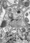
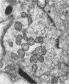
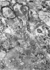
Images in this article
Selected References
These references are in PubMed. This may not be the complete list of references from this article.
- DE LORENZO A. J. Electron microscopic observations on the taste buds of the rabbit. J Biophys Biochem Cytol. 1958 Mar 25;4(2):143–150. doi: 10.1083/jcb.4.2.143. [DOI] [PMC free article] [PubMed] [Google Scholar]
- ECCLES J. C. The mechanism of synaptic transmission. Ergeb Physiol. 1961;51:299–430. [PubMed] [Google Scholar]
- ENGSTROM H., WERSALL J. Myelin sheath structure in nerve fibre demyelinization and branching regions. Exp Cell Res. 1958 Apr;14(2):414–425. doi: 10.1016/0014-4827(58)90200-3. [DOI] [PubMed] [Google Scholar]
- GRAY E. G. A morphological basis for pre-synaptic inhibition? Nature. 1962 Jan 6;193:82–83. doi: 10.1038/193082a0. [DOI] [PubMed] [Google Scholar]
- GRAY E. G. Axo-somatic and axo-dendritic synapses of the cerebral cortex: an electron microscope study. J Anat. 1959 Oct;93:420–433. [PMC free article] [PubMed] [Google Scholar]
- GRAY E. G. Electron microscopy of synaptic contacts on dendrite spines of the cerebral cortex. Nature. 1959 Jun 6;183(4675):1592–1593. doi: 10.1038/1831592a0. [DOI] [PubMed] [Google Scholar]
- GRAY E. G., GUILLERY R. W. The basis for silver staining of synapses of the mammalian spinal cord: a light and electron microscope study. J Physiol. 1961 Aug;157:581–588. doi: 10.1113/jphysiol.1961.sp006744. [DOI] [PMC free article] [PubMed] [Google Scholar]
- GRAY E. G. The granule cells, mossy synapses and Purkinje spine synapses of the cerebellum: light and electron microscope observations. J Anat. 1961 Jul;95:345–356. [PMC free article] [PubMed] [Google Scholar]
- KIDD M. Electron microscopy of the inner plexiform layer of the retina in the cat and the pigeon. J Anat. 1962 Apr;96:179–187. [PMC free article] [PubMed] [Google Scholar]
- LUFT J. H. Permanganate; a new fixative for electron microscopy. J Biophys Biochem Cytol. 1956 Nov 25;2(6):799–802. doi: 10.1083/jcb.2.6.799. [DOI] [PMC free article] [PubMed] [Google Scholar]
- LUSE S. A. Electron microscopic observations of the central nervous system. J Biophys Biochem Cytol. 1956 Sep 25;2(5):531–542. doi: 10.1083/jcb.2.5.531. [DOI] [PMC free article] [PubMed] [Google Scholar]
- PALAY S. L. The morphology of synapses in the central nervous system. Exp Cell Res. 1958;14(Suppl 5):275–293. [PubMed] [Google Scholar]
- SINGER M., SALPETER M. M. The bodies of Eberth and associated structures in the skin of the frog tadpole. J Exp Zool. 1961 Jun;147:1–19. doi: 10.1002/jez.1401470102. [DOI] [PubMed] [Google Scholar]
- TAXI J. [Study of the ultrastkucture of the synaptic zones in the sympathetic ganglia of the frog]. C R Hebd Seances Acad Sci. 1961 Jan 4;252:174–176. [PubMed] [Google Scholar]
- UZMAN B. G., NOGUEIRA-GRAF G. Electron microscope studies of the formation of nodes of Ranvier in mouse sciatic nerves. J Biophys Biochem Cytol. 1957 Jul 25;3(4):589–598. doi: 10.1083/jcb.3.4.589. [DOI] [PMC free article] [PubMed] [Google Scholar]
- WYCKOFF R. W., YOUNG J. Z. The motorneuron surface. Proc R Soc Lond B Biol Sci. 1956 Mar 13;144(917):440–450. doi: 10.1098/rspb.1956.0002. [DOI] [PubMed] [Google Scholar]


