Full text
PDF
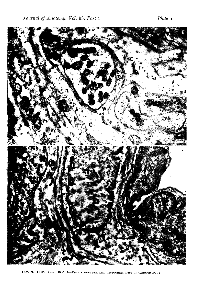
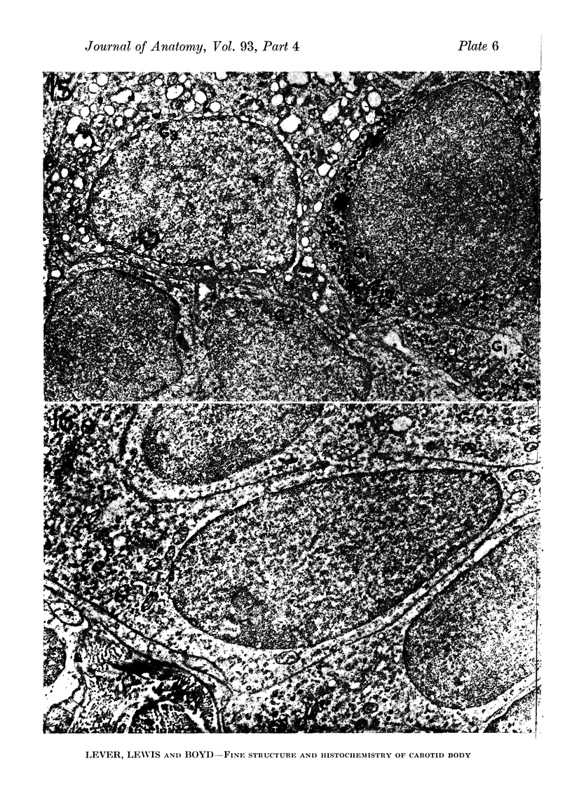
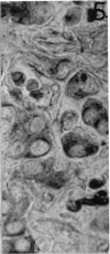
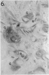
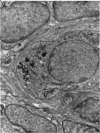
Images in this article
Selected References
These references are in PubMed. This may not be the complete list of references from this article.
- BLASCHKO H., HAGEN P., WELCH A. D. Observations on the intracellular granules of the adrenal medulla. J Physiol. 1955 Jul 28;129(1):27–49. doi: 10.1113/jphysiol.1955.sp005336. [DOI] [PMC free article] [PubMed] [Google Scholar]
- CHIFFELLE T. L., PUTT F. A. Propylene and ethylene glycol as solvents for Sudan IV and Sudan black B. Stain Technol. 1951 Jan;26(1):51–56. doi: 10.3109/10520295109113178. [DOI] [PubMed] [Google Scholar]
- COMLINE R. S., SILVER M. Response of the adrenal medulla of the sheep foetus to asphyxia. Nature. 1958 Jan 24;181(4604):283–284. doi: 10.1038/181283a0. [DOI] [PubMed] [Google Scholar]
- DE CASTRO F. Sur la structure de la synapse dans les chemocepteurs; leur mécanisme d'excitation et rôle dans la circulation sanguine locale. Acta Physiol Scand. 1951 Feb 21;22(1):14–43. doi: 10.1111/j.1748-1716.1951.tb00747.x. [DOI] [PubMed] [Google Scholar]
- DE KOCK L. L. The intra-glomerular tissues of the carotid body. Acta Anat (Basel) 1954;21(2):101–116. doi: 10.1159/000140922. [DOI] [PubMed] [Google Scholar]
- DONTAS A. S. Effects of reserpine and hydralazine on carotid and splanchnic nerve activity and blood pressure. J Pharmacol Exp Ther. 1957 Sep;121(1):1–7. [PubMed] [Google Scholar]
- ERSPAMER V. Pharmacology of indole-alkylamines. Pharmacol Rev. 1954 Dec;6(4):425–487. [PubMed] [Google Scholar]
- FARQUHAR M. G., WELLINGS S. R. Electron microscopic evidence suggesting secretory granule formation within the Golgi apparatus. J Biophys Biochem Cytol. 1957 Mar 25;3(2):319–322. doi: 10.1083/jcb.3.2.319. [DOI] [PMC free article] [PubMed] [Google Scholar]
- GARNER C. M., DUNCAN D. Observations on the fine structure of the carotid body. Anat Rec. 1958 Apr;130(4):691–709. doi: 10.1002/ar.1091300406. [DOI] [PubMed] [Google Scholar]
- HILLARP N. A., HOKFELT B. Histochemical demonstration of noradrenaline and adrenaline in the adrenal medulla. J Histochem Cytochem. 1955 Jan;3(1):1–5. doi: 10.1177/3.1.1. [DOI] [PubMed] [Google Scholar]
- HOFFMAN H., BIRRELL J. H. The carotid body in normal and anoxic states: an electron microscopic study. Acta Anat (Basel) 1958;32(4):297–311. doi: 10.1159/000141332. [DOI] [PubMed] [Google Scholar]
- KOELLE G. B. The histochemical differentiation of types of cholinesterases and their localizations in tissues of the cat. J Pharmacol Exp Ther. 1950 Oct;100(2):158–179. [PubMed] [Google Scholar]
- KRAMER H., WINDRUM G. M. Sulphation techniques in histochemistry with special reference to metachromasia. J Histochem Cytochem. 1954 May;2(3):196–208. doi: 10.1177/2.3.196. [DOI] [PubMed] [Google Scholar]
- LACY P. E. Electron microscopic identification of different cell types in the islets of Langerhans of the guinea pig, rat, rabbit and dog. Anat Rec. 1957 Jun;128(2):255–267. doi: 10.1002/ar.1091280209. [DOI] [PubMed] [Google Scholar]
- LEVER J. D., BOYD J. D. Osmiophile granules in the glomus cells of the rabbit carotid body. Nature. 1957 May 25;179(4569):1082–1083. doi: 10.1038/1791082b0. [DOI] [PubMed] [Google Scholar]
- LEVER J. D. Cytological appearances in the normal and activated parathyroid of the rat: a combined study by electron and light microscopy with certain quantitative assessments. J Endocrinol. 1958 Sep;17(3):210–217. doi: 10.1677/joe.0.0170210. [DOI] [PubMed] [Google Scholar]
- LEVER J. D. Electron microscopic observations on the normal and denervated adrenal medulla of the rat. Endocrinology. 1955 Nov;57(5):621–635. doi: 10.1210/endo-57-5-621. [DOI] [PubMed] [Google Scholar]
- LEVER J. D. Fine structural appearances in the rat parathyroid. J Anat. 1957 Jan;91(1):73–81. [PMC free article] [PubMed] [Google Scholar]
- LILLIE R. D., BURTNER H. J., HENSON J. P. G. Diazo-safranin for staining enterochromaffin. J Histochem Cytochem. 1953 May;1(3):154–159. doi: 10.1177/1.3.154. [DOI] [PubMed] [Google Scholar]
- LILLIE R. D., BURTNER H. J. The ferric ferricyanide reduction test in histochemistry. J Histochem Cytochem. 1953 Mar;1(2):87–92. doi: 10.1177/1.2.87. [DOI] [PubMed] [Google Scholar]
- MILES A. E., LINDER J. E. Polyethylene glycols as histological embedding media: with a note on the dimensional change of tissue during embedding in various media. J R Microsc Soc. 1952 Dec;72(4):199–213. doi: 10.1111/j.1365-2818.1952.tb02336.x. [DOI] [PubMed] [Google Scholar]
- NORDENSTAM H., ADAMS-RAY J. Chromaffin granules and their cellular location in human skin. Z Zellforsch Mikrosk Anat. 1957;45(4):435–443. doi: 10.1007/BF00338886. [DOI] [PubMed] [Google Scholar]
- PALADE G. E. A small particulate component of the cytoplasm. J Biophys Biochem Cytol. 1955 Jan;1(1):59–68. doi: 10.1083/jcb.1.1.59. [DOI] [PMC free article] [PubMed] [Google Scholar]
- PALADE G. E., PORTER K. R. Studies on the endoplasmic reticulum. I. Its identification in cells in situ. J Exp Med. 1954 Dec 1;100(6):641–656. doi: 10.1084/jem.100.6.641. [DOI] [PMC free article] [PubMed] [Google Scholar]
- RINEHART J. F., FARQUHAR M. G. The fine vascular organization of the anterior pituitary gland; an electron microscopic study with histochemical correlations. Anat Rec. 1955 Feb;121(2):207–239. doi: 10.1002/ar.1091210206. [DOI] [PubMed] [Google Scholar]
- ROSS L. L. A cytological and histochemical study of the carotid body of the cat. Anat Rec. 1957 Dec;129(4):433–455. doi: 10.1002/ar.1091290407. [DOI] [PubMed] [Google Scholar]


