Summary
Patellar tuberculosis (TB) is a rare manifestation of musculoskeletal tuberculosis, accounting for a small proportion of cases. This case report presents a detailed analysis of a female patient in her early 30s who presented with isolated TB of the patella without concurrent pulmonary involvement. The patient received antitubercular chemotherapy, consisting of a 4-month intensive phase followed by an 8-month continuation phase. This case report underscores the rarity and diagnostic complexities associated with patellar TB. The condition often presents with non-specific symptoms, often mimicking prepatellar bursitis, necessitating a high level of clinical suspicion, particularly in patients from the endemic areas. Radiographic imaging and histopathological examination play crucial roles in establishing an accurate diagnosis. Antitubercular chemotherapy forms the cornerstone of treatment while surgical intervention is reserved for cases of extensive bone destruction or treatment failure.
Keywords: Tuberculosis, Orthopaedics
Background
The knee joint is the third most common site involved in musculoskeletal tuberculosis (TB), preceded by the spine and the hip joint. It constitutes around 10% of all cases of musculoskeletal TB.1 2 The spread of infection is haematogenous, often starting in either the synovium or the subchondral bone. When TB begins in the synovium, it typically remains confined to the synovial membrane for a long time before affecting the bone or joint, presenting as tubercular synovitis. Usually, the subchondral lesion around the knee begins in the distal femur or proximal tibia, with the patella being the rare location. Patellar TB is exceedingly rare, with only a limited number of cases reported in the medical literature and a reported incidence of as low as 0.1%.3 The condition presents diagnostic challenges due to its low incidence and non-specific clinical features, often leading to delayed or missed diagnoses. Therefore, raising awareness about patellar TB is crucial to improve early recognition and appropriate management.
In this case report, we present a rare case of patellar TB without concomitant pulmonary TB in an immunocompetent woman in her 30s, highlighting the diagnosis, clinical manifestations, radiological findings and management approach. Through this report, we aim to enhance clinicians’ knowledge about this unusual presentation of TB and emphasise the importance of early identification for optimal patient outcomes.
Case presentation
A woman in her early 30s presented with the chief complaint of persistent pain and swelling in her right knee over the preceding 4 months (figure 1). 4 months earlier, she was symptom-free but then began to experience pain and noticed a small swelling on the front of her knee. The pain was dull-aching and constant, worsened with weight-bearing activities. The patient sought healthcare locally and was treated symptomatically. Despite seeking prior medical attention and receiving symptomatic treatment with analgesics, topical treatments and a brief course of antibiotics, her symptoms failed to resolve. The pain was insidious in onset and gradually worsened over time. There was no history of cough, fever, haemoptysis, low back ache or significant trauma. Additionally, no notable family or medical history was reported, and constitutional symptoms were absent, except for decreased appetite.
Figure 1. Clinical image showing swelling over the suprapatellar area of the right knee (arrow).
On clinical examination, there was swelling over the proximal pole of the patella, which was fluctuant. Local temperature was not raised, there was no sinus scar or dilated veins over the swelling and overlying skin appeared normal. Movements of the knee were terminally painful, with restriction of terminal flexion (ROM 0–110). Blood investigations revealed an elevated erythrocyte sedimentation rate (ESR) and a haemoglobin level of 9.6 g/L, indicating potential chronic inflammation and anaemia.
Investigations
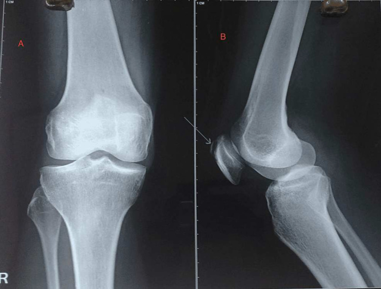
Radiographic imaging of the affected knee revealed a lytic lesion localised in the proximal pole of the patella, predominantly visible on lateral projection (figure 2). Notably, the articular margin of the patella remained intact, with no evidence of cortical breach. The possible differentials at this stage were tumorous conditions of the patella (giant cell tumour, aneurysmal bone cyst, etc), non-specific osteomyelitis and TB of the patella. Chest X-ray findings were unremarkable, ruling out pulmonary involvement. Sputum examination for acid-fast bacilli (AFB) yielded negative results, and limitations of sputum examination include its variable sensitivity, particularly in extrapulmonary TB, where bacillary load may be low. False-negative results can occur due to inadequate sample collection, intermittent shedding of bacilli or the presence of non-acid-fast mycobacteria. A needle biopsy was done from the affected site (proximal pole of patella) under fluoroscopic control. Histopathological examination of the tissue obtained via needle biopsy revealed epithelial cell granulomas with central caseous necrosis, indicative of TB, along with the presence of Langerhans-type giant cells, suggestive of TB. Despite negative Ziehl–Neelsen staining for AFB, PCR testing confirmed the presence of Mycobacterium tuberculosis DNA, solidifying the diagnosis of TB. Due to financial constraints, MRI of the knee could not be obtained.
Figure 2. Anteroposterior (A) and lateral projection (B) of X-ray of the right knee. The anteroposterior view (A) seems innocuous, while the lateral projection (B) shows a lytic lesion over the proximal pole of the patella (arrow).
Differential diagnosis
In cases of persistent knee pain particularly in a TB-endemic area, a broad range of differential diagnoses must be considered. Key differentials include tumorous conditions such as giant cell tumour, aneurysmal bone cyst, which can present with osteolytic lesions, and localised pain. Infectious conditions like non-specific osteomyelitis and TB of the patella are also crucial to consider, especially given the patient’s symptoms and elevated ESR and normal leucocyte counts, indicating a chronic inflammatory process. Inflammatory conditions such as prepatellar bursitis and rheumatoid arthritis can present with joint pain and swelling, potentially mimicking the symptoms of this case. Additionally, miscellaneous conditions like gout and pigmented villonodular synovitis should be considered due to their potential to cause similar clinical presentations. A thorough evaluation through clinical examination, imaging studies and laboratory investigations is essential to differentiate these conditions and arrive at an accurate diagnosis. In this particular case, the combination of radiographic findings, persistent symptoms despite prior treatment and elevated ESR warranted further investigation, leading to a biopsy that confirmed the diagnosis of TB of the patella.
Treatment
Prompt initiation of antitubercular therapy (ATT) was deemed imperative for the patient’s management. The intensive phase of treatment comprised rifampicin (10 mg/kg), isoniazid (5 mg/kg), pyrazinamide (20–40 mg/kg) and ethambutol (15 mg/kg) for the initial 4 months, followed by continuation with rifampicin and isoniazid for an additional 8 months.
Outcome and follow-up
The patient exhibited a favourable response to antitubercular treatment, with noticeable improvement noted within 8–10 weeks of therapy initiation. Pain and swelling subsided progressively, and at final follow-up, complete resolution of symptoms was observed. Radiographic evaluation demonstrated healing of the osseous lesion in the patella (figure 3). Moreover, the patient achieved significant improvement in knee range of motion, with flexion extending to 130°, indicative of successful disease management and functional recovery at 2-year follow-up.
Figure 3. Radiograph of the affected knee taken at 12-week follow-up showing anteroposterior (A) and lateral views (B). The anteroposterior view (A) is normal, and the lateral view (B) shows healing of the osteolytic lesion (white arrow).
Discussion
Skeletal TB is a significant health problem in most developing countries and constitutes 2–3% of all extrapulmonary TB cases.1 3 The knee joint is the third most common site to be affected in skeletal TB, preceded by the spine and the hip joint. TB of the patella is very rare, with a reported incidence of 0.09–0.15% in different case series.1,5 Tuli et al reported a single case of patellar TB in their series of 980 patients with 1074 lesions.2 The disease can be diagnosed if the suspicion is high (patient from an endemic area) as the signs and symptoms are generally vague.
The condition is difficult to diagnose in the early stage and is often diagnosed late. It most commonly affects young males and adolescents. There are reports available in the literature showing cases of TB of the patella in children, which is even rare and poses challenges in diagnosis owing to immature bones.6 7 Reactive joint effusion is frequently seen. Clinical features of this disease are non-specific, and often there is an irrelevant history of trauma as well. Constitutional symptoms may or may not be seen. Laboratory markers like C reactive protein, ESR and TLC appear normal in most cases. Definitive diagnosis can be made only when Mycobacterium tuberculi are seen on culture, but the disease being of paucibacillary culture may be negative in nearly half of the patients. The histopathological section reveals inflammatory granulomas with caseation and Langerhans giant cells.8 An X-ray showing osteolysis of a bone with a central sequestrum surrounded by peripheral sclerosis is suggestive of TB (chronic osteomyelitis). MRI can be done to locate the lesion and its extension to the joint. It also detects early bone marrow changes and soft-tissue abnormalities, whereas CT helps detect calcification.
Three cases of patellar TB were reported by Hartofiladikis Garofalidis, which were treated by patellectomy and ATT, and one of which had associated involvement of tibial condyle.9 The treatment mainly relies on ATT. Although the duration of treatment varies, there are at least 2 months of the intensive phase (four drug therapies), followed by the continuation phase (two drug therapies) as per WHO.3 Surgery is mainly required for biopsy to diagnose the condition or for debridement and curettage if fistula formation is there with persistent drainage of the cold abscess.
Patellar TB, although rare, poses unique diagnostic and management challenges. The patella’s distinct anatomical and physiological characteristics make it an unusual site for TB infection, and its involvement requires a comprehensive approach to ensure accurate diagnosis and effective treatment. This discussion focuses on the clinical features, diagnostic modalities, treatment strategies and patient outcomes associated with patellar TB.
Patellar TB often presents with non-specific symptoms such as knee pain, swelling and limited range of motion. These symptoms can mimic other common knee conditions, leading to diagnostic delays. A high index of suspicion is crucial, especially in patients from TB-endemic regions or with a history of TB infection.10 In this case report, the patient exhibited progressive knee pain and swelling, leading to further investigation.
Accurate diagnosis of patellar TB requires a combination of radiological imaging, laboratory investigations and histopathological examination. Radiographic findings may include lytic or sclerotic lesions, cortical destruction and periosteal reactions. MRI can provide detailed information about soft-tissue involvement and aid in differentiating TB from other pathologies. Laboratory investigations such as AFB staining, culture and PCR for M. tuberculosis can contribute to a definitive diagnosis.11 12 In our case, radiographic imaging and histopathological examination were crucial in confirming the diagnosis.
The management of patellar TB typically involves a multidisciplinary approach, including pharmacological interventions and surgical measures. Antitubercular chemotherapy with a combination of appropriate drugs is the cornerstone of treatment. Surgical intervention may be necessary in cases of extensive bone destruction, joint instability or failure to respond to medical therapy. Surgical options range from curettage and debridement to patellectomy or arthroplasty.9 11 12 The prognosis for patellar TB depends on early diagnosis and the initiation of appropriate treatment. Delayed diagnosis and inadequate treatment can lead to significant morbidity, including joint destruction and functional impairment. Prompt intervention, coupled with adherence to ATT, can improve outcomes and prevent long-term complications.10,12 In our case, early detection and a comprehensive treatment approach resulted in satisfactory clinical outcomes.
Patient’s perspective.
I started experiencing persistent knee pain and swelling, which was tough due to the lack of good healthcare in my rural area. I was diagnosed with tuberculosis of the patella. This was scary but learning that it was treatable gave me hope. I began taking antitubercular medicine, and over time, I gradually noticed significant improvements—the pain reduced, the swelling decreased and my knee’s mobility improved. At my final check-up, the complete resolution of symptoms and the healing of the lesion as confirmed by my doctors filled me with gratitude and relief.
Learning points.
Patellar tuberculosis (TB) is a rare form of skeletal TB that necessitates a thorough diagnostic evaluation and a multidisciplinary treatment approach.
Patellar TB should be considered in patients with chronic knee pain and swelling, especially from TB-endemic areas.
Recognition of the unique clinical presentation, combined with the appropriate use of radiological imaging, laboratory investigations and histopathological examination, is essential for accurate diagnosis.
Prompt initiation of antitubercular chemotherapy and consideration of surgical intervention, when indicated, can optimise patient outcomes and prevent long-term complications.
Footnotes
Funding: The authors have not declared a specific grant for this research from any funding agency in the public, commercial or not-for-profit sectors.
Case reports provide a valuable learning resource for the scientific community and can indicate areas of interest for future research. They should not be used in isolation to guide treatment choices or public health policy.
Provenance and peer review: Not commissioned; externally peer reviewed.
Patient consent for publication: Consent obtained directly from patient(s).
References
- 1.Mittal R, Trikha V, Rastogi S. Tuberculosis of patella. Knee. 2006;13:54–6. doi: 10.1016/j.knee.2005.09.006. [DOI] [PubMed] [Google Scholar]
- 2.Tuli SM. Tuberculosis of the Skeletal System: Bone, Joints, Spine and Bursal Sheaths. 3rd. New Delhi: Jaypee Brothers Medical Publishers; 2004. edn. [Google Scholar]
- 3.World Health Organization . World Health Organization; 2017. Treatment of tuberculosis: guidelines for treatment of drug-susceptible tuberculosis and patient care, 2017 update. [Google Scholar]
- 4.Khan A, Amedu OS, Kumar P, et al. Tuberculosis of Patella Complicated by Synovitis of Knee Joint: A Case Report. Cureus. 2022;PMCID:e24618. doi: 10.7759/cureus.24618. [DOI] [PMC free article] [PubMed] [Google Scholar]
- 5.Dharmshaktu GS. Isolated Tuberculosis of Patella in A Child: A Case Report and Literature Review. Int J Adv Integr Med Sci. 2017;2:101–3. doi: 10.5005/jp-journals-10050-10085. [DOI] [Google Scholar]
- 6.Demni K. Tuberculosis of the patella in children – case report. J Orthop. 2007;4:e23 [Google Scholar]
- 7.Galois L, Chary-Valckenaere I, Mainard D, et al. Tuberculosis of the patella. Arch Orthop Trauma Surg. 2003;123:192–4. doi: 10.1007/s00402-003-0495-y. [DOI] [PubMed] [Google Scholar]
- 8.Hartofilakidis-Garofalidi G. Cystic tuberculosis of the patella. Report of three cases. J Bone Joint Surg Am. 1969;51:582–5. [PubMed] [Google Scholar]
- 9.Agrawal VK, Agrawal RG. Tuberculosis of patella in an immunocompetent patient--a case report. Ind Tuberc. 2011;58:29–31. [PubMed] [Google Scholar]
- 10.MacLean S, Kulkarni S. Tuberculosis of the patella masquerading as prepatellar bursitis. Ann R Coll Surg Engl. 2013;95:e17–9. doi: 10.1308/003588413x13511609955490. [DOI] [PMC free article] [PubMed] [Google Scholar]
- 11.Prakash J, Vijay V. Tuberculosis of the patella imitating chronic knee synovitis. BMJ Case Rep. 2014;2014:bcr2014203971. doi: 10.1136/bcr-2014-203971. [DOI] [PMC free article] [PubMed] [Google Scholar]
- 12.Khezami K, Bennour MA, Basssalah E, et al. Primary patella tuberculosis mimicking pre-patellar bursitis: A case report and review of the literature. Int J Surg Case Rep. 2021;86:106367. doi: 10.1016/j.ijscr.2021.106367. [DOI] [PMC free article] [PubMed] [Google Scholar]


