Abstract
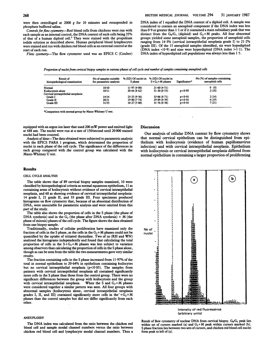
Cervical biopsy samples were taken from 79 patients who had various grades of cervical intraepithelial neoplasia or who showed evidence, in the form of koilocytosis, of human papillomavirus infection of the uterine cervix and from 10 women with normal cervices. The DNA content of the cells in the samples was analysed by flow cytometry. Analysis of the data obtained showed that the biopsy samples from women with cervical intraepithelial neoplasia and human papillomavirus lesions contained significantly more dividing cells (31.2% of cells from human papillomavirus lesions with no cervical intraepithelial neoplasia and 33.06%, 29.89%, and 31.76% of cells from cervical intraepithelial neoplasia grades I, II, and III, respectively) than those from women with normal cervices (21.6%). The proportion of aneuploid samples from the group who showed evidence of human papillomavirus infection only (18.2%) did not differ significantly from the group with cervical intraepithelial neoplasia grade III (21.2%). Aneuploidy and an increased rate of cellular proliferation are recognised characteristics of malignancy. These results therefore support the view that human papillomavirus plays an important part in the aetiology of cervical carcinoma and are relevant to the clinical management of patients.
Full text
PDF


Selected References
These references are in PubMed. This may not be the complete list of references from this article.
- Buckley C. H., Butler E. B., Fox H. Cervical intraepithelial neoplasia. J Clin Pathol. 1982 Jan;35(1):1–13. doi: 10.1136/jcp.35.1.1. [DOI] [PMC free article] [PubMed] [Google Scholar]
- Fletcher S. Histopathology of papilloma virus infection of the cervix uteri: the history, taxonomy, nomenclature and reporting of koilocytic dysplasias. J Clin Pathol. 1983 Jun;36(6):616–624. doi: 10.1136/jcp.36.6.616. [DOI] [PMC free article] [PubMed] [Google Scholar]
- Jakobsen A., Bichel P., Sell A. DNA distribution in biopsy specimens from human cervical carcinoma investigated by flow cytometry. Virchows Arch B Cell Pathol. 1979 Feb 6;29(4):337–342. doi: 10.1007/BF02899364. [DOI] [PubMed] [Google Scholar]
- Jakobsen A., Kristensen P. B., Poulsen H. K. Flow cytometric classification of biopsy specimens from cervical intraepithelial neoplasia. Cytometry. 1983 Sep;4(2):166–169. doi: 10.1002/cyto.990040210. [DOI] [PubMed] [Google Scholar]
- Laerum O. D., Farsund T. Clinical application of flow cytometry: a review. Cytometry. 1981 Jul;2(1):1–13. doi: 10.1002/cyto.990020102. [DOI] [PubMed] [Google Scholar]
- Reid R., Crum C. P., Herschman B. R., Fu Y. S., Braun L., Shah K. V., Agronow S. J., Stanhope C. R. Genital warts and cervical cancer. III. Subclinical papillomaviral infection and cervical neoplasia are linked by a spectrum of continuous morphologic and biologic change. Cancer. 1984 Feb 15;53(4):943–953. doi: 10.1002/1097-0142(19840215)53:4<943::aid-cncr2820530421>3.0.co;2-x. [DOI] [PubMed] [Google Scholar]
- Reid R. Genital warts and cervical cancer. II. Is human papillomavirus infection the trigger to cervical carcinogenesis? Gynecol Oncol. 1983 Apr;15(2):239–252. doi: 10.1016/0090-8258(83)90080-x. [DOI] [PubMed] [Google Scholar]
- Reid R., Stanhope C. R., Herschman B. R., Booth E., Phibbs G. D., Smith J. P. Genital warts and cervical cancer. I. Evidence of an association between subclinical papillomavirus infection and cervical malignancy. Cancer. 1982 Jul 15;50(2):377–387. doi: 10.1002/1097-0142(19820715)50:2<377::aid-cncr2820500236>3.0.co;2-a. [DOI] [PubMed] [Google Scholar]
- Vindeløv L. L., Christensen I. J., Nissen N. I. Standardization of high-resolution flow cytometric DNA analysis by the simultaneous use of chicken and trout red blood cells as internal reference standards. Cytometry. 1983 Mar;3(5):328–331. doi: 10.1002/cyto.990030504. [DOI] [PubMed] [Google Scholar]


