

A 44-year-old man presented with exertional dyspnea of 2 years' duration. He was in sinus rhythm. The mean jugular venous pressure was elevated, with a prominent “a” wave. The 1st heart sound was loud, and the 2nd heart sound was normally split, with an accentuated pulmonary component. There were opening snaps in the apex and in the lower left sternal border. Middiastolic murmurs of grade 4 at the apex and grade 3 at the lower left sternal border were audible. There was a grade-3 systolic ejection murmur at the upper left sternal border that was conducted to the carotid arteries.
Transthoracic echocardiography showed stenosis of all 4 cardiac valves. Figure 1 shows severe stenosis of the mitral and tricuspid valves. The mitral valve area was 1 cm2 and the tricuspid valve area was 1.5 cm2. The mean gradient was 17 mmHg across the mitral valve and 8 mmHg across the tricuspid valve. The peak systolic gradient was 72 mmHg across the aortic valve and 58 mmHg across the pulmonary valve.
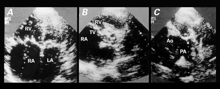
Fig. 1 On transthoracic echocardiography, severe stenosis of the mitral and tricuspid valves is shown in the 4-chamber view (A), stenosis of the aortic and tricuspid valves in the short-axis view (B), and stenosis of the pulmonary valve in the short-axis view (C).
AO = aorta; AV = aortic valve; LA = left ventricle; LV = left ventricle; MV = mitral valve;
PA = pulmonary artery; PV = pulmonary valve; RA = right atrium; RV = right ventricle;
TV = tricuspid valve
Rheumatic involvement of all 4 cardiac valves is rare. 1 Stenosis of all 4 valves is rarer still; only a few case reports are available. 2 Rheumatic quadrivalvular damage has been found on autopsy (1 of 586 patients with valve deformities) 3 and by cardiac catheterization, but reports of echocardiographic diagnosis are much rarer. 4 There is a high incidence of multivalvular damage when Aschoff bodies are identified at necropsy. 5
Of note, it is possible that by the time echocardiography came into common use, the incidence as well as the severity of rheumatic heart disease had already been drastically reduced. This could explain the rarity of echocardiographic detection of quadrivalvular rheumatic involvement, which seems to be the case even in developing countries.
Footnotes
Address for reprints: Dr. K.M. Krishnamoorthy, Assistant Professor of Cardiology, Sree Chitra Tirunal Institute for Medical Sciences & Technology, Trivandrum – 695 011, India
References
- 1.Paraskos JA. Combined valvular disease. In: Dalen JE, Alpert JS, editors. Valvular heart disease. 2nd ed. Boston: Little, Brown; 1987. p. 439–508.
- 2.Gialloreto O, Aerichide N, Allard PP. Stenotic involvement of all four heart valves. Report of three cases. Am J Cardiol 1961;7:865–73. [DOI] [PubMed]
- 3.Clawson BJ. Rheumatic heart disease. An analysis of 796 cases. Am Heart J 1940;20:454–74.
- 4.Bandin MA, Vargas-Barron J, Keirns C, Romero-Cardenas A, Villegas M, Buendia A. Echocardiographic diagnosis of rheumatic cardiopathy affecting all four cardiac valves. Am Heart J 1990;120:1004–7. [DOI] [PubMed]
- 5.Roberts WC, Virmani R. Aschoff bodies at necropsy in valvular heart disease. Evidence from an analysis of 543 patients over 14 years of age that rheumatic heart disease, at least anatomically, is a disease of the mitral valve. Circulation 1978;57:803–7. [DOI] [PubMed]