

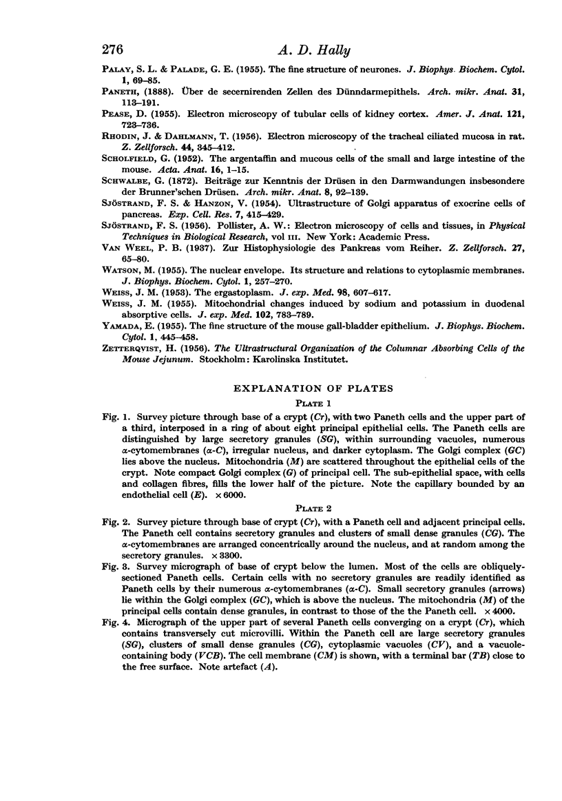
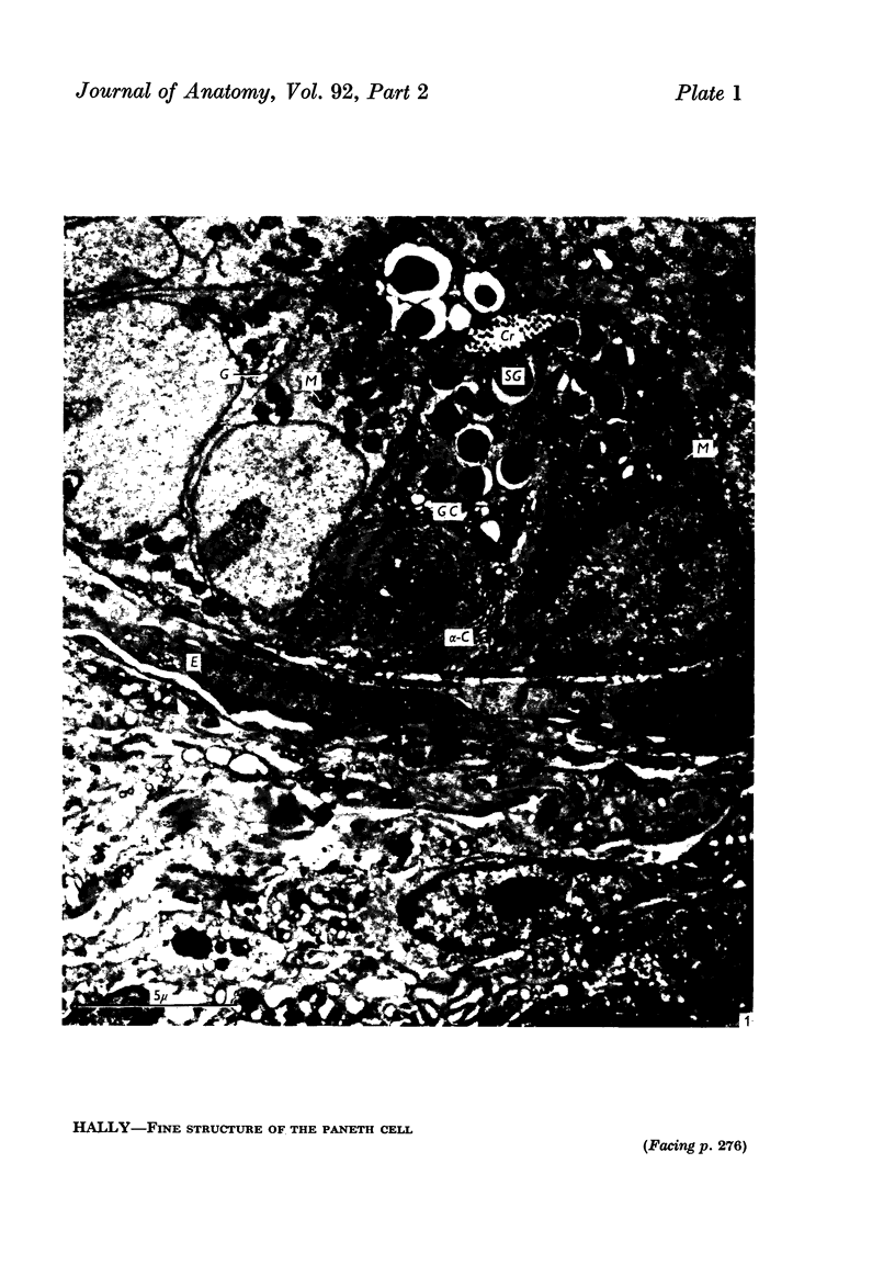
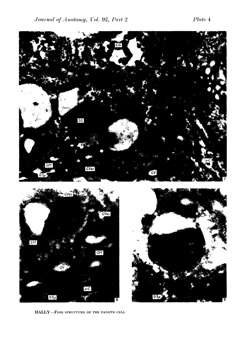
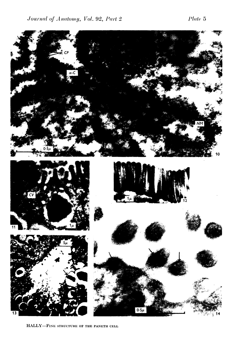
Full text
PDF
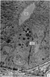
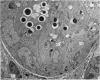
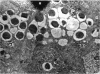
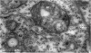
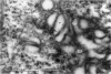
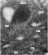
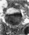
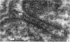
Images in this article

Selected References
These references are in PubMed. This may not be the complete list of references from this article.
- BURGOS M. H., FAWCETT D. W. Studies on the fine structure of the mammalian testis. I. Differentiation of the spermatids in the cat (Felis domestica). J Biophys Biochem Cytol. 1955 Jul 25;1(4):287–300. doi: 10.1083/jcb.1.4.287. [DOI] [PMC free article] [PubMed] [Google Scholar]
- DALTON A. J., FELIX M. D. Cytologic and cytochemical characteristics of the Golgi substance of epithelial cells of the epididymis in situ, in homogenates and after isolation. Am J Anat. 1954 Mar;94(2):171–207. doi: 10.1002/aja.1000940202. [DOI] [PubMed] [Google Scholar]
- DAWSON I. M., HOSSACK J., WYBURN G. M. Observations on the Nissl's substance, cytoplasmic filaments and the nuclear membrane of spinal ganglion cells. Proc R Soc Lond B Biol Sci. 1955 Aug 16;144(914):132–142. doi: 10.1098/rspb.1955.0039. [DOI] [PubMed] [Google Scholar]
- LATTA H., HARTMANN J. F. Use of a glass edge in thin sectioning for electron microscopy. Proc Soc Exp Biol Med. 1950 Jun;74(2):436–439. doi: 10.3181/00379727-74-17931. [DOI] [PubMed] [Google Scholar]
- PALADE G. E. A small particulate component of the cytoplasm. J Biophys Biochem Cytol. 1955 Jan;1(1):59–68. doi: 10.1083/jcb.1.1.59. [DOI] [PMC free article] [PubMed] [Google Scholar]
- PALADE G. E. The endoplasmic reticulum. J Biophys Biochem Cytol. 1956 Jul 25;2(4 Suppl):85–98. doi: 10.1083/jcb.2.4.85. [DOI] [PMC free article] [PubMed] [Google Scholar]
- PALAY S. L., PALADE G. E. The fine structure of neurons. J Biophys Biochem Cytol. 1955 Jan;1(1):69–88. doi: 10.1083/jcb.1.1.69. [DOI] [PMC free article] [PubMed] [Google Scholar]
- PEASE D. C. Electron microscopy of the tubular cells of the kidney cortex. Anat Rec. 1955 Apr;121(4):723–743. doi: 10.1002/ar.1091210403. [DOI] [PubMed] [Google Scholar]
- RHODIN J., DALHAMN T. Electron microscopy of the tracheal ciliated mucosa in rat. Z Zellforsch Mikrosk Anat. 1956;44(4):345–412. doi: 10.1007/BF00345847. [DOI] [PubMed] [Google Scholar]
- SCHOFIELD G. The argentaffin and mucous cells of the small and large intestines of the mouse. Acta Anat (Basel) 1952;16(1-2):1–15. doi: 10.1159/000140760. [DOI] [PubMed] [Google Scholar]
- SJOSTRAND F. S., HANZON V. Ultrastructure of Golgi apparatus of exocrine cells of mouse pancreas. Exp Cell Res. 1954 Nov;7(2):415–429. doi: 10.1016/s0014-4827(54)80087-5. [DOI] [PubMed] [Google Scholar]
- WATSON M. L. The nuclear envelope; its structure and relation to cytoplasmic membranes. J Biophys Biochem Cytol. 1955 May 25;1(3):257–270. doi: 10.1083/jcb.1.3.257. [DOI] [PMC free article] [PubMed] [Google Scholar]
- WEISS J. M. Mitochondrial changes induced by potassium and sodium in the duodenal absorptive cell as studied with the electron microscope. J Exp Med. 1955 Dec 1;102(6):783–788. doi: 10.1084/jem.102.6.783. [DOI] [PMC free article] [PubMed] [Google Scholar]
- WEISS J. M. The ergastoplasm; its fine structure and relation to protein synthesis as studied with the electron microscope in the pancreas of the Swiss albino mouse. J Exp Med. 1953 Dec;98(6):607–618. doi: 10.1084/jem.98.6.607. [DOI] [PMC free article] [PubMed] [Google Scholar]
- YAMADA E. The fine structure of the gall bladder epithelium of the mouse. J Biophys Biochem Cytol. 1955 Sep 25;1(5):445–458. doi: 10.1083/jcb.1.5.445. [DOI] [PMC free article] [PubMed] [Google Scholar]