Abstract
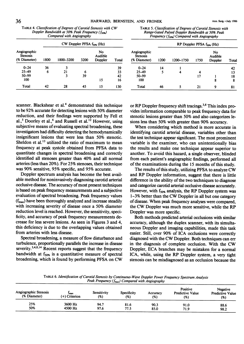
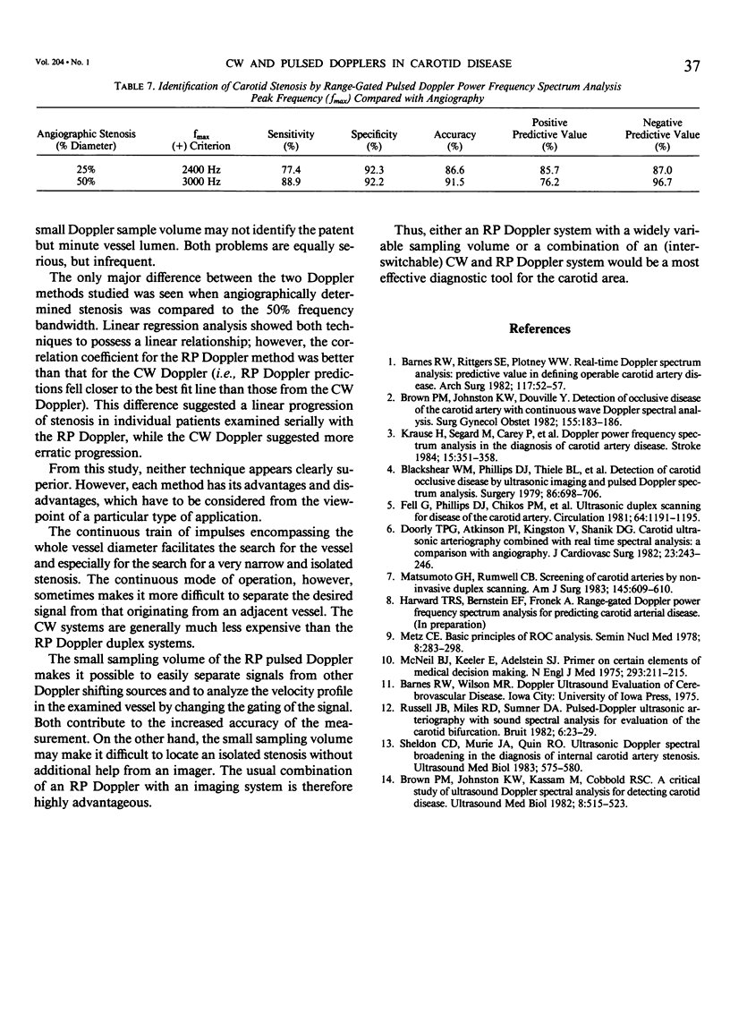
Two types of ultrasonic Doppler velocity metering devices currently used in the detection of extracranial carotid artery disease, the continuous-wave (CW) and the range-gated pulsed (RP) Doppler systems, were compared in the present study. Power frequency spectrum analysis (PFSA) was performed on 130 carotid arterial bifurcations with a CW Doppler and 81 carotid arteries with an RP Doppler system. All results were compared with angiographic findings. The frequency bandwidth at 50% peak power (f50%), a quantitative index for defining spectral broadening, detected stenoses equal to or greater than 50% diameter reduction with 93% sensitivity, 92% specificity, and 92% accuracy with the CW system. With the RP Doppler, the same degree of stenosis was identified with 94% sensitivity, 93% specificity, and 93% accuracy. Compared with angiographic classification into 0-24%, 25-49%, and 50-99% diameter reduction categories, CW Doppler PFSA and an 85% overall accuracy, and the RP Doppler overall accuracy was 86%. CW Doppler also correctly identified 15 of 16 internal carotid artery (ICA) occlusions; 8 of 8 ICA occlusions were correctly identified with the RP Doppler. Thus, both techniques detected carotid artery disease with comparable results. For research and ease of operation, an RP Doppler system with a variable sampling volume appears to be most desirable. However, a standard CW system is superior if utility and cost-effectiveness are of prime importance.
Full text
PDF
Selected References
These references are in PubMed. This may not be the complete list of references from this article.
- Barnes R. W., Rittgers S. E., Putney W. W. Real-time Doppler spectrum analysis: predictive value in defining operable carotid artery disease. Arch Surg. 1982 Jan;117(1):52–57. doi: 10.1001/archsurg.1982.01380250034008. [DOI] [PubMed] [Google Scholar]
- Blackshear W. M., Jr, Phillips D. J., Thiele B. L., Hirsch J. H., Chikos P. M., Marinelli M. R., Ward K. J., Strandness D. E., Jr Detection of carotid occlusive disease by ultrasonic imaging and pulsed Doppler spectrum analysis. Surgery. 1979 Nov;86(5):698–706. [PubMed] [Google Scholar]
- Brown P. M., Johnston K. W., Douville Y. Detection of occlusive disease of the carotid artery with continuous wave Doppler spectral analysis. Surg Gynecol Obstet. 1982 Aug;155(2):183–186. [PubMed] [Google Scholar]
- Brown P. M., Johnston K. W., Kassam M., Cobbold R. S. A critical study of ultrasound Doppler spectral analysis for detecting carotid disease. Ultrasound Med Biol. 1982;8(5):515–523. doi: 10.1016/0301-5629(82)90083-7. [DOI] [PubMed] [Google Scholar]
- Doorly T. P., Atkinson P. I., Kingston V., Shanik D. G. Carotid ultrasonic arteriography combined with real time spectral analysis: a comparison with angiography. J Cardiovasc Surg (Torino) 1982 May-Jun;23(3):243–246. [PubMed] [Google Scholar]
- Fell G., Phillips D. J., Chikos P. M., Harley J. D., Thiele B. L., Strandness D. E., Jr Ultrasonic duplex scanning for disease of the carotid artery. Circulation. 1981 Dec;64(6):1191–1195. doi: 10.1161/01.cir.64.6.1191. [DOI] [PubMed] [Google Scholar]
- Krause H., Segard M., Carey P., Bernstein E. F., Fronek A. Doppler power frequency spectrum analysis in the diagnosis of carotid artery disease. Stroke. 1984 Mar-Apr;15(2):351–358. doi: 10.1161/01.str.15.2.351. [DOI] [PubMed] [Google Scholar]
- Matsumoto G. H., Rumwell C. B. Screening of carotid arteries by noninvasive duplex scanning. Am J Surg. 1983 May;145(5):609–610. doi: 10.1016/0002-9610(83)90103-4. [DOI] [PubMed] [Google Scholar]
- McNeil B. J., Keller E., Adelstein S. J. Primer on certain elements of medical decision making. N Engl J Med. 1975 Jul 31;293(5):211–215. doi: 10.1056/NEJM197507312930501. [DOI] [PubMed] [Google Scholar]
- Metz C. E. Basic principles of ROC analysis. Semin Nucl Med. 1978 Oct;8(4):283–298. doi: 10.1016/s0001-2998(78)80014-2. [DOI] [PubMed] [Google Scholar]
- Sheldon C. D., Murie J. A., Quin R. O. Ultrasonic doppler spectral broadening in the diagnosis of internal carotid artery stenosis. Ultrasound Med Biol. 1983 Nov-Dec;9(6):575–580. doi: 10.1016/0301-5629(83)90001-7. [DOI] [PubMed] [Google Scholar]


