Abstract
Telomerase transgenics are an important tool to assess the role of telomerase in cancer, as well as to evaluate the potential use of telomerase for gene therapy of age-associated diseases. Here, we have targeted the expression of the catalytic component of mouse telomerase, mTERT, to basal keratinocytes using the bovine keratin 5 promoter. These telomerase-transgenic mice are viable and show histologically normal stratified epithelia with high levels of telomerase activity and normal telomere length. Interestingly, the epidermis of these mice is highly responsive to the mitogenic effects of phorbol esters, and it is more susceptible than that of wild-type littermates to the development skin tumors upon chemical carcinogenesis. The epidermis of telomerase-transgenic mice also shows an increased wound-healing rate compared with wild-type littermates. These results suggest that, contrary to the general assumption, telomerase actively promotes proliferation in cells that have sufficiently long telomeres and unravel potential risks of gene therapy for age-associated diseases based on telomerase upregulation.
Keywords: epithelial tumors/gene therapy/mTERT/skin carcinogenesis/telomerase-transgenic mouse
Introduction
Telomeres consist of tandem DNA repeats and specific proteins and protect the chromosome ends from degradation, recombination and DNA repair activities (reviewed in Blackburn, 1991; Greider, 1996). In this regard, loss of telomeric function, either by loss of telomeric repeats or by mutation of telomeric proteins, is associated with increased chromosomal instability and loss of cell viability (Counter et al., 1992; Blasco et al., 1997; van Steensel et al., 1998; Samper et al., 2000).
Telomerase is the cellular reverse transcriptase that synthesizes telomeric repeats de novo (Greider and Blackburn, 1985; reviewed in Nugent and Lundblad, 1998). It is composed of a catalytic subunit known as TERT (telomerase reverse transcriptase) (Harrington et al., 1997a; Kilian et al., 1997; Lingner et al., 1997; Meyerson et al., 1997; Nakamura et al., 1997; Greenberg et al., 1998; Martín-Rivera et al., 1998), an RNA molecule or TERC (telomerase RNA component), which is used as template for the addition of new telomeric repeats (Greider and Blackburn, 1989; Singer and Gottschling, 1994; Blasco et al., 1995; Feng et al., 1995), and associated proteins (Collins et al., 1995; Harrington et al., 1997b; Nakayama et al., 1997; Gandhi and Collins, 1998; Mitchell et al., 1999).
Telomerase activity is upregulated in the vast majority of human tumors as compared with normal somatic tissues. It has been shown that expression of the catalytic subunit of telomerase, TERT, in cultured human primary cells reconstitutes telomerase activity and allows immortal growth (Bodnar et al., 1998; Kiyono et al., 1998; Jiang et al., 1999; Morales et al., 1999). Furthermore, TERT-mediated telomerase activation is able to cooperate with oncogenes in transforming cultured primary human cells into neoplastic cells (Hahn et al., 1999). In addition, it has been shown recently that TERT-driven cell proliferation results in activation of the c-myc oncogene (Wang et al., 2000). These findings in cultured cells have opened up the possibility that telomerase upregulation, which occurs in >90% of all human tumors (reviewed in Shay and Bacchetti, 1997), may contribute actively to tumor growth (reviewed in Weitzman and Yaniv, 1999). On the other hand, telomerase activity reconstitution in adult somatic cells or tissues is envisaged as a potential approach for gene therapy of age-related diseases. To address directly if tissue-specific telomerase overexpression has an impact on tumor susceptibility or normal tissue biology, we targeted expression of the catalytic component of mouse telomerase, mTERT (Greenberg et al., 1998; Martín-Rivera et al., 1998), to basal keratinocytes of stratified epithelia. Here, we describe the construction and characterization of these mice.
Results and discussion
In vivo reconstitution of telomerase activity in transgenic K5-mTERT mice
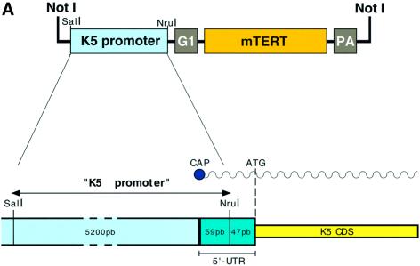
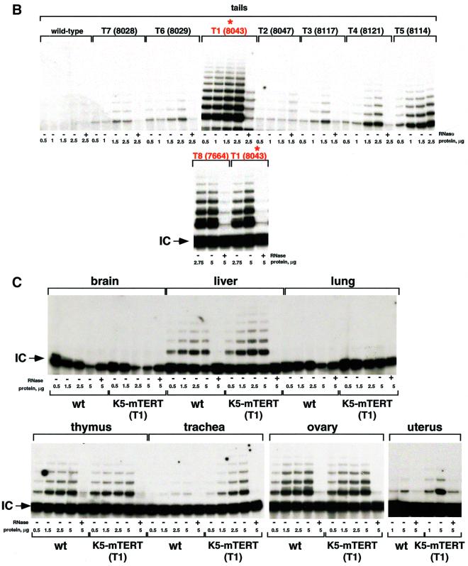
We used the 5′-regulatory region of the bovine keratin K5 gene (Ramírez et al., 1994; Murillas et al., 1995) to target mTERT expression to basal keratinocytes (see Figure 1A for details of the bovine K5 promoter region used to express mTERT) (Materials and methods). Eight different founder K5-mTERT transgenic mice were identified (T1–T8) that showed increased telomerase activity in the skin of the tail compared with wild-type littermates (Figure 1B). Two of these founders, T1 = 8043 and T8 = 7664 (highlighted in red in Figure 1B) showed the highest in vivo reconstitution of telomerase activity in the tail skin. All the results described here were obtained using the K5-mTERT mice derived from the T1 founder, K5-mTERT (T1) (asterisk in Figure 1B), and were confirmed, where indicated, with the T8 founder-derived K5-mTERT transgenics, K5-mTERT (T8), to rule out possible non-specific effects due to transgene insertion.
Fig. 1. Generation of K5-mTERT mice. (A) A scheme of the K5-mTERT transgene construct. The functional elements include a SalI–NruI fragment with the bovine K5 regulatory sequences (K5 promoter box), the rabbit β-globin intron 2 (G1 box), the coding sequence of the mTERT gene (mTERT box) and the SV40 early gene poly(A) addition signal (PA box). (B) Telomerase TRAP activity in the tail of wild-type and K5-mTERT founders (T1–T8). The two founders chosen for the study (T1 and T8) are highlighted in red. The asterisk highlights founder mouse T1, which was used for most of the experiments described here. (C) Telomerase TRAP activity in wild-type and K5-mTERT (T1) tissues. The indicated protein concentrations of the S-100 extract were used. Extracts were pre-treated (+) or not (–) with RNase A.
In agreement with the known expression pattern of keratin K5 (Ramírez et al., 1994; Murillas et al., 1995), the stratified epithelia of K5-mTERT (T1) mice tested, including uterus, trachea and skin, showed significant upregulation of telomerase activity as compared with the corresponding littermate wild-type tissues (Figure 1B and C). This telomerase upregulation coincided with increased mTERT mRNA levels in K5-mTERT transgenic skin (between 25- and 50-fold) compared with wild-type controls (data not shown). Other tissues of the K5-mTERT (T1) mice where the transgene is not expressed (i.e. liver, brain, lung, ovary and whole thymus) did not show upregulation of telomerase activity as compared with wild-type littermates (Figure 1C).
Telomere length in wild-type and K5-mTERT transgenic basal keratinocytes
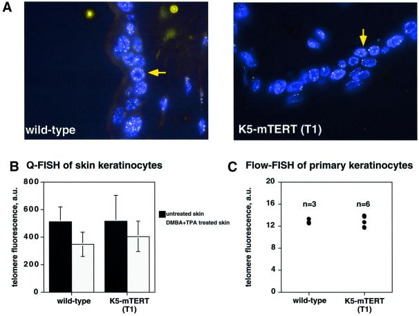
Previous studies using cultured human cells showed that TERT-mediated telomerase activity reconstitution resulted in extension or maintenance of telomere length (Bodnar et al., 1998; Kiyono et al., 1998; Hahn et al., 1999). We measured telomere length in K5-mTERT (T1) skin keratinocytes by quantitative fluorescence in situ hybridization (Q-FISH) on skin sections as previously described (González-Suárez et al., 2000). Figure 2A shows Q-FISH images of interphase nuclei in the skin of age-matched (8-week-old) littermate wild-type and K5-mTERT (T1) mice; the basal layer of skin keratynocytes is indicated with a yellow arrow. Quantification of the fluorescence intensity of telomere dots showed that telomeres in K5-mTERT (T1) epidermis are similar in length to those of age-matched littermate wild-type mice (Figure 2B; black bars). Primary keratinocyte cultures derived from newborn wild-type and K5-mTERT (T1) mice also showed similar telomere length using Flow-FISH, an independent quantitative FISH technique to measure telomere length that is based on flow cytometry (see Materials and methods) (Rufer et al., 1998) (Figure 2C). All together, these results suggest that TERT-driven telomerase upregulation in basal skin keratinocytes does not result in a significant extension of telomeres in the skin as compared with age-matched wild-type mice.
Fig. 2. Telomere fluorescence in skin sections. (A) Illustrative images showing telomere fluorescence in wild-type and K5-mTERT (T1) skin sections. The basal layer of skin keratinocytes is indicated (yellow arrow). (B) Quantification of telomere fluorescence on untreated skin sections or on skin sections treated with DMBA + TPA, as indicated. More than 50 keratinocyte nuclei of each genotype were analyzed by Q-FISH. (C) Average telomere fluorescence as determined by Flow-FISH of three wild-type and six K5-mTERT primary keratinocyte cultures. Between 2000 and 3000 nuclei were analyzed by flow cytometry for each culture.
K5-mTERT stratified epithelia are histologically normal
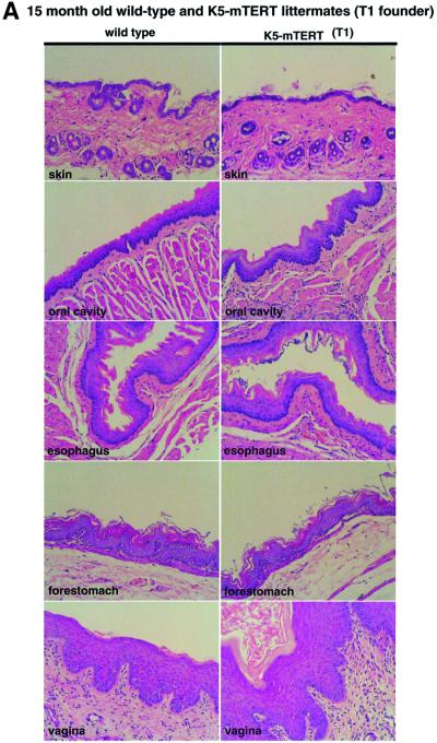
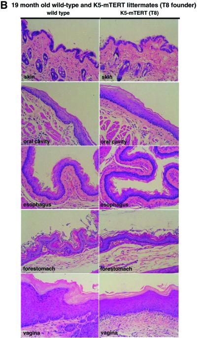
K5-mTERT (T1 and T8) mice, ranging from newborn to 19 months old, were phenotypically indistinguishable from the corresponding age-matched wild-type littermates and showed no pathologies, no spontaneous development of epithelial tumors and no loss of viability with age (not shown). In agreement with this, histopathological analysis of stratified epithelia (skin, oral cavity, esophagus, forestomach and vagina) from 15- to 19-month-old K5-mTERT (T1 and T8, as indicated) and the corresponding age-matched littermate wild-type controls revealed no macroscopic or histological differences between genotypes, indicating that transgenic telomerase expression per se does not alter the stratified epithelium structure (Figure 3). These results suggest that potential telomerase reconstitution for gene therapy of epithelial aging-associated diseases would not have any deleterious effects on the structure of stratified epithelia.
Fig. 3. (A and B) Histology of stratified epithelia in littermate 15- to 19-month-old wild-type and K5-mTERT (T1 or T8, as indicated) transgenics. The epithelia studied were skin, oral cavity, esophagus, forestomach and vagina. No significant histological differences between genotypes were observed. Magnification ×20.
Increased tumor susceptibility and mortality of K5-mTERT transgenics produced by chemical carcinogens
To study further the effects of telomerase overexpression on the normal biology and function of stratified epithelia, we first addressed whether transgenic telomerase expression impacts on epithelial tumor formation upon exposure to chemical carcinogens. The stages of initiation, promotion and tumor progression in the classical skin chemical carcinogenesis model are well characterized (Heckner et al., 1982). In wild-type mice, initiation using 7,12- dimethylbenz[a]anthracene (DMBA) and subsequent promotion with 12-o-tetradecanoylphorbol 13-acetate (TPA) leads to papillomas that are hyperplastic, well-differentiated, skin lesions. H-ras activation, loss of the p53 tumor suppressor and telomerase upregulation are reported in the majority of DMBA-initiated papillomas (Balmain et al., 1984, 1992; Quintanilla et al., 1986; Bednarek et al., 1997). Furthermore, when telomeres are critically short, as in late generation mice deficient for the RNA component of mouse telomerase, Terc (Blasco et al., 1997; Lee et al., 1998), the absence of telomerase activity has a dramatic inhibitory impact on skin tumorigenesis (González-Suárez et al., 2000), indicating that tumor formation in skin requires telomere maintenance above a threshold length.
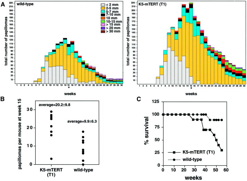
Age-matched (8- to 12-week-old) wild-type and K5-mTERT (T1) littermate mice received a single DMBA treatment followed by TPA–acetone treatment twice a week for 15 weeks (Materials and methods). Six weeks after the start of DMBA + TPA treatment, papillomas appeared in both wild-type and K5-mTERT (T1) mice, and these papillomas continued to grow in size throughout treatment (Figure 4A). The number of papillomas in K5-mTERT (T1) mice was significantly increased compared with wild-type littermates (Figure 4A and B). In particular, at week 15 (the end of TPA treatment), a total of 222 and 109 papillomas for K5-mTERT (T1) and wild-type mice, respectively, were counted (Figure 4A); this corresponds to 20.0 ± 9.8 and 9.9 ± 6.3 papillomas per mouse for K5-mTERT (T1) and wild-type mice, respectively (Figure 4B; Student’s t-test P = 0.0091). After termination of TPA treatment at week 15 (asterisk in Figure 4A), many of the papillomas regressed in both genotypes, and a similar percentage of them progressed to larger lesions, maintaining the difference in total number of papillomas between wild-type and K5-mTERT (T1) littermates. In particular, 6.4 and 7.2% of the papillomas at week 15 progressed to lesions >1 cm at week 30, in wild-type and K5-mTERT (T1) mice, respectively (Figure 4A). Despite the difference in total number of papillomas, no histological differences were found between wild-type and K5-mTERT (T1) papillomas (Figure 6A; papilloma in wild-type and K5-mTERT T1 mice).
Fig. 4. (A) The total numbers of papillomas are plotted versus the number of weeks after the start of carcinogen treatment. Termination of TPA treatment (week 15) is indicated by an asterisk. (B) Average number of papillomas per mouse at week 15 after the start of carcinogen treatment. Wild-type and K5-mTERT (T1) transgenics showed an average of 9.9 ± 6.3 and 20.0 ± 9.8 papillomas per mouse, respectively. (C) Survival of DMBA + TPA-treated wild-type and K5-mTERT (T1) mice during the multistage chemical carcinogenesis experiment.
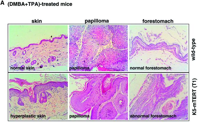
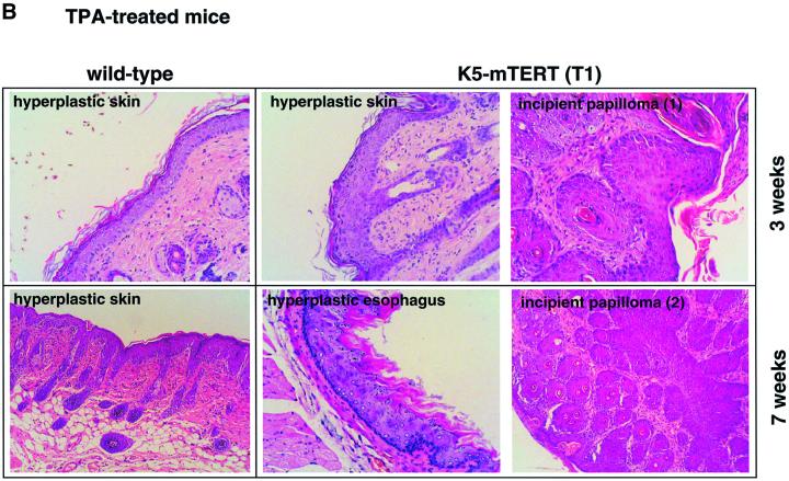
Fig. 6. Histopathology of skin lesions. (A) Both DMBA + TPA-treated wild-type and K5-mTERT (T1) mice show typical papilloma lesions in the skin (see ‘papilloma’). Seven weeks after termination of DMBA + TPA treatment, the DMBA + TPA-treated K5-mTERT (T1) mice show skin with severe hyperplasia (see ‘hyperplastic skin’); this is not observed in the skin of similarly treated wild-type mice (see ‘normal skin’). A forestomach lesion showing marked hyperplasia and hyperkeratosis corresponding to a DMBA + TPA-treated K5-mTERT (T1) mouse is shown (see ‘abnormal forestomach’); these lesions were never detected in similary treated wild-type mice (see ‘normal forestomach’). Magnification ×20. (B) Skin lesions in wild-type and K5-mTERT (T1) trangenics after repeated weekly exposure to TPA (no DMBA). Incipient papilloma lesions are shown for TPA-treated K5-mTERT (T1) mice (see ‘incipient papilloma’); these lesions were never detected in similarly treated wild-type mice. A hyperplastic esophagus lesion is shown for TPA-treated K5-mTERT (T1) mice (see ‘hyperplastic esophagus’); these lesions were never found in similarly treated wild-type mice. Magnification ×20.
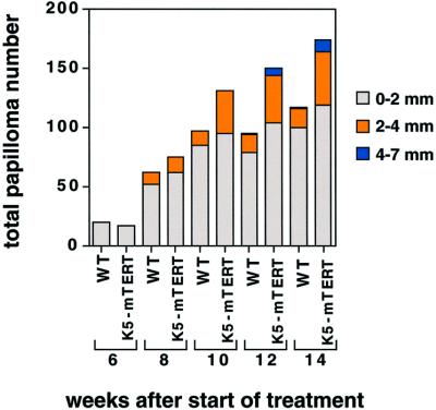
The increased susceptibility of transgenic K5-mTERT (T1) skin to develop papillomas due to chemical carcinogenesis was confirmed using K5-mTERT mice derived from a different founder, T8 (Figure 1B). K5-mTERT (T8) transgenics also showed an increased number of papillomas upon DMBA + TPA treatment as compared with the corresponding wild-type littermates (Figure 5).
Fig. 5. The total numbers of papillomas of different sizes are plotted versus the number of weeks after the start of DMBA + TPA treatment in wild-type and K5-mTERT (T8) transgenics. The total number of mice is nine each for wild-type and K5-mTERT genotypes (T8).
Strikingly, 80% of the DMBA + TPA-treated K5-mTERT (T1) mice (eight out of 10) died before week 60 after the start of treatment, whereas 90% of the similarly treated wild-type cohorts survived during this time (nine out of 10; Figure 4C). Death of the DMBA + TPA-treated K5-mTERT (T1) mice was associated with several pathologies (see Table I for quantifications) including the following. (i) Severe gastric hemorrhages resulting from an abnormal forestomach epithelium, which showed a marked hyperplasia and hyperkeratosis (Figure 6A; compare the normal forestomach epithelium in DMBA + TPA-treated wild-type mice with the abnormal forestomach in DMBA + TPA-treated K5-mTERT mice). This observation suggests that other K5-mTERT (T1) stratified epithelia besides the skin are prone to tumorigenesis upon DMBA + TPA treatment. (ii) Benign epidermal lessions: numerous keratoacanthomas and blue nevi (melanocyte accumulations in the skin). (iii) Pre-malignant epidermal lesions: severe dysplastic papillomas. (iv) Malignant epidermal tumors: basal cell carcinomas, malignant keratoacanthomas. (v) Tumors of non-epithelial origin: subcutaneous fibrosarcoma, uterus histiocytic sarcoma, liver histiocytic sarcoma, cecum lymphoma, as well as infiltrations of tumoral lymphoid cells in various tissues (multicentric lymphomas).
Table I. Non-papilloma lesions detected in DMBA + TPA-treated wild-type and K5-mTERT transgenics that died before week 60 after the start of treatment.
| Total no. of lesions | No. of micea | Weeks after start of treatmentb | |
|---|---|---|---|
| Non-papilloma epithelial lesionsc | |||
| keratoacanthoma | 9 | 4 | 38–50 |
| blue nevus | 17 | 6 | 38–58 |
| hyperplastic forestomach | 3 | 3 | 28–38 |
| basal cell tumor | 3 | 3 | 38–58 |
| severely dysplastic papilloma | 1 | 1 | 28–50 |
| malignant keratoacanthoma | 3 | 1 | 50 |
| Non-epithelial lesionsc | |||
| uterus histiocytic sarcoma | 1 | 1 | 48 |
| liver histiocytic sarcoma | 1 | 1 | 52 |
| lymphoma of cecum | 1 | 1 | 38 |
| multicentric lymphoma | 1 | 1 | 50 |
| subcutaneus fibrosarcoma | 1 | 1 | 48 |
| lymphoid hyperplasia | 1 | 1 | 44 |
| Non-epithelial lesionsd | |||
| subcutaneus fibrosarcoma | 1 | 1 | 44 |
aNumber of mice that showed the lesion at the time of death before week 60 after the start of DMBA + TPA treatment. The lesions were counted in eight K5-mTERT transgenics and one wild-type littermate that died after the start of the DMBA + TPA treatment.
bTime at which lesions appeared (in weeks) after the start of DMBA + TPA treatment.
cK5-mTERT transgenics.
dWild-type mice.
Hence, the increased mortality of K5-mTERT mice upon DMBA + TPA carcinogenesis coincides with a higher susceptibility of these mice to develop neoplasias.
Telomeric Q-FISH analysis on skin sections from DMBA + TPA-treated mice showed that telomeres were similar or slightly elongated in K5-mTERT (T1) mice as compared with wild-type controls (Figure 2B; gray bars).
K5-mTERT skins are more sensitive to the mitogenic effects of phorbol esters
Curiously, whereas wild-type mice showed areas of histo logically normal skin surrounding papillomas 7 weeks after termination of DMBA + TPA treatment (Figure 6A; normal skin), the DMBA + TPA-treated K5-mTERT (T1) mice maintained a marked hyperplasia of the skin, with regions showing up to 10 layers of keratinocytes (Figure 6A; hyperplastic skin). These observations suggested an increased proliferative response of K5-mTERT (T1) basal keratinocytes to repetitive TPA treatment. To study this further, wild-type and K5-mTERT (T1) mice were treated weekly with TPA (5 µg of TPA in 200 µl of acetone) in the absence of previous treatment with the carcinogen DMBA. After 3 weeks of repetitive TPA exposure, K5-mTERT (T1) mice showed a more marked hyperplasia of the skin than similarly treated wild-type mice (Figure 6B; hyperplastic skin at week 3 of TPA treatment). Strikingly, after prolonged (3 and 7 weeks) repetitive TPA exposure, the K5-mTERT (T1) mice showed incipient papilloma lesions in the skin, which were never detected in similarly treated wild-type mice (Figure 6B; incipient papillomas at weeks 3 and 7 of TPA treatment). Some of these incipient papilloma lesions in the K5-mTERT (T1) transgenics progressed to macroscopic papillomas upon further TPA treatment; again, this was never observed in the similarly treated wild-type littermates (not shown). Furthermore, probably due to licking of the skin-treated region, TPA-treated K5-mTERT mice also showed areas of focal hyperplasia in the esophagus and forestomach epithelia, which were absent in similarly treated wild-type mice (Figure 6B; hyperplastic esophagus at week 7 of TPA treatment).
Taken together, these results suggest that constitutive telomerase activity in stratified epithelia results in increased proliferation of basal keratinocytes in response to TPA treatment. This could be a way in which constitutive telomerase expression helps to promote tumor formation and progression in the K5-mTERT transgenic mice subjected to chemical carcinogenesis of the skin.
Increased wound healing in K5-mTERT epidermis as compared with wild-type epidermis
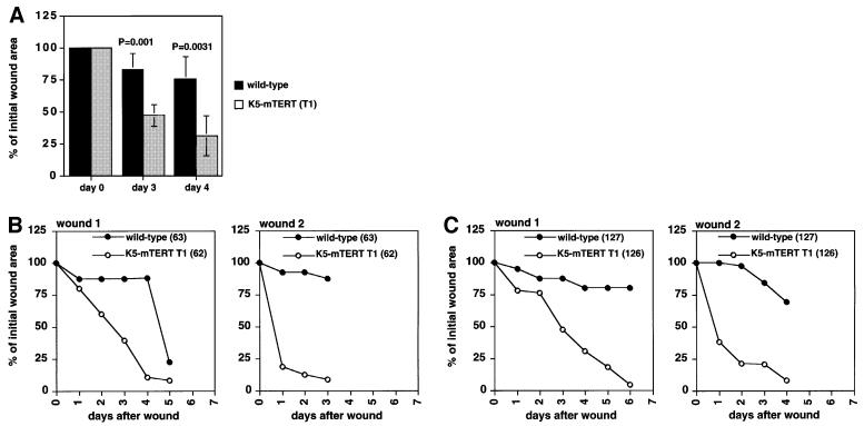
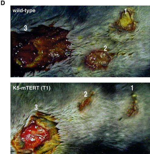
As an independent way to assay the functionality of K5-mTERT (T1) trangenic epithelia, we carried out wound-healing experiments in the skin of 2- to 4-month-old littermate wild-type and K5-mTERT (T1) transgenics. Wound healing is a complex process involving cellular proliferation, growth factor production and an immune system reaction. Two consecutive 13 mm2 (4 mm diameter) circular punch biopsies were performed on the back skin of six wild-type and six K5-mTERT (T1) littermate mice. The rate of wound healing was monitored as the percentage of the initial wound area left open with time after the punch was made (see Materials and methods). One wound was made 2 days after the other and the animals were killed 7 days after the first wound was made. K5-mTERT (T1) transgenics showed faster wound healing compared with wild-type littermates (Figure 7A–C). Figure 7A shows the percentage of the initial wound area open in wild-type and K5-mTERT (T1) mice (six mice in each group) 3 and 4 days after each wound was created. The average wound areas at day 3 were 82.85 ± 12.38 and 46.95 ± 8.3% for wild-type and K5-mTERT (T1) mice, respectively (Student’s t-tests P = 0.001); and 75.45 ± 17.11 and 30.8 ± 15.5% for wild-type and K5-mTERT (T1) mice, respectively, at day 4 (Student’s t-tests P = 0.0031) (Figure 7A). These differences in wound healing between genotypes were highly significant, as indicated by the Student’s t-test values (P <0.01). As examples, Figure 7B and C shows the rate of healing of the two wounds (wound 1 and 2) in two representative pairs of wild-type and K5-mTERT (T1) littermates. Figure 7D shows representative images of wild-type and K5-mTERT (T1) wounds 4 days after the first wound was made (1 and 2 in Figure 7D correspond to the first and second wound, respectively); the two wild-type wounds remained opened whereas the two K5-mTERT (T1) wounds were already closed. As a standard for wound size, fresh punch biopsies were also made prior to sacrifice of the mice (marked with a 3 in Figure 7D). Histology of the wounds confirmed a faster rate of re-epithelilization of the K5-mTERT (T1) wounds as compared with wild-type littermates (not shown).
Fig. 7. Wound healing of wild-type and K5-mTERT (T1) skin. (A) Percentage of initial wound area left 3 and 4 days after the punch was created in a group of six wild-type and six K5-mTERT (T1) mice. Statistical analysis showing that the healing rate is significantly different between genotypes is also shown (P-values). (B and C) Rate of wound healing in two wild-type and K5-TERT (T1) littermate pairs; the results are presented as the percentage of open wound area versus time after wound creation. Closed circles, wild-type mice; open circles, K5-mTERT (T1) transgenics. (D) Gross appearance of healing wounds in a wild-type and a K5-mTERT (T1) mouse 5 and 2 days, for wounds 1 and 2, respectively, after wound creation. Just prior to sacrifice of the mice, a third wound (wound 3) was created as an indicator of initial wound areas.
Taken together, these results indicate that the skin of K5-mTERT (T1) mice has a faster wound-healing rate than that of the corresponding wild-type littermates. This may reflect a proliferative advantage of telomerase-transgenic skins as compared with controls in response to proliferative signals associated with wound healing, in agreement with the greater hyperplasia seen in K5-mTERT epithelia as compared with wild-type littermates upon treatment with the mitogen TPA.
Normal p53, Ras and c-Myc levels in K5-mTERT skin
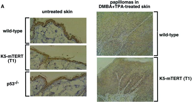
p53 loss of function and H-ras upregulation are described as occurring in multistage chemical carcinogenesis of the skin, and are thought to favor the appearance of tumors (Balmain et al., 1984, 1992; Quintanilla et al., 1986). To determine whether constitutive telomerase expression induces changes associated with transformation in the skin, we studied several tumor markers such as p53, Ras and c-Myc in the skin of wild-type and littermate K5-mTERT (T1) mice. To analyze p53 status in skin keratinocytes, we performed immunohistochemistry of the skin with anti-p53 antibody, as previously described (González-Suárez et al., 2000) (see Materials and methods). There is no significant variation in p53 levels between wild-type and K5-mTERT (T1) untreated skin or DMBA + TPA-induced papillomas (Figure 8A), suggesting that transgenic telomerase expression does not affect normal p53 levels; as a control, we show the skin of a p53-deficient mouse (Figure 8A). Furthermore, there was no significant difference in Ras protein levels in nuclear extracts prepared from wild-type and K5-mTERT (T1) primary keratinocyte cultures, as determined by western blot (Figure 8B). Detection of Ku70 nuclear protein was used as a loading control (Figure 8B).
Fig. 8. (A) p53 levels in wild-type and K5-mTERT (T1) skin keratinocytes. Immunohistochemistry with p53 antibody of untreated and DMBA + TPA-treated wild-type and K5-mTERT (T1) skin. The control untreated wild-type and K5-mTERT (T1) skin show similar p53 positivity in basal keratinocyte nuclei; p53–/– mouse skin is shown as a negative control. Wild-type and K5-mTERT (T1) papillomas show decreased p53 staining of basal keratinocytes compared with wild-type skin. Magnification ×20. (B) Ras protein levels detected by western blot in wild-type and K5-mTERT (T1) primary keratinocytes. No significant differences in Ras protein levels were detected between genotypes. As a control for loading, nuclear protein Ku70 was also detected. Different numbers indicate different primary keratinocyte cultures. The arrows indicate the positions of theRas- and Ku86-specific bands. (C) c-myc mRNA levels detected by northern blot in the skin of four K5-mTERT (T1 or T8, as indicated) and the corresponding wild-type littermate skins. Mice 30, 32, 34 and 35 are littermates derived from founder T8. Mice 130, 131, 132 and 134 are littermates derived from founder T1. As a positive control for c-myc detection, total RNA from interleukin-2-stimulated Ba/FO3 cells was used (Hatakeyama et al., 1989). As control for loading, actin was also detected in the blots. Table II shows quantification of c-myc levels.
It has been shown recently that TERT-driven cell proliferation in cultured cells can result in the activation of the c-myc oncogene (Wang et al., 2000). We studied c-myc mRNA levels in the skin from several K5-mTERT (T1 and T8, as indicated) mice and corresponding wild-type littermate controls. No significant differences in the c-myc mRNA levels were observed between genotypes (see Materials and methods; Figure 8C and Table II for quantitation of c-Myc levels using a phosphoimager), suggesting that transgenic mTERT expression in the skin does not result in c-myc upregulation.
Table II. Quantification of c-myc mRNA levels in the skin of K5-mTERT (T1 and T8, as indicated) and corresponding wild-type littermates.
| c-myc (AU) | Actin (AU) | Relative c-myc abundance (%)a | |
|---|---|---|---|
| Wild-type 34 (T8) | 57 050.67 | 409 571.97 | 127 |
| Wild-type 35 (T8) | 72 780.37 | 558 146.15 | 118 |
| K5-mTERT 30 (T8) | 73 277,28 | 661 005.82 | 101 |
| K5-mTERT 32 (T8) | 80 397.53 | 622 336.42 | 117 |
| Wild-type 130 (T1) | 84 549.74 | 675 757.15 | 114 |
| Wild-type 134 (T1) | 53 872.73 | 491 477.81 | 100 |
| K5-mTERT 131 (T1) | 61 120.25 | 478 277,03 | 116 |
| K5-mTERT 132 (T1) | 83 926.68 | 595 155,99 | 128 |
| Control c-myc | 200 557.77 | 503 331.87 | 363 |
AU, arbitrary units after quantification with the phosphoimager; T8, mice derived from the T8 founder; T1, mice derived from the T1 founder.
Mice 30,32, 34 and 35 are littermates, and mice 130, 131, 132 and 134 are littermates.
aRelative abundance of c-myc corrected by actin levels and expressed as a percentage. Levels were considered 100% in one of the wild-type mice showing the lowest c-myc levels (indicated in bold).
Conclusions
We show here that constitutive high levels of telomerase activity in stratified epithelia do not alter the normal epithelium structure and are not associated with changes in p53, Ras or c-Myc levels, in agreement with previous reports showing that TERT immortalized human cultured cells do not show changes associated with transformation (Jiang et al., 1999; Morales et al., 1999). However, follow ing DMBA + TPA treatment of the skin, K5-mTERT (T1 and T8) mice are significantly more susceptible to the development of neoplasias than the corresponding wild-type littermates. This increased tumor susceptibility of K5-mTERT transgenic skin is accompanied by a greater proliferation of basal keratinocytes upon TPA treatment, as well as by an increased would-healing rate of the K5-mTERT skin compared with wild-type mice. All these observations are in agreement with a direct or indirect role for telomerase in signaling proliferation under mitogenic conditions. These results agree with previous observations that indicated that first generation Terc–/– mice, which lack telomerase activity but still have long telomeres, show a lower incidence of papillomas than wild-type mice (González-Suárez et al., 2000). These findings unravel the potential risks of gene therapy for age-associated diseases based on telomerase upregulation, and indicate that telomerase upregulation, which occurs in the vast majority of mouse and human tumors, actively promotes tumor growth even in the presence of long telomeres. Since the average length of telomeres in wild-type and K5-mTERT skin was similar, it is tempting to speculate that telomere conformation must be different whether or not telomerase activity is present in the cell, and that telomeres that are being ‘maintained’ by telomerase could be signaling proliferation and/or survival under mitogenic conditions.
Finally, K5-mTERT mice constitute a new experimental model to study the direct impact of transgenic telomerase expression in both skin tumorigenesis and aging in vivo. Tissue-specific telomerase-transgenic mice are essential to study the impact of potential use of telomerase for gene therapy of age-related diseases. K5-mTERT mice also constitute a new animal model system to study the effectiveness of anti-telomerase therapies in cancer.
Materials and methods
Generation and genotyping of transgenic K5-mTERT mice
An EcoRI fragment containing the full-length mTERT cDNA and 3′-untranslated sequences was cloned 3′ downstream of 5 kb of bovine keratin K5 regulatory sequences (see Figure 1A) (Ramírez et al., 1994; Martín-Rivera et al., 1998). The transgene was digested with NotI, purified using Genclean (Bio 101 Inc.), adjusted to a final concentration of ∼2 µg/ml and microinjected into (C57BL/6 × DBA/2) mouse embryos, as described (Hogan et al., 1986). Eight different founder mice were identified by Southern blot analysis of tail DNA, using the 5 kb fragment corresponding to the bovine keratin K5 promoter as a probe (not shown). The experiments described here were carried out with founder T1 and were reproduced, as indicated, with founder T8, which shows a similar upregulation of telomerase activity (T1 = 8043 and T8 = 7664; highlighted in red in Figure 1B). The other founders showing lower reconstitution of telomerase activity in the skin were not included in this study.
Handling of mice
Mice were housed in our barrier area, where pathogen-free procedures are employed in all mouse rooms. Quarterly health monitoring reports have been negative for all pathogens in accordance with FELASA recommendations (Federation of European Laboratory Animal Science Associations).
Telomerase assays
Telomerase activity was measured with a modified telomeric repeat amplification protocol (TRAP), as described (Blasco et al., 1997).
Tumor induction experiments
Ten and 12 age-matched (8- to 12-week-old) mice of each genotype, wild-type and K5-mTERT (T1), respectively, were shaved and treated with a single dose of DMBA (0.1 µg/µl in acetone; Sigma). Nine age-matched (8-week-old) mice of each genotype, wild-type and K5-mTERT (T8), were shaved and treated with a single dose of DMBA (0.1 µg/µl in acetone; Sigma). In all cases, subsequently mice were treated twice weekly with TPA (12.5 µg in 200 µl acetone each treatment; Sigma), for 15 weeks. Four control mice of each genotype were treated with acetone only. In samples analyzed after weekly cumulative TPA doses, the TPA concentration used was 12.3 µg in 200 µl of acetone.
Histopathological analyses and immunochemistry
Papilloma, skin, esophagus, forestomach, oral mucosa and vagina sections from 15- to 19-month-old wild-type and K5-mTERT (T1 or T8, as indicated) mice were fixed in 10% buffered formalin and stained with hematoxylin–eosin. Images were captured with an Olympus-Vanox microscope at a 20× magnification.
For the DMBA + TPA-treated mice that died, necropsy of animals was performed and all organs were examined macroscopically. Tumoral and normal tissues were fixed in 10% buffered formalin and embedded in paraffin. Sections of 4 µm were stained with hematoxylin–eosin. Images were captured with an Olympus-Vanox microscope at a 20× magnification.
Immunohistochemistry for p53 was performed on deparaffinated sections (Vectabond slides) after pressure cooker processing. For p53 detection, we used a streptavidin–biotin complex technique, with a polyclonal rabbit anti-mouse p53 antibody, CM5 (Novocastra Laboratory Ltd; 1:1500 dilution, overnight, 4°C), and a biotinylated anti-rabbit IgG (Vector Laboratory; 1:400 dilution, 30 min, room temperature), followed by streptavidin peroxidase (Zymed Laboratory, Inc.; 1:20 dilution, 30 min, room temperature); the chromogen was developed with diaminobenzidine, and slides were counterstained with hematoxylin. Control slides were obtained by replacing the primary antibody with TBS (not shown) (see also González-Suárez et al., 2000).
Wound-healing experiments
Two to three full-thickness punch biopsies extending through the epidermis and dermis (punch diameter 4 mm; PFM, Köln, Germany) were performed in six wild-type and six K5-mTERT mice (2–4 months of age) after depilation. Mice were anesthesized prior to wound creation. The wound-healing rate was calculated as the percentage of initial wound area with time. Wound areas were calculated with the formula (area = πr2; where r is the ratio of the wound).
Telomere length measurements
For Q-FISH, paraffin-embedded skin sections (untreated or treated with DMBA + TPA) were hybridized with a PNA-tel probe, and telomere length was determined as described (Zijlmans et al., 1997; González-Suárez et al., 2000). Slides were deparaffinated in three xylene washes (3 min each), then treated for 3 min with a 100, 95 and 70% ethanol series. More than 50 keratinocyte nuclei from each genotype were captured at a 100× magnification and the telomere fluorescence was integrated using spot IOD analysis in the TFL-TELO program. For Flow-FISH, primary mouse keratinocytes (105) were prepared (Hennings, 1994) and hybridized using the Flow-FISH protocol (Rufer et al., 1998). Telomere fluorescence of 2000–3000 nuclei gated at the G1–G0 cell cycle stage was measured using a Coulter EPICS XL flow cytometer with SYSTEM 2 software. The same cells were hybridized in parallel without the PNA–fluorescein isothiocyanate (FITC) probe to obtain background fluorescence values, which were subtracted from the fluorescence intensity of each sample. We used three wild-type and six K5-mTERT (T1) primary keratinocyte cultures.
Western blots
Nuclear extracts were prepared from three primary wild-type and four K5-mTERT (T1) keratinocyte cultures (Hennings, 1994), and western blots were performed as previously described (González-Suárez et al., 2000). The protein concentration per well was 20 µg. For Ras detection, pan-ras antibody (Ab-3; Calbiochem) at a 1:1000 dilution was used. For Ku86 detection, anti-human p70 (Hamiya Biomedical Company, 1:000 dilution) was used. The percentage of the acrylamide gel was 15%.
Northern blots
Total RNA was extracted from the skin of a total of four wild-type and four K5-mTERT (T1 or T8, as indicated) littermate mice essentially as described (Chomczynski and Sacchi, 1987). A 50 µg aliquot of RNA was used per lane in the northern blot. A 1500 nucleotide fragment of the human c-myc gene was used as probe for murine c-myc mRNA detection; as a positive control, total RNA from murine lymphocyte cell line Ba/FO3 upon interleukin-2 stimulation was used (Hatakeyama et al., 1989). As control for loading, actin was also detected in the blots.
Acknowledgments
Acknowledgements
We are indebted to F.Larcher for help in isolating primary keratinocytes. We thank E.Santos and Rosa Serrano for mouse care and genotyping, P.Jurado for her contribution to the construction of the K5-mTERT transgene, and M.Serrano and C.Mark for critical reading of the manuscript. E.G.-S. is a predoctoral fellow from the Fondo de Investigaciones Sanitarias. E.S. is a predoctoral fellow from the Regional Government of Madrid (CAM). Research at the laboratory of M.A.B. is funded by Swiss Bridge Award 2000, by grants PM97-0133 from the Ministry of Science and Technology (MCT), Spain, 08.1/0030/98 from the CAM, FIGH-CT1999-00009, FIGH-CT-1999-00002 and QLG1-1999-01341 from the European Union, and by the Department of Immunology and Oncology (DIO). Research at the laboratory of J.L.J. was funded by grant PM98-0039 from the MCT. The DIO was founded and is supported by the Spanish Research Council (CSIC) and by the Pharmacia Corporation.
References
- Balmain A., Ramsden,M., Bowden,G.T. and Smith,J. (1984) Activation of the mouse Harvey-ras gene in chemically induced benign skin papillomas. Nature, 307, 658–660. [DOI] [PubMed] [Google Scholar]
- Balmain A. et al. (1992) Functional loss of tumor suppressor genes in multistage chemical carcinogenesis. In Harris,C.C. et al. (eds), Multistage Carcinogenesis. Japan Scientific Society Press/CRC Press, Boca Raton, FL, pp. 97–108.
- Bednarek A.K., Chu,Y., Slaga,T.J. and Aldaz,C.M. (1997). Telomerase and cell proliferation in mouse skin papillomas. Mol. Carcinog., 20, 329–310. [DOI] [PubMed] [Google Scholar]
- Blackburn E.H. (1991) Structure and function of telomeres. Nature, 350, 569–573. [DOI] [PubMed] [Google Scholar]
- Blasco M.A., Funk,W.D., Villeponteau,B. and Greider,C.W. (1995) Functional characterization and developmental regulation of mouse telomerase RNA. Science, 269, 1267–1270. [DOI] [PubMed] [Google Scholar]
- Blasco M.A., Lee,H.-W., Hande,P., Samper,E., Lansdorp,P., DePinho,R. and Greider,C.W. (1997) Telomere shortening and tumor formation by mouse cells lacking telomerase RNA. Cell, 91, 25–34. [DOI] [PubMed] [Google Scholar]
- Bodnar A.G. et al. (1998) Extension of life-span by introduction of telomerase into normal human cells. Science, 279, 349–352. [DOI] [PubMed] [Google Scholar]
- Chomczynski P and Sacchi,N. (1987) Single-step method of RNA isolation by acid guanidinium thiocyanate–phenol–chloroform extraction. Anal. Biochem., 162, 156–159. [DOI] [PubMed] [Google Scholar]
- Collins K., Kobayashi,R. and Greider,C.W. (1995) Purification of Tetrahymena telomerase and cloning of the genes for the two protein components of the enzyme. Cell, 81, 677–686. [DOI] [PubMed] [Google Scholar]
- Counter C.M., Avilion,A.A., LeFeuvre,C.E., Stewart,N.G., Greider,C.W., Harley,C.B. and Bacchetti,S. (1992) Telomere shortening associated with chromosome instability is arrested in immortal cells which express telomerase activity. EMBO J., 11, 1921–1929. [DOI] [PMC free article] [PubMed] [Google Scholar]
- Feng J. et al. (1995) The RNA component of human telomerase. Science, 269, 1236–1241. [DOI] [PubMed] [Google Scholar]
- Gandhi L. and Collins,K. (1998) Interaction of recombinant Tetrahymena telomerase proteins p80 and p95 with telomerase RNA and telomeric DNA substrates. Genes Dev., 12, 721–733. [DOI] [PMC free article] [PubMed] [Google Scholar]
- González-Suárez E., Samper,E., Flores,J.M. and Blasco,M.A. (2000) Telomerase-deficient mice with short telomeres are resistant to skin tumorigenesis. Nature Genet., 26, 114–117 [DOI] [PubMed] [Google Scholar]
- Greenberg R.A., Allsopp,R.C., Chin,L., Morin,G. and DePinho,R. (1998) Expression of mouse telomerase reverse transcriptase during develop ment, differentiation and proliferation. Oncogene, 16, 1723–1730. [DOI] [PubMed] [Google Scholar]
- Greider C.W. (1996) Telomere length regulation. Annu. Rev. Biochem., 65, 337–365. [DOI] [PubMed] [Google Scholar]
- Greider C.W. and Blackburn,E.H. (1985) Identification of a specific telomere terminal transferase activity in Tetrahymena extracts. Cell, 43, 405–413. [DOI] [PubMed] [Google Scholar]
- Greider C.W. and Blackburn,E.H. (1989) A telomeric sequence in the RNA of Tetrahymena telomerase required for telomere repeat synthesis. Nature, 337, 331–337. [DOI] [PubMed] [Google Scholar]
- Hahn W.C., Counter,C.M., Lundberg,A.S., Beijersbergen,R.L., Brooks,M.W. and Weinberg,R.A. (1999) Creation of human tumour cells with defined genetic elements. Nature, 400, 464–468. [DOI] [PubMed] [Google Scholar]
- Harrington L., Zhou,W., McPhail,T., Oulton,R., Yeung,D., Mar,V., Bass,M.B. and Robinson,M.O. (1997a) Human telomerase contains evolutionarily conserved catalytic and structural subunits. Genes Dev., 11, 3109–3115. [DOI] [PMC free article] [PubMed] [Google Scholar]
- Harrington L., McPhail,T., Mar,V., Zhou,W., Oulton,R., Bass,M.B., Arruda,I. and Robinson,M.O. (1997b) A mammalian telomerase-associated protein. Science, 275, 973–977. [DOI] [PubMed] [Google Scholar]
- Hatakeyama M., Mori,H., Doi,T. and Taniguchi,T. (1989) A restricted cytoplasmic region of IL-2 receptor β chain is essential for growth signal transduction but not for ligand binding and internalization. Cell, 59, 837–845. [DOI] [PubMed] [Google Scholar]
- Heckner E., Fusenig,N.E., Kunz,W., Marks,F. and Thielmann,H.W. (1982) Carcinogenesis: A Comprehensive Survey, Vol. 7. Co-carcinogenesis and Biological Effects of Tumor Promoters. Raven Press, New York, NY.
- Hennings H. (1994) Primary culture of keratinocytes from newborn mouse epidermis in medium with lowered levels of Ca2+. In Leigh,I.M. and Watt,F.M. (eds), Keratinocyte Methods. Cambridge University Press, Cambridge, UK, pp. 21–23.
- Hogan B., Constantini,F. and Lacy,E. (1986) Manipulating the Mouse Embryo: A Laboratory Manual. Cold Spring Harbor Laboratory Press, Cold Spring Harbor, NY.
- Jiang X.-R. et al. (1999) Telomerase expression in human somatic cells does not induce changes associated with a transformed phenotype. Nature Genet., 21, 111–114. [DOI] [PubMed] [Google Scholar]
- Kilian A., Bowtell,D.D.L., Abud,H.E., Hime,G.R., Venter,D.J., Keese,P.K., Duncan,E.L., Reddel,R.R. and Jefferson,R.A. (1997) Isolation of a candidate human telomerase catalytic subunit gene, which reveals complex splicing patterns in different cell types. Hum. Mol. Genet., 6, 2011–2019. [DOI] [PubMed] [Google Scholar]
- Kiyono T., Foster,S.A., Koop,J.I., McDougall,J.K., Galloway,D.A. and Klingelhutz,A.J. (1998) Both Rb/p16INK4a inactivation and telomerase activity are required to immortalize human epithelial cells. Nature, 396, 84–88. [DOI] [PubMed] [Google Scholar]
- Lee H.-W., Blasco,M.A., Gottlieb,G.J., Horner,J.W., Greider,C.W. and DePinho,R.A. (1998) Essential role of mouse telomerase in highly proliferative organs. Nature, 392, 569–574. [DOI] [PubMed] [Google Scholar]
- Lingner J., Hughes,T.R., Schevchenko,A., Mann,M., Lundblad,V. and Cech T. (1997) Reverse transcriptase motifs in the catalytic subunit of telomerase. Science, 276, 561–567. [DOI] [PubMed] [Google Scholar]
- Martín-Rivera L., Herrera,E., Albar,J.P. and Blasco,M.A. (1998) Expression of mouse telomerase catalytic subunit in embryos and adult tissues. Proc. Natl Acad. Sci. USA, 95, 10471–10476. [DOI] [PMC free article] [PubMed] [Google Scholar]
- Meyerson M. et al. (1997) hEST2, the putative human telomerase catalytic subunit gene, is upregulated in tumor cells and during immortalization. Cell, 90, 785–795. [DOI] [PubMed] [Google Scholar]
- Mitchell J.R., Wood,E. and Collins,K. (1999) A telomerase component is defective in the human disease dyskeratosis congenita. Nature, 402, 551–555. [DOI] [PubMed] [Google Scholar]
- Morales C.P., Holt,S.E., Ouellette,M., Kaur,K.J., Yan,Y., Wilson,K.S., White,M.A., Wright,W.E. and Shay,J.W. (1999) Absence of cancer-associated changes in human fibroblasts immortalized with telomerase. Nature Genet., 21, 115–118. [DOI] [PubMed] [Google Scholar]
- Murillas R., Larcher,F., Conti,C.J., Santos,M., Ullrich,A. and Jorcano,J.L. (1995) Expression of a dominant negative mutant of epidermal growth factor receptor in the epidermis of transgenic mice elicits striking alterations on hair follicle development and skin structure. EMBO J., 14, 5216–5223. [DOI] [PMC free article] [PubMed] [Google Scholar]
- Nakamura T.M., Morin,G.B., Chapman,K.B., Weinrich,S.L., Andrews,W.H., Lingner,J., Harley,C.B. and Cech,T. (1997) Telomerase catalytic subunit homologs from fission yeast and human. Science, 277, 955–959. [DOI] [PubMed] [Google Scholar]
- Nakayama J., Saito,M., Nakamura,H., Matsuura,A. and Ishikawa,F. (1997) TLP1: a gene encoding a protein component of mammalian telomerase is a novel member of WD repeat family. Cell, 88, 875–884. [DOI] [PubMed] [Google Scholar]
- Nugent C.I. and Lundblad,V. (1998) The telomerase reverse transcriptase: components and regulation. Genes Dev., 12, 1073–1085. [DOI] [PubMed] [Google Scholar]
- Quintanilla M., Brown,K., Ramsden,M. and Balmain,A. (1986) Carcinogen-specific mutation and amplification of Ha-ras during mouse skin carcinogenesis. Nature, 322, 78–80. [DOI] [PubMed] [Google Scholar]
- Ramírez A., Bravo,A., Jorcano,J.L and Vidal,M. (1994) Sequences 5′ of the bovine keratin 5 gene direct tissue- and cell-type-specific expression of a LacZ gene in the adult and during development. Differentiation, 58, 53–64. [DOI] [PubMed] [Google Scholar]
- Rufer N., Dragowska,W., Thornbury,G., Roosnek,E. and Lansdorp,P.M. (1998) Telomere length dynamics in human lymphocyte subpopulations measured by flow cytometry. Nature Biotechnol., 16, 743–147. [DOI] [PubMed] [Google Scholar]
- Samper E., Goytisolo,F., Slijepcevic,P., van Buul,P. and Blasco,M.A. (2000) Mammalian Ku86 prevents telomeric fusions independently of the length of TTAGGG repeats and the G-strand overhang. EMBO Rep., 1, 244–252. [DOI] [PMC free article] [PubMed] [Google Scholar]
- Shay J.W. and Bacchetti,S. (1997) A survey of telomerase activity in human cancer. Eur. J. Cancer, 33, 787–791. [DOI] [PubMed] [Google Scholar]
- Singer M.S. and Gottschling,D.E. (1994) TLC1: template RNA component of the Saccharomyces cerevisiae telomerase. Science, 266, 387–388. [DOI] [PubMed] [Google Scholar]
- van Steensel B., Smogorzewska,A. and de Lange,T. (1998) TRF2 protects human telomeres from end-to-end fusions. Cell, 92, 401–413 [DOI] [PubMed] [Google Scholar]
- Wang J., Hannon,G.J. and Beach,D.H. (2000) Risky immortalization by telomerase. Nature, 405, 755–756. [DOI] [PubMed] [Google Scholar]
- Weitzman J.B. and Yaniv,M. (1999) Rebuilding the road to cancer. Nature, 400, 401–402. [DOI] [PubMed] [Google Scholar]
- Zijlmans J.M., Martens,U.M., Poon,S., Raap,A.K., Tanke,H.J., Ward,R.K. and P.M. Lansdorp. (1997) Telomeres in the mouse have large inter-chromosomal variations in the number of T2AG3 repeats. Proc. Natl Acad. Sci. USA, 94, 7423–7428. [DOI] [PMC free article] [PubMed] [Google Scholar]


