

Abstract
Introduction
Hydatid liver disease is endemic in many regions and, though often curable with surgery, can lead to rare long-term complications.
Case presentation
We report a 57-year-old woman with a history of hydatid cyst excision and cholecystectomy 40 years prior, presenting with right hypochondrial parietal gangrene and fever. Imaging revealed hepatic subcapsular collections fistulizing into the abdominal wall, complicated by necrotizing fasciitis. Emergency debridement and drainage were performed, followed by broad-spectrum antibiotics. ERCP with sphincterotomy was later performed to remove bile duct stones.
Clinical discussion
This case highlights a rare and severe late complication of hydatid liver surgery. The delayed fistulization and secondary infection emphasize the need to consider remote surgical history in atypical soft tissue infections.
Conclusion
Necrotizing fasciitis due to a delayed hepatic hydatid fistula is exceptional. Early recognition, imaging, surgical intervention, and multidisciplinary care are vital for favorable outcomes.
Keywords: Hydatid liver disease, Necrotizing fasciitis, Abdominal wall fistula, Delayed complication, Case report
Highlights
-
•
Delayed hepatic hydatid fistula can manifest decades after surgery with life-threatening complications.
-
•
Abdominal wall necrotizing fasciitis may reveal hidden hepatobiliary pathology.
-
•
A staged management combining emergency debridement and delayed ERCP ensures better outcomes in complex hydatid disease sequelae.
1. Introduction
Necrotizing fasciitis of the abdominal wall is a rare but life-threatening condition that requires prompt diagnosis and surgical intervention [1]. It is often associated with immunosuppression, trauma, or previous surgical procedures that create a potential entry point for infection [2]. Hydatid liver disease remains highly prevalent in Tunisia and other endemic regions, where exposure to Echinococcus granulosus is common. While surgical management is often curative, long-term sequelae such as biliary fistula, secondary infection of residual cavities, or fistulization to adjacent structures may occur. Chronic hepatic collections communicating with the abdominal wall are extremely uncommon, particularly decades after initial surgery.
We present an unusual case of delayed parietal necrotizing fasciitis due to a subcapsular hepatic collection fistulizing through the abdominal wall, 40 years after hydatid cyst excision and cholecystectomy. This case has been reported in line with the SCARE criteria [3].
2. Case presentation
We report the case of a 57-year-old woman with a surgical history of right subcostal laparotomy for hepatic hydatid cyst excision and concomitant cholecystectomy performed 40 years ago. Unfortunately, operative records from that time are no longer available, and details of the cyst type or surgical technique (radical vs. conservative) could not be retrieved.
She presented to our emergency department with complaints of upper abdominal pain and a foul-smelling parietal gangrene in the right hypochondrium, evolving over the previous month. The symptoms were accompanied by fever but without jaundice or other associated signs.
On clinical examination, the patient was febrile with a temperature of 40 °C, heart rate of 80 beats/min, and blood pressure of 120/70 mmHg. She was not icteric. Abdominal examination revealed a 20 cm area of parietal gangrene in the right hypochondrium with purulent discharge [Fig. 1, Fig. 2].
Fig. 1.

Clinical photograph showing extensive necrotizing fasciitis of the right abdominal wall with marked parietal gangrene and purulent discharge.
Fig. 2.
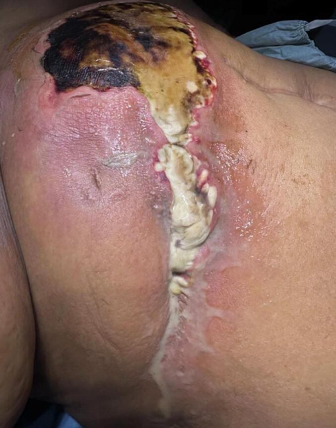
Close-up view of the necrotic abdominal wall tissue demonstrating subcutaneous emphysema and skin changes consistent with necrotizing soft tissue infection.
Laboratory investigations showed a white blood cell count of 9000/μL, hemoglobin of 7.9 g/dL (normal: 12.5–15.5 g/dL), and a markedly elevated C-reactive protein level at 295 mg/L (normal: <0.5 mg/L). Liver function tests and coagulation profile were within normal limits.
Contrast-enhanced abdominal CT revealed two subcapsular collections in segment III of the liver, measuring 28 × 12 mm and 27 × 14 mm, each with fistulous tracts to the anterior abdominal wall. These extended over 29 mm and 34 mm, respectively, and were associated with extensive subcutaneous soft tissue infiltration, poorly defined necrotic areas spanning more than 10 cm in height, and the presence of subcutaneous emphysematous bullae suggestive of necrotizing fasciitis [Fig. 3, Fig. 4]. Additionally, intra- and extrahepatic biliary dilatation was noted upstream of multiple stones in the common bile duct.
Fig. 3.
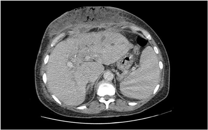
Contrast-enhanced abdominal CT scan (axial view) revealing subcapsular hepatic collections in segment III with fistulous tracts extending to the anterior abdominal wall.
Fig. 4.

CT image (sagittal view) illustrating extensive soft tissue gas infiltration and necrosis involving the abdominal wall consistent with necrotizing fasciitis.
The patient was taken urgently to the operating room. Intraoperatively, extensive parietal gangrene (approximately 20 cm) with necrosis and purulent discharge was observed. Wide surgical debridement was performed, followed by copious irrigation with normal saline. A deep necrotic cavity with active purulent drainage was identified and drained using a Redon drain after lavage and suction [Fig. 5]. Unfortunately, intraoperative samples for culture were not obtained.
Fig. 5.

Postoperative clinical image showing the debrided abdominal wall wound with a Redon drain in situ and evidence of healthy granulation tissue during recovery.
Postoperative recovery was uneventful. The patient was treated with intravenous antibiotics including a third-generation cephalosporin, metronidazole, and gentamicin, alongside regular local wound care.
One month later, endoscopic retrograde cholangiopancreatography (ERCP) with sphincterotomy was performed to extract the bile duct stones. The procedure was successful, and the patient had an uneventful recovery. She was discharged with advice for long-term outpatient follow-up and surveillance imaging.
3. Discussion
Necrotizing fasciitis is an uncommon and often initially insidious soft tissue infection characterized by rapid inflammation and destruction of the fascia, which may extend to involve the overlying muscle and skin [1]. It affects individuals of all ages and both sexes equally. However, it is more commonly observed in patients with diabetes, chronic alcoholism, immunosuppression, intravenous drug use, or underlying peripheral vascular disease [4].
Our case is unusual in that necrotizing fasciitis developed in a previously operated field four decades after an open hydatid cyst excision and cholecystectomy. Chronic hepatic hydatid disease can lead to late complications such as residual cavities, secondary bacterial infection, and, rarely, fistula formation to the abdominal wall [7]. In this patient, two subcapsular hepatic collections developed a fistulous tract to the anterior abdominal wall, likely serving as a persistent source of infection and ultimately resulting in parietal necrosis and soft tissue gas formation.
The clinical presentation of necrotizing fasciitis can be nonspecific in its early stages, often mimicking cellulitis or abscess. However, the presence of rapidly spreading erythema, crepitus, disproportionate pain, systemic toxicity, and soft tissue gas on imaging should raise strong suspicion [8,9]. In our case, contrast-enhanced CT provided key diagnostic information, revealing hepatic subcapsular abscesses with fistulous communication, soft tissue necrosis, and emphysematous changes consistent with necrotizing fasciitis.
Surgical debridement remains the cornerstone of treatment, supported by broad-spectrum intravenous antibiotics and intensive supportive care [10]. In our patient, wide excision of devitalized tissue and drainage of the infected cavity led to clinical improvement. The decision to delay management of the biliary obstruction was strategic, allowing initial sepsis control before proceeding to endoscopic retrograde cholangiopancreatography (ERCP) and sphincterotomy for stone removal. This staged approach contributed to the favorable outcome.
4. Limitations
-
•
Microbiological cultures from pus and necrotic tissue were not performed, which limited identification of the exact microbial flora (polymicrobial vs. anaerobic origin).
-
•
Details regarding the type of initial hydatid cyst and the surgical approach (radical vs. conservative) could not be retrieved.
-
•
Long-term follow-up beyond post-ERCP recovery was not available at the time of submission, and recurrence surveillance remains necessary.
This case highlights the importance of considering late infectious complications in patients with a history of hepatic hydatid disease, particularly when presenting with abdominal wall infections of unclear origin. It also underscores the diagnostic utility of CT imaging and the need for timely surgical intervention in suspected necrotizing fasciitis.
5. Conclusion
This case illustrates a rare and delayed complication of hepatic hydatid surgery, presenting as necrotizing fasciitis of the abdominal wall due to fistulization of a chronic subcapsular hepatic collection. It highlights the importance of considering prior surgical history in the diagnostic workup of unusual soft tissue infections. Early imaging, prompt surgical debridement, and appropriate antibiotic therapy are critical to improving outcomes. A staged approach to managing underlying biliary pathology, once the infection is controlled, can further optimize recovery.
Author contribution
Ghazi Lâamiri: Data curation, Writing – original draft preparation Hazem Alouani: Conceptualization, methodology Khadija Yaich: Visualization, Investigation Houda Gazzah: Investigation Mehdi Bouassida: Writing – Reviewing and Editing Hassen Touinsi: Supervision.
Patient consent
Written informed consent was obtained from the patient for publication of this case report and accompanying images and is available for reviewing if needed.
Ethical approval
This is a case report and, according to the guidelines of the Faculty of Medicine of Tunis, it does not constitute human subject research and therefore did not require approval from an ethics committee. The Institutional Review Board of Mohamed Taher Maamouri University Hospital of Nabeul, Tunisia determined that ethical approval was not required for single-patient case reports.
Guarantor
Hazem Alouani accepts full responsibility for the integrity of the content presented in this article. As the guarantor, Hazem Alouani affirms that the research, data, and conclusions presented are accurate, complete, and represent the work performed. The guarantor has overseen the project to ensure that all ethical guidelines and research standards were followed and that all co-authors have approved the final version of the manuscript.
Research registration number
Not applicable.
Author agreement statement
We the undersigned declare that this manuscript is original, has not been published before and is not currently being considered for publication elsewhere. We confirm that the manuscript has been read and approved by all named authors and that there are no other persons who satisfied the criteria for authorship but are not listed. We further confirm that the order of authors listed in the manuscript has been approved by all of us. We understand that the Corresponding Author is the sole contact for the Editorial process. He is responsible for communicating with the other authors about progress, submissions of revisions and final approval of proofs.
Funding
This research received no specific grant from any funding agency in the public, commercial, or not-for-profit sectors.
Conflict of interest statement
The authors declare no conflict of interest.
Data availability
The data that support the findings of this study are available from the corresponding author upon reasonable request.
References
- 1.Heidelberg Laura S., et al. An atypical case of necrotizing fasciitis secondary to perforated cecal cancer. J. Surg. Case Rep. 10 Nov. 2020;2020(11) doi: 10.1093/jscr/rjaa371. [DOI] [PMC free article] [PubMed] [Google Scholar]
- 2.Bahl Nicholas, et al. A case of necrotizing soft tissue infection secondary to perforated colon cancer. Cureus. 2 Sep. 2021;13(9) doi: 10.7759/cureus.17663. [DOI] [PMC free article] [PubMed] [Google Scholar]
- 3.Kerwan A., Al-Jabir A., Mathew G., Sohrabi C., Rashid R., Franchi T., Nicola M., Agha M., Agha R.A. Revised Surgical CAse REport (SCARE) guideline: an update for the age of Artificial Intelligence. Prem. J. Sci. 2025;10 [Google Scholar]
- 4.Green R.J., et al. Necrotizing fasciitis. Chest. 1996;110(1):219–229. doi: 10.1378/chest.110.1.219. [DOI] [PubMed] [Google Scholar]
- 7.Dziri Chadli, et al. Treatment of hydatid cyst of the liver: where is the evidence? World J. Surg. 2004;28(8):731–736. doi: 10.1007/s00268-004-7516-z. [DOI] [PubMed] [Google Scholar]
- 8.Morgan M.S. Diagnosis and management of necrotising fasciitis: a multiparametric approach. J. Hosp. Infect. 2010;75(4):249–257. doi: 10.1016/j.jhin.2010.01.028. [DOI] [PubMed] [Google Scholar]
- 9.Becker M., et al. Necrotizing fasciitis of the head and neck: role of CT in diagnosis and management. Radiology. 1997;202(2):471–476. doi: 10.1148/radiology.202.2.9015076. [DOI] [PubMed] [Google Scholar]
- 10.Anaya Daniel A., Dellinger E. Patchen. Necrotizing soft-tissue infection: diagnosis and management. Clin. Infect. Dis. 2007;44(5):705–710. doi: 10.1086/511638. [DOI] [PubMed] [Google Scholar]
Associated Data
This section collects any data citations, data availability statements, or supplementary materials included in this article.
Data Availability Statement
The data that support the findings of this study are available from the corresponding author upon reasonable request.