Abstract
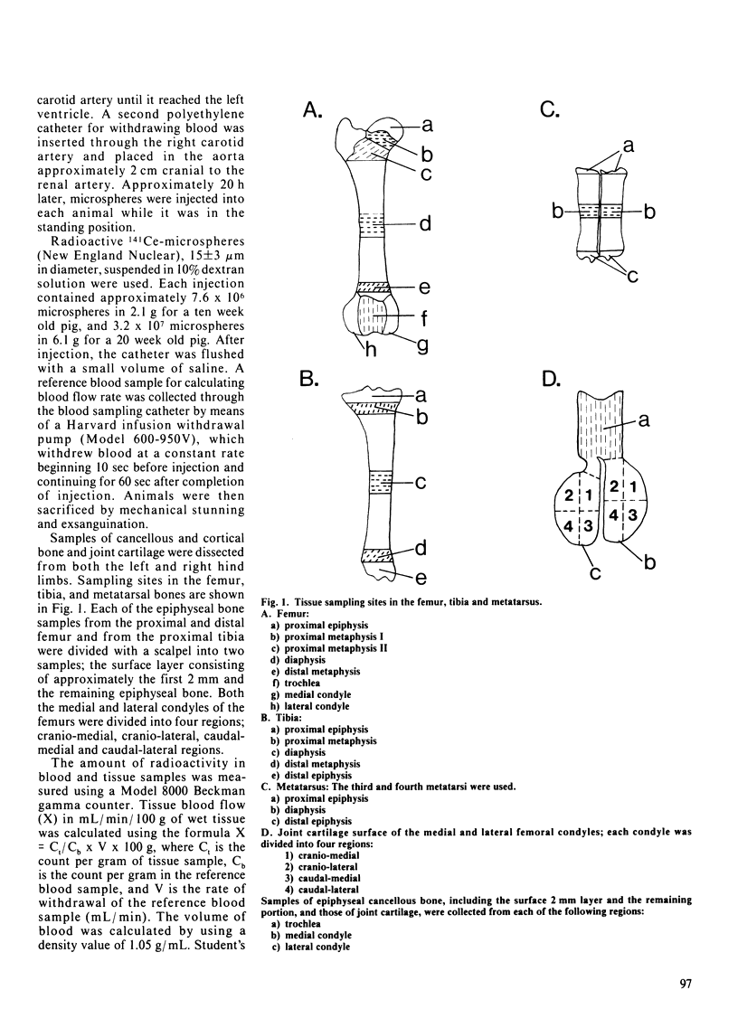
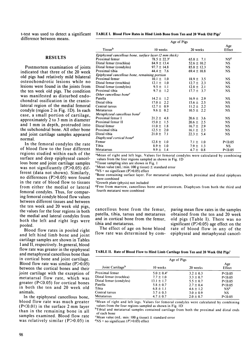
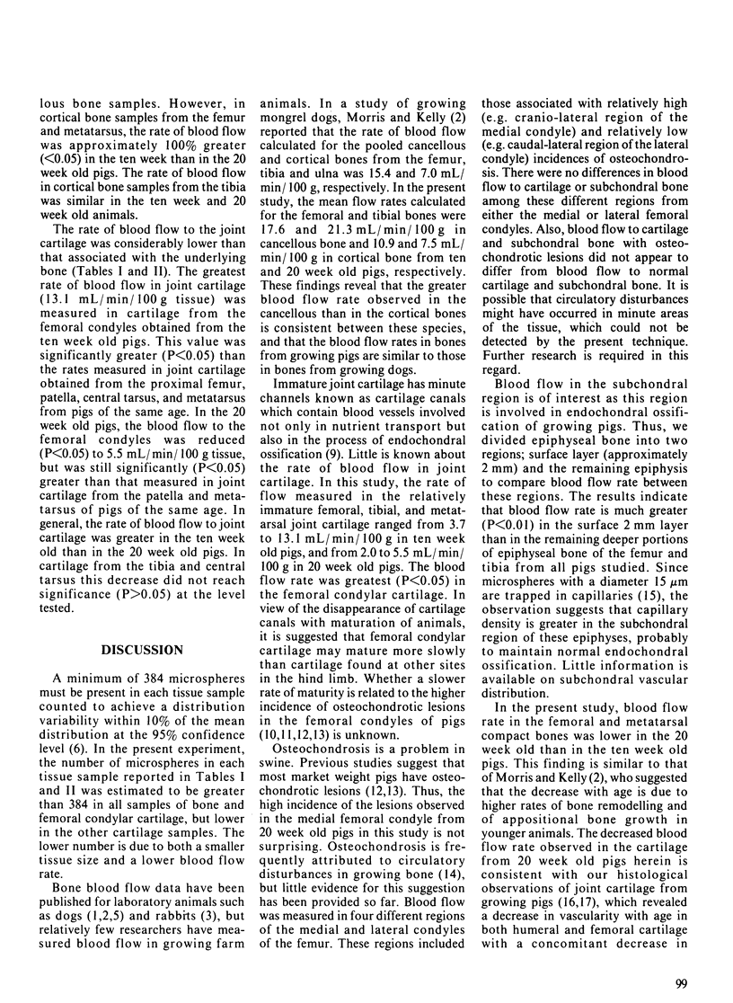
Blood flow distribution was studied in hind limb bones and joint cartilage from four ten week old and four 20 week old pigs using 141Ce-microspheres. In general, blood flow rate was greater in the epiphyseal and metaphyseal cancellous bone than in the cortical bone and joint cartilage. In the epiphyseal cancellous bone, blood flow rate was three to tenfold greater (P less than 0.01) in the surface 2 mm layer than in the remaining deeper bone in all femoral and tibial samples examined. In the joint cartilage, blood flow rate in the femoral condyle was greater (P less than 0.05) than in the proximal femur, patella, central tarsus and metatarsus in ten week old pigs, and was greater (P less than 0.05) in the femoral condyle than in the patella and metatarsus in 20 week old pigs. A significant (P less than 0.05) age-associated decrease in the rate of blood flow was observed in the femoral, patellar and metatarsal cartilage. Within the femoral condyle, no differences in blood flow were found between areas associated with relatively high versus relatively low incidences of osteochondrosis.
Full text
PDF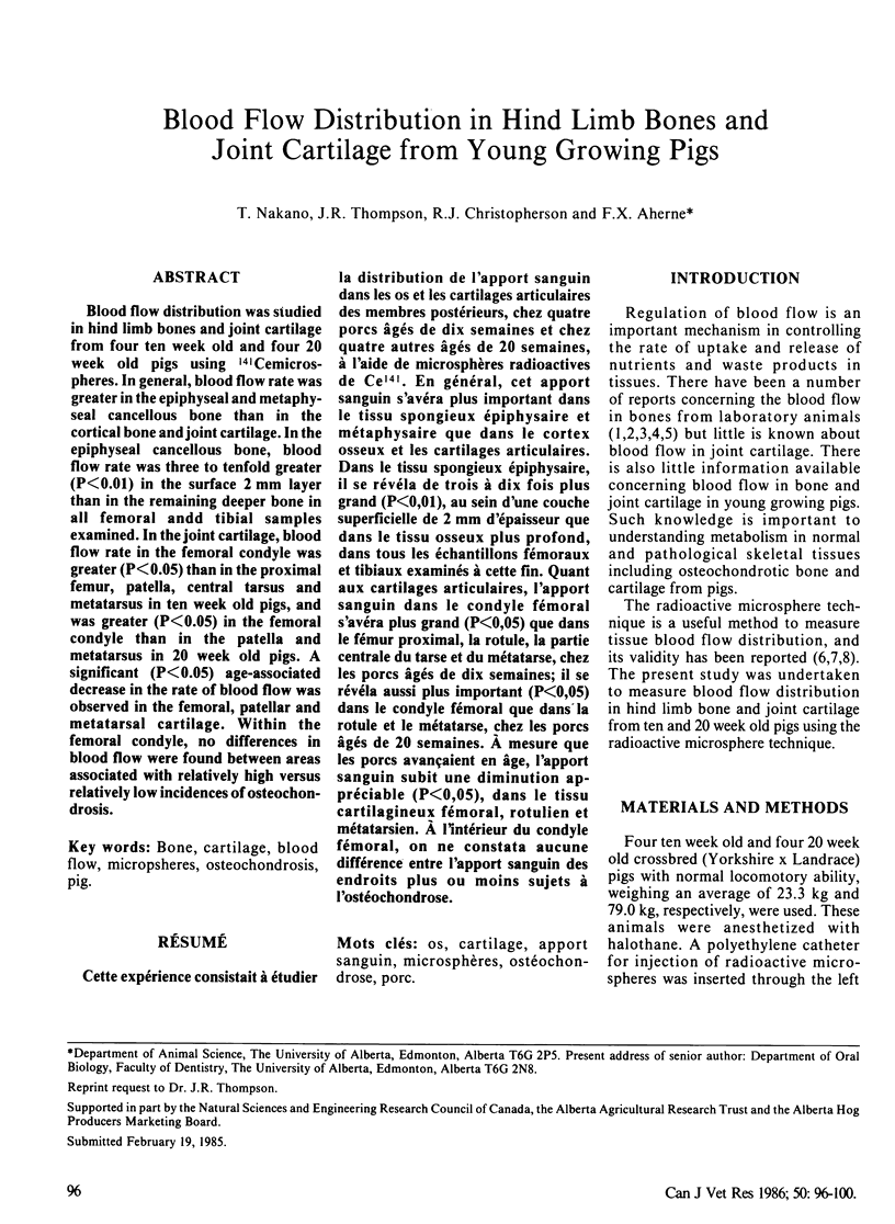
Selected References
These references are in PubMed. This may not be the complete list of references from this article.
- Bhatnagar R., Christian R. G., Nakano T., Aherne F. X., Thompson J. R. Age related changes and osteochondrosis in swine articular and epiphyseal cartilage: light ane electron microscopy. Can J Comp Med. 1981 Apr;45(2):188–195. [PMC free article] [PubMed] [Google Scholar]
- Buckberg G. D., Luck J. C., Payne D. B., Hoffman J. I., Archie J. P., Fixler D. E. Some sources of error in measuring regional blood flow with radioactive microspheres. J Appl Physiol. 1971 Oct;31(4):598–604. doi: 10.1152/jappl.1971.31.4.598. [DOI] [PubMed] [Google Scholar]
- Goedegebuure S. A., Häni H. J., van der Valk P. C., van der Wal P. G. Osteochondrosis in six breeds of slaughter pigs. I. A morphological investigation of the status of osteochondrosis in relation to breed and level of feeding. Tijdschr Diergeneeskd. 1980 Jan 15;105(2):28–41. [PubMed] [Google Scholar]
- Grondalen T. Osteochondrosis and arthrosis in pigs. I. Incidence in animals up to 120 kg live weight. Acta Vet Scand. 1974;15(1):1–25. doi: 10.1186/BF03547490. [DOI] [PMC free article] [PubMed] [Google Scholar]
- Gross P. M., Marcus M. L., Heistad D. D. Measurement of blood flow to bone and marrow in experimental animals by means of the microsphere technique. J Bone Joint Surg Am. 1981 Jul;63(6):1028–1031. [PubMed] [Google Scholar]
- Heymann M. A., Payne B. D., Hoffman J. I., Rudolph A. M. Blood flow measurements with radionuclide-labeled particles. Prog Cardiovasc Dis. 1977 Jul-Aug;20(1):55–79. doi: 10.1016/s0033-0620(77)80005-4. [DOI] [PubMed] [Google Scholar]
- Kane W. J., Grim E. Blood flow to canine hind-limb bone, muscle, and skin. A quantitative method and its validation. J Bone Joint Surg Am. 1969 Mar;51(2):309–322. [PubMed] [Google Scholar]
- Lunde P. K., Michelsen K. Determination of cortical blood flow in rabbit femur by radioactive microspheres. Acta Physiol Scand. 1970 Sep;80(1):39–44. doi: 10.1111/j.1748-1716.1970.tb04767.x. [DOI] [PubMed] [Google Scholar]
- MacPherson J. N., Tothill P. Bone blood flow and age in the rat. Clin Sci Mol Med. 1978 Jan;54(1):111–113. doi: 10.1042/cs0540111. [DOI] [PubMed] [Google Scholar]
- Morris M. A., Kelly P. J. Use of tracer microspheres to measure bone blood flow in conscious dogs. Calcif Tissue Int. 1980;32(1):69–76. doi: 10.1007/BF02408523. [DOI] [PubMed] [Google Scholar]
- Okubo M., Kinoshita T., Yukimura T., Abe Y., Shimazu A. Experimental study of measurement of regional bone blood flow in the adult mongrel dog using radioactive microspheres. Clin Orthop Relat Res. 1979 Jan-Feb;(138):263–270. [PubMed] [Google Scholar]
- Reiland S. Morphology of osteochondrosis and sequelae in pigs. Acta Radiol Suppl. 1978;358:45–90. [PubMed] [Google Scholar]
- Siffert R. S. Classification of the osteochondroses. Clin Orthop Relat Res. 1981 Jul-Aug;(158):10–18. [PubMed] [Google Scholar]
- Warren D. J., Ledingham J. G. Measurement of cardiac output distribution using microspheres. Some practical and theoretical considerations. Cardiovasc Res. 1974 Jul;8(4):570–581. doi: 10.1093/cvr/8.4.570. [DOI] [PubMed] [Google Scholar]
- Wilsman N. J., Van Sickle D. C. The relationship of cartilage canals to the initial osteogenesis of secondary centers of ossification. Anat Rec. 1970 Nov;168(3):381–391. doi: 10.1002/ar.1091680305. [DOI] [PubMed] [Google Scholar]
- Wolf J. Blood supply and nutrition of articular cartilage. Folia Morphol (Praha) 1975;23(3):197–209. [PubMed] [Google Scholar]


