

Abstract
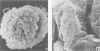
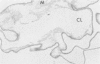
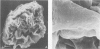
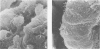
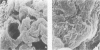
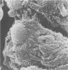
Glomeruli isolated from three male dogs affected with Samoyed hereditary glomerulopathy were compared by scanning electron microscopy with glomeruli of one carrier female and six unaffected dogs. Scanning electron microscopy was performed before and after removal of podocytes and endothelial cells with enzyme and detergent, producing cellular and acellular glomeruli respectively. Cellular glomeruli of unaffected dogs showed podocytes with normally arranged foot processes, while in acellular glomeruli, the subepithelial surface of glomerular capillary basement membranes appeared smooth to finely granular. In contrast, cellular glomeruli of affected males showed microvilli, globular cytoplasmic projections from podocytes, and effacement of foot processes; acellular glomeruli demonstrated ridges and plaque-like irregularities on the subepithelial surface of glomerular capillary basement membranes. Changes in the glomeruli of the carrier female were intermediate between those of unaffected and affected male dogs. The appearance of the subepithelial surface of glomerular capillary basement membranes of acellular glomeruli seen by scanning electron microscopy correlated with the extent of multilaminar splitting of glomerular capillary basement membranes seen by transmission electron microscopy.
Full text
PDF


Images in this article


Selected References
These references are in PubMed. This may not be the complete list of references from this article.
- Arakawa M. A scanning electron microscopy of the glomerulus of normal and nephrotic rats. Lab Invest. 1970 Nov;23(5):489–496. [PubMed] [Google Scholar]
- Arakawa M., Tokunaga J. A scanning electron microscope study of the glomerulus. Further consideration of the mechanism of the fusion of podocyte terminal processes in nephrotic rats. Lab Invest. 1972 Oct;27(4):366–371. [PubMed] [Google Scholar]
- Bonsib S. M. Glomerular basement membrane discontinuities. Scanning electron microscopic study of acellular glomeruli. Am J Pathol. 1985 Jun;119(3):357–360. [PMC free article] [PubMed] [Google Scholar]
- Bonsib S. M. Scanning electron microscopy of acellular glomeruli in human glomerulonephritis: application of a technique. Ultrastruct Pathol. 1984;7(2-3):215–217. doi: 10.3109/01913128409141479. [DOI] [PubMed] [Google Scholar]
- Bonsib S. M. Scanning electron microscopy of acellular glomeruli in nephrotic syndrome. Kidney Int. 1985 Apr;27(4):678–684. doi: 10.1038/ki.1985.64. [DOI] [PubMed] [Google Scholar]
- Bonsib S. M. Segmental subepithelial deposits in primary glomerulonephritis: scanning electron microscopic examination of acellular glomeruli. Hum Pathol. 1985 Nov;16(11):1115–1121. doi: 10.1016/s0046-8177(85)80179-9. [DOI] [PubMed] [Google Scholar]
- Carlson E. C., Chatterjee S. N. Ultrastructure of isolated basement membranes in the acellular human renal cortex. Ren Physiol. 1983 Jul-Aug;6(4):197–208. doi: 10.1159/000172901. [DOI] [PubMed] [Google Scholar]
- Carlson E. C., Kenney M. C. An ultrastructural analysis of isolated basement membranes in the acellular renal cortex: a comparison study of human and laboratory animals. J Morphol. 1982 Feb;171(2):195–211. doi: 10.1002/jmor.1051710207. [DOI] [PubMed] [Google Scholar]
- Jansen B., Thorner P. S., Singh A., Patterson J. M., Lumsden J. H., Valli V. E., Baumal R., Basrur R. K. Animal model of human disease: hereditary nephritis in Samoyed dogs. Am J Pathol. 1984 Jul;116(1):175–178. [PMC free article] [PubMed] [Google Scholar]
- Jansen B., Thorner P., Baumal R., Valli V., Maxie M. G., Singh A. Samoyed hereditary glomerulopathy (SHG). Evolution of splitting of glomerular capillary basement membranes. Am J Pathol. 1986 Dec;125(3):536–545. [PMC free article] [PubMed] [Google Scholar]
- Jansen B., Tryphonas L., Wong J., Thorner P., Maxie M. G., Valli V. E., Baumal R., Basrur P. K. Mode of inheritance of Samoyed hereditary glomerulopathy: an animal model for hereditary nephritis in humans. J Lab Clin Med. 1986 Jun;107(6):551–555. [PubMed] [Google Scholar]
- Jones D. B. Correlative scanning and transmission electron microscopy of glomeruli. Lab Invest. 1977 Dec;37(6):569–578. [PubMed] [Google Scholar]
- KRAKOWER C. A., GREENSPON S. A. Localization of the nephrotoxic antigen within the isolated renal glomerulus. AMA Arch Pathol. 1951 Jun;51(6):629–639. [PubMed] [Google Scholar]
- Kondo Y., Kubosawa H., Akikusa B., Sugano I. A scanning electron microscopic study of crescentic Masugi nephritis in the rabbit. Virchows Arch B Cell Pathol Incl Mol Pathol. 1986;50(4):345–353. doi: 10.1007/BF02889913. [DOI] [PubMed] [Google Scholar]
- Martinez-Hernandez A., Amenta P. S. The basement membrane in pathology. Lab Invest. 1983 Jun;48(6):656–677. [PubMed] [Google Scholar]
- Ng W. L., Chan K. W., Ma L. A scanning electron microscope study of isolated glomeruli in glomerulonephritis. Pathology. 1983 Apr;15(2):139–146. doi: 10.3109/00313028309084701. [DOI] [PubMed] [Google Scholar]
- Tarpey P. A., Williams G. Scanning electron microscope studies of various glomerulonephropathies. Med Lab Sci. 1980 Jan;37(1):57–80. [PubMed] [Google Scholar]
- Timpl R., Wiedemann H., van Delden V., Furthmayr H., Kühn K. A network model for the organization of type IV collagen molecules in basement membranes. Eur J Biochem. 1981 Nov;120(2):203–211. doi: 10.1111/j.1432-1033.1981.tb05690.x. [DOI] [PubMed] [Google Scholar]
- Weidner N., Lorentz W. B., Jr Scanning electron microscopy of the acellular glomerular and tubular basement membrane in lupus nephritis. Am J Clin Pathol. 1986 Feb;85(2):135–145. doi: 10.1093/ajcp/85.2.135. [DOI] [PubMed] [Google Scholar]
- Weidner N., Lorentz W. B., Jr Scanning electron microscopy of the acellular glomerular basement membranes in idiopathic membranous glomerulopathy. Lab Invest. 1986 Jan;54(1):84–92. [PubMed] [Google Scholar]
- Wieslander J., Langeveld J., Butkowski R., Jodlowski M., Noelken M., Hudson B. G. Physical and immunochemical studies of the globular domain of type IV collagen. Cryptic properties of the Goodpasture antigen. J Biol Chem. 1985 Jul 15;260(14):8564–8570. [PubMed] [Google Scholar]