

Abstract
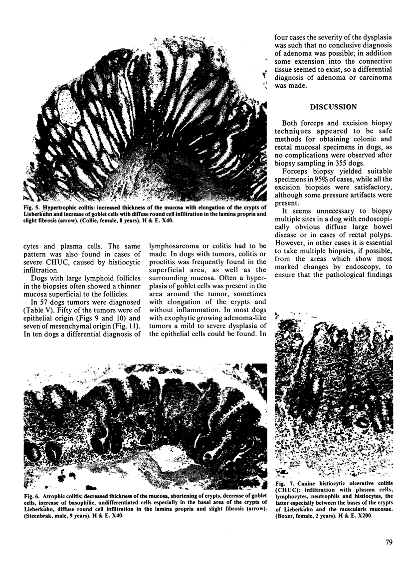
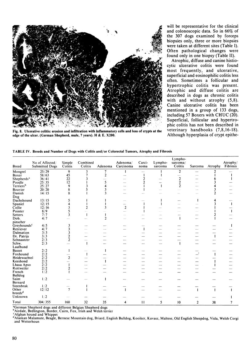
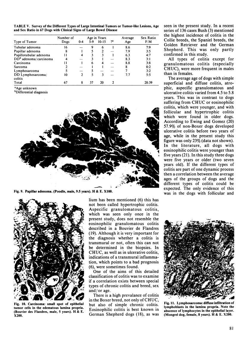
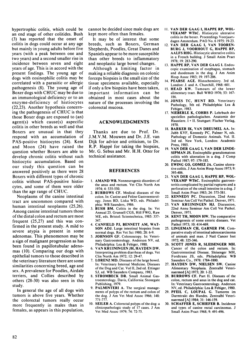
Colonic and rectal forceps and excision biopsies of 355 dogs with clinical signs of large bowel disease were investigated. Five percent of the forceps biopsies were unsuitable for examination; all excision biopsies were of good quality. Forceps biopsies were obtained from one to eight sites, up to 60 cm cranial from the anus, while excision biopsies, mostly from tumors, were from the rectoanal region. Slight to severe colitis and/or proctitis was found in 192 dogs (54%). A single type of colitis was seen in 160 dogs; in 53 cases the lesions were local, in 107 cases multiple. A combination of different types of colitis was found in 32 dogs. Atrophic colitis, diffuse colitis and canine histiocytic ulcerative colitis were the most prominent findings, followed by ulcerative, superficial and eosinophilic colitis. Follicular, hypertrophic and aspecific granulomatous colitis were found in only a few cases. Tumors were diagnosed in 57 dogs (16%). Of these tumors 50 were of epithelial and seven were of mesenchymal origin. A high percentage (61%) of the epithelial tumors consisted of adenomas of the rectoanal region. In ten other dogs (3%) a differential diagnosis of lymphosarcoma or colitis had to be made. Colitis and colorectal tumors were more prevalent in Boxers, German Shepherds, Poodles, Great Danes and Spaniels. In the Boxers simple chronic colitis, as well as canine histiocytic ulcerative colitis were more frequently found, the latter especially in females. Other biopsy findings were edema, crypt cysts, hemorrhages, an increased number of intraepithelial lymphocytes and an increased or decreased number of goblet cells.
Full text
PDF
Images in this article
Selected References
These references are in PubMed. This may not be the complete list of references from this article.
- Amand W. B. Nonneurogenic disorders of the anus and rectum. Vet Clin North Am. 1974 Aug;4(3):535–550. doi: 10.1016/s0091-0279(74)50056-5. [DOI] [PubMed] [Google Scholar]
- Canfield P. J., Bennett A. M., Watson A. D. Large intestinal biopsies from normal dogs. Res Vet Sci. 1980 Jan;28(1):6–9. [PubMed] [Google Scholar]
- Hayden D. W., Nielsen S. W. Canine alimentary neoplasia. Zentralbl Veterinarmed A. 1973 Jan;20(1):1–22. [PubMed] [Google Scholar]
- Head K. W. Tumours of the lower alimentary tract. Bull World Health Organ. 1976;53(2-3):167–186. [PMC free article] [PubMed] [Google Scholar]
- Lingeman C. H., Garner F. M. Comparative study of intestinal adenocarcinomas of animals and man. J Natl Cancer Inst. 1972 Feb;48(2):325–346. [PubMed] [Google Scholar]
- Palminteri A. The surgical management of polyps of the rectum and colon of the dog. J Am Vet Med Assoc. 1966 Apr 1;148(7):771–777. [PubMed] [Google Scholar]
- Pfeil C., Loupal G. Tumoren im Darmtrakt des Hundes. Zentralbl Veterinarmed A. 1984 Mar;31(2):146–159. [PubMed] [Google Scholar]
- Seiler R. J. Colorectal polyps of the dog: a clinicopathologic study of 17 cases. J Am Vet Med Assoc. 1979 Jan 1;174(1):72–75. [PubMed] [Google Scholar]
- Van der Gaag I., Van Toorenburg J. V., Voorhout G., Happe R. P., Aalfs R. H. Histiocytic ulcerative colitis in a French Bulldog. J Small Anim Pract. 1978 May;19(5):283–290. doi: 10.1111/j.1748-5827.1978.tb05493.x. [DOI] [PubMed] [Google Scholar]
- van der Gaag I., van der Linde-Sipman J. S. Eosinophilic granulomatous colitis with ulceration in a dog. J Comp Pathol. 1987 Mar;97(2):179–185. doi: 10.1016/0021-9975(87)90038-7. [DOI] [PubMed] [Google Scholar]