

Abstract
A 3.5-year-old, spayed female, chow chow presented with a pruritic, ulcerative, butterfly lesion on the nasal planum, typical of the pemphigus complex. Pemphigus erythematosus was diagnosed by histopathologic examination. Treatment consisted of enrofloxacin and immunosuppressive therapy with prednisone and azathioprine. Within 5 wk of treatment, the lesions had resolved almost completely.
Résumé
Pemphigus érythémateux chez un Chow-chow. Une femelle Chow-chow stérilisée, âgée de 3,5 ans a été présentée avec une lésion pruritique, ulcérative et en forme de papillon sur la truffe, typique d’un pemphigus. Un pemphigus érythémateux a été diagnostiqué par examen histopathologique. Le traitement comprenait de l’enrofloxacine et une thérapie immunosuppressive à la prednisone et à l’azathioprime. En 5 semaines de traitement, les lésions s’étaient presque complètement résorbées.
(Traduit par Docteur André Blouin)
A 3.5-year-old, spayed female, chow chow presented on day 1 with pruritic, oozing, ulcerative lesions on the nasal planum, as well as at the medial canthus of both eyes. The differential diagnoses included superficial pyoderma, pemphigus foliaceus, pemphigus erythematosus, discoid lupus erythematosus, demodicosis, dermatophytosis, and erythema multiform. At this time, no diagnostic tests were done. Initially, the dog was administered cephalexin (Novolexin; Novopharm, Toronto, Ontario), 25 mg/kg bodyweight (BW), PO, q12h for 21 d as empirical treatment for pyoderma, and meloxicam (Metacam; Boehringer Ingelheim, Burlington, Ontario), 0.1 mg/kg BW, PO, q24h for 7 d to reduce inflammation. By day 7, she was less pruritic and there was some improvement in the lesion on the nasal planum. However, when examined on day 14, the lesions had spread up the bridge of the nose and along the lower eyelids in the periocular region, and there were new lesions involving the tips of the pinnae of both ears. On day 21 (Figure 1a), skin biopsies were obtained, by using a 5-mm biopsy punch (Dermal Biopsy Punch; Miltex Instrument, Bethpage, New York, USA), for routine culture, sensitivity testing, and histopathologic examination. Samples were collected into liquid media swabs (BBL CultureSwab; Becton Dickinson, Sparks, Maryland, USA) and 10% buffered formalin and submitted to the AHL (University of Guelph, Guelph, Ontario). Six skin scrapings were taken from the lesions on the nasal planum, periocular region, and pinnae to rule out demodicosis on day 21 by using a # 10 scalpel blade. One Demodex canis mite was found on 1 of 6 scrapings. One mite is not significant, as Demodex mites in low numbers are natural inhabitants of canine skin. The skin culture revealed 3+ Pseudomonas spp. and 3+ Bacillus cereus. The Pseudomonas sp. was considered opportunistic in this case and the bacillus was likely a normal flora contaminant. The histopathologic diagnosis was pemphigus erythematosus. The epidermis was variably hyperplastic, with multifocal erosions and ulcerations, and was covered by massively thick serocellular crusts containing well-preserved neutrophils. Some hair follicles contained small distinct intraspinous pustules in the outer root sheath consisting of intact neutrophils surrounding acantholytic cells. Although these histological changes are typical of pemphigus erythematosus, pemphigus foliaceus could not be ruled out as a diagnosis. These 2 diseases are distinguished by the presence of interface activity and single cell necrosis in the basal epithelial cells.
Figure 1a.

Pemphigus erythematosus in a chow chow at day 21 showing severe superficial erosions and crusts on the nasal planum, bridge of nose and periocular region.
Chow chows are not only predisposed to pemphigus complex, but many are also refractory to treatment (Dr. Jan Hall, personal communication); therefore, aggressive treatment was initiated. This consisted of prednisone (Apo-prednisone; Apotex, Toronto, Ontario), 2 mg/kg BW, PO, q12h, along with azathioprine (Imuran; Glaxo Smith Kline, Mississauga, Ontario) 2 mg/kg BW, PO, q48h. Based on sensitivity testing results, the dog was also prescribed enrofloxacin (Baytril; Bayer, Etobicoke, Ontario), 10 mg/kg BW, PO, q24h for 4 wk. By day 28, there was some improvement in the skin lesions and pruritus, and no side effects as a result of the large dosage of prednisone were noted. The lesions had continued to show improvement by day 35, evidenced by some regrowth of hair along the muzzle and marked improvement in the scabs on the nasal planum, over the bridge of the nose, and in the periocular region. The dog had, however, started to show signs of polyuria, polydipsia, urinary incontinence, poor appetite, and weight loss. Poor appetite and weight loss are not typical side effects of treatment with prednisone; however, the owner reported that the dog had experienced a seasonal decrease in appetite over the past few years. The dosage of prednisone was decreased to 1.25 mg/kg BW, q12h.
The lesions continued to improve and pigmentation in the nose was returning by day 42. However, the side effects of treatment were still present. The dosage of prednisone was further decreased to 0.625 mg/kg BW, q12h, and the dosing regime of azathioprine continued unchanged. The lesions on the bridge of the nose were almost completely resolved and the scabs on the pinnae were no longer present by day 49. The dog was still polyuric and polydypsic, although less so than before, and was eating better. The dosage of prednisone was further decreased to 0.625 mg/kg BW, q24h, with plans to maintain at this dose for 2 to 3 wk, if there was a resolution of side effects. A CBC was performed in order to monitor azathioprine treatment. All parameters were within normal limits. Azathioprine can also lead to hepatotoxic changes and pancreatitis; however, a biochemical panel was not performed at this time. By day 79, the polyuria and polydipsia had resolved, and there had been continued improvement in the skin lesions. A thin band of nonepithelialized tissue at the nasal planum was still present and bled periodically when bumped. A CBC at this time revealed a mild anemia. The dog was maintained on the same dose of prednisone and azathioprine. By day 96, all scabs were gone and there was good hair regrowth. The dosage of prednisone was decreased to 0.625 mg/kg BW, q48h. By day 129, (Figure 1b), the skin lesions had almost completely resolved and only mild depigmentation and a thin line of epithelialization was noted on the nasal planum. A CBC at this time revealed a mild anemia and a low normal platelet count. The owner was advised that lifelong treatment would likely be necessary to maintain the dog lesion-free.
Figure 1b.
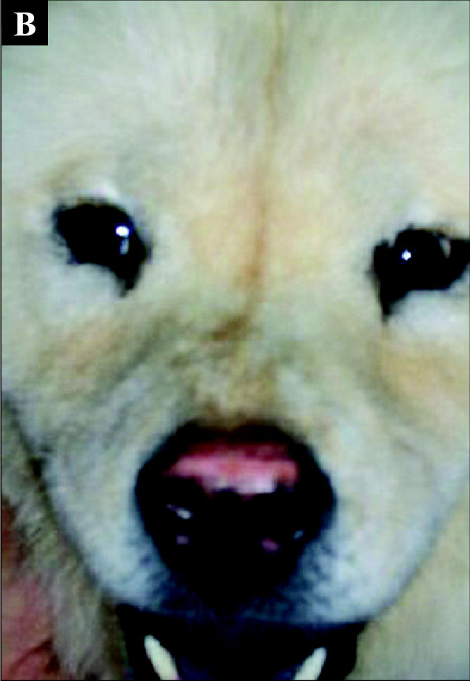
Day 129: Note the lesions have almost completely resolved. However, mild nasal depigmentation and a thin line of superficial erosion were still present on the nasal planum.
Pemphigus is a complex of vesiculopustular autoimmune diseases that affect the skin and mucous membranes of dogs. In dogs, the autoimmune disease is caused by circulating antibodies against desmosomal proteins present on the surface of keratinocytes (4). The development of pemphigus may be affected by endogenous and exogenous factors: The former include breed predilection and the latter include drugs, nutrition, and viral infections. In addition, paraneoplastic pemphigus and drug-induced pemphigus have also been reported in dogs.
Pemphigus can be classified into 4 types: pemphigus vulgaris (PV), pemphigus foliaceus (PF), pemphigus erythematosus (PE), and pemphigus vegetans (Pveg). Pemphigus vulgaris is rare in dogs and tends to cause systemic illness. Pemphigus vegetans is extremely rare and thought to be the more benign version of PV. Although pemphigus is considered an uncommon disease in veterinary medicine, pemphigus foliaceus is the most commonly reported dermatologic autoimmune disease in dogs. Three types of PF have been described in dogs: spontaneous, drug-induced, and a form that occurs in dogs with a history of chronic dermatologic disease. Chow chows and akitas show a breed predilection for spontaneous PF (4,7). There is no sex predilection in dogs, but middle-aged dogs tend to be affected more often (5,7). Pemphigus erythematosus is thought to be a rare variant of PF and, as in the case reported here, is localized to the face (bridge of nose and around eyes) and pinnae of ears (1,6). It is more benign that PF and is thought to be a cross-over between PF and discoid lupus erythematosus (4,8). German shepherds, collies, and Shetland sheepdogs are predisposed to PE (8). Pemphigus erythematosus is usually diagnosed on the basis of the clinical history: PE is usually characterized by a waxing and waning course, and typical physical examination findings include pustular and crusting lesions, butterfly lesions on nasal planum and bridge of nose, and characteristic cytologic and histopathologic changes (5).
Pemphigus foliaceus and PE are often distinguished by using clinical signs and histopathologic findings. Although PE is localized to the head, PF can also start out as a localized disease, with lesions on the face, ears, and feet (clawbeds and footpads), and may become generalized or multifocal within 6 mo (7). Pemphigus erythematosus is almost identical to PF histologically, except that there is often lichenoid cellular infiltrate of mononuclear cells, plasma cells, and neutrophils or eosinophils, or both. It carries a much better prognosis than PF. Treatment for mild PE usually includes minimizing exposure to the sun and to the use of topical glucocorticoids. Severe cases, such as this one, require immunosuppressive therapy (6). We instituted aggressive therapy because this dog had severe lesions and chow chows are difficult to get into remission (Dr. Jan Hall, personal communication, 2003). It was important to weigh the pros and cons of treatment in this case, because immunosuppressive treatment can be very dangerous. Azathioprine has reported side effects, such as bone marrow suppression, especially anemia, leukopenia, and thrombocytopenia; hepatotoxicity; pancreatitis; and gastrointestinal toxicity (3). As in this case, animals on azathioprine should have their bone marrow function monitored closely during initial treatment to avoid severe myelosuppression. Serum biochemical profiles should also be performed periodically to monitor liver and pancreatic parameters. Initially CBCs and platelet counts should be done every 2 wk. However, once the animal is in remission and the condition is stable, monitoring can be decreased to once every 4 mo. The dog in this case showed evidence of a mild anemia, and based on platelet trends, decreased platelet numbers. Periodic monitoring of these cells will be continued to avoid complications of treatment. Adverse effects of prednisone treatment include iatrogenic hyperadrenocorticism, gastrointestinal ulceration, recurrent urinary tract infections, and pancreatitis (2). In addition, side effects of prednisone treatment, such as polyuria, polydipsia, and polyphagia, often contribute to complicating an already difficult situation. The side effects of the treatment can be very frustrating for owners, who may demand euthanasia due to the side effects of the drug(s) rather than the disease. Other possible treatments for PE include the use of topical steroids, as well as topical tacrolimus ointment (Protopic 0.1%; Fujisawa Healthcare, Deerfield, Illinois, USA). These treatments can be used in milder cases of PE or as an adjunct to systemic therapy in more severe cases.
Acknowledgments
The author thanks Drs. Brian Crabbe, Jan Hall, Josepha Delay (AHL), and the staff at Port Elgin Veterinary Clinic for their advice, guidance, and support. CVJ
Footnotes
Dr. Gonsalves-Hubers’ current address is Burgess Veterinary Emergency Clinic, 1–775 Woodview Road, Burlington, Ontario L7N 3S1.
Dr. Gonsalves-Hubers will receive 50 free reprints of her article, courtesy of The Canadian Veterinary Journal.
References
- 1.Carlotti D. Autoimmune mediated skin diseases. J Small Anim Pract. 1989;30:223–227. [Google Scholar]
- 2.Couto C. Use and misuse of immunosuppressants. Proc Am Coll Vet Intern Med 2002.
- 3.Kidd L, Trepanier L, Salavaggione E, Szumlanski C, Perez A, Weinshilboum R. Azathioprine metabolism in dogs: Implications for drug dosing. Proc Am Coll Vet Intern Med 2002.
- 4.Marsella R. Canine pemphigus complex: Pathogenesis and clinical presentation. Compend Contin Educ Pract Vet. 2000;22:568–572. [Google Scholar]
- 5.Marsella R. Canine pemphigus complex: Diagnosis and therapy. Compend Contin Educ Pract Vet. 2000;22:680–685. [Google Scholar]
- 6.Medlau L, Hnilica KA. Small Animal Dermatology: A Color Atlas and Therapeutic Guide. Philadelphia: WB Saunders 2000:130–135.
- 7.Scott DW, Miller WH, Griffin CE. Muller and Kirk’s Small Animal Dermatology. 6th ed. Philadelphia: WB Saunders, 2001:686–693.
- 8.Werner AH. Recognizing and treating discoid lupus erythematosus and pemphigus foliaceus in dogs. Vet Med. 1999;94:955–966. [Google Scholar]