Abstract
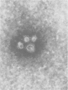
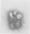
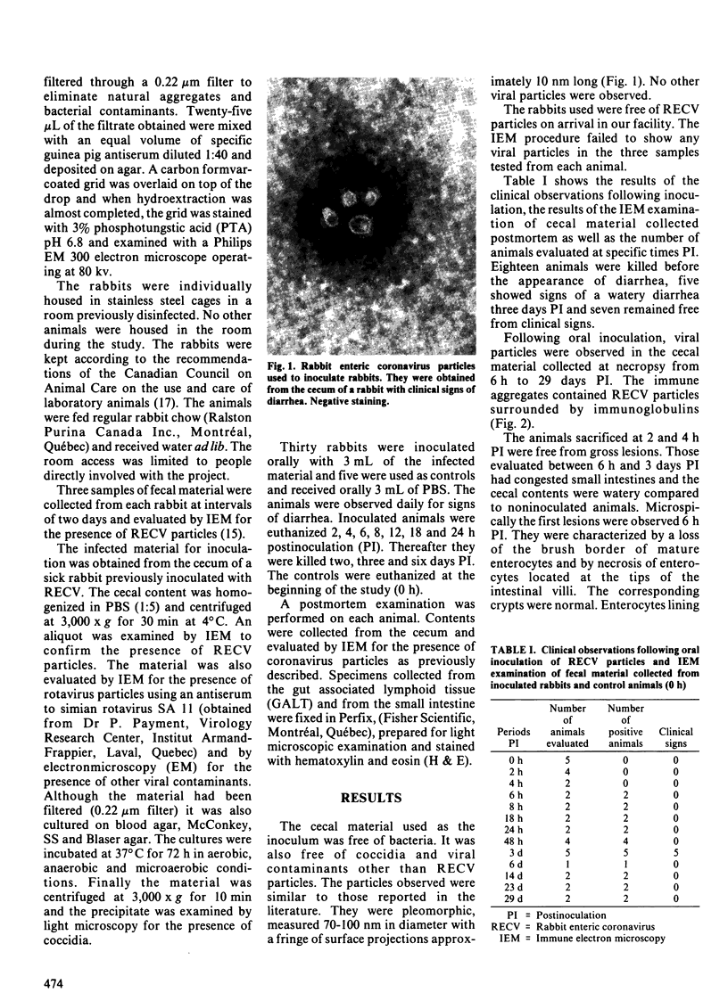
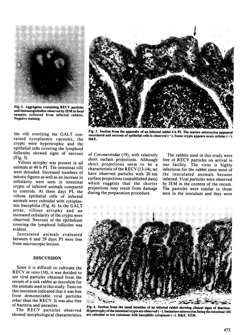
The clinical signs and lesions caused by the rabbit enteric coronavirus (RECV) were studied in young rabbits orally inoculated with a suspension containing RECV particles. The inoculated animals were observed daily for evidence of diarrhea. Fecal samples and specimens from the small intestine and from the gut associated lymphoid tissue (GALT) were collected from 2 h to 29 days postinoculation (PI) and processed for immune electron microscopy (IEM) and light microscopy. Coronavirus particles were detected in the cecal contents of most inoculated animals from 6 h to 29 days PI. Lesions were first observed 6 h PI and were characterized by a loss of the brush border of mature enterocytes located at the tips of intestinal villi and by necrosis of these cells. At 48 h PI, short intestinal villi and hypertrophic crypts were noted. In the GALT, complete necrosis of the M cells as well as necrosis of the enterocytes lining the villi above the lymphoid follicules with hypertrophy of the corresponding crypts were observed in all the animals. Five inoculated rabbits had diarrhea three days PI. The presence of RECV particles in the feces of the sick animals and the microscopic lesions observed in the small intestine suggested that the virus was responsible for the clinical signs. A few inoculated rabbits remained free of diarrhea. Fecal material collected at postmortem examination contained RECV particles. The results suggest that the virus could also produce a subclinical infection.
Full text
PDF
Images in this article
Selected References
These references are in PubMed. This may not be the complete list of references from this article.
- Aelterman E. O., Hooper B. E. Transmissible gastroenteritis of swine as a model for the study of enteric disease. Gastroenterology. 1967 Jul;53(1):109–113. [PubMed] [Google Scholar]
- Bryden A. S., Thouless M. E., Flewett T. H. Rotavirus and rabbits. Vet Rec. 1976 Oct 16;99(16):323–323. doi: 10.1136/vr.99.16.323-a. [DOI] [PubMed] [Google Scholar]
- Chu R. M., Glock R. D., Ross R. F. Changes in gut-associated lymphoid tissues of the small intestine of eight-week-old pigs infected with transmissible gastroenteritis virus. Am J Vet Res. 1982 Jan;43(1):67–76. [PubMed] [Google Scholar]
- Chu R. M., Li N. J., Glock R. D., Ross R. F. Applications of peroxidase-antiperoxidase staining technique for detection of transmissible gastroenteritis virus in pigs. Am J Vet Res. 1982 Jan;43(1):77–81. [PubMed] [Google Scholar]
- Collins J. K., Riegel C. A., Olson J. D., Fountain A. Shedding of enteric coronavirus in adult cattle. Am J Vet Res. 1987 Mar;48(3):361–365. [PubMed] [Google Scholar]
- Descôteaux J. P., Lussier G., Berthiaume L., Alain R., Seguin C., Trudel M. An enteric coronavirus of the rabbit: detection by immunoelectron microscopy and identification of structural polypeptides. Arch Virol. 1985;84(3-4):241–250. doi: 10.1007/BF01378976. [DOI] [PMC free article] [PubMed] [Google Scholar]
- DiGiacomo R. F., Thouless M. E. Epidemiology of naturally occurring rotavirus infection in rabbits. Lab Anim Sci. 1986 Apr;36(2):153–156. [PubMed] [Google Scholar]
- Eaton P. Preliminary observations on enteritis associated with a coronavirus-like agent in rabbits. Lab Anim. 1984 Jan;18(1):71–74. doi: 10.1258/002367784780864938. [DOI] [PubMed] [Google Scholar]
- Lapierre J., Marsolais G., Pilon P., Descôteaux J. P. Preliminary report on the observation of a coronavirus in the intestine of the laboratory rabbit. Can J Microbiol. 1980 Oct;26(10):1204–1208. doi: 10.1139/m80-201. [DOI] [PubMed] [Google Scholar]
- Matsunaga Y., Matsuno S., Mukoyama J. Isolation and characterization of a parvovirus of rabbits. Infect Immun. 1977 Nov;18(2):495–500. doi: 10.1128/iai.18.2.495-500.1977. [DOI] [PMC free article] [PubMed] [Google Scholar]
- Ononiwu J. C., Julian R. J. An outbreak of Tyzzer's disease in an Ontario rabbitry. Can Vet J. 1978 Apr;19(4):107–109. [PMC free article] [PubMed] [Google Scholar]
- Osterhaus A. D., Teppema J. S., van Steenis G. Coronavirus-like particles in laboratory rabbits with different syndromes in the Netherlands. Lab Anim Sci. 1982 Dec;32(6):663–665. [PubMed] [Google Scholar]
- Patton N. M., Holmes H. T., Riggs R. J., Cheeke P. R. Enterotoxemia in rabbits. Lab Anim Sci. 1978 Oct;28(5):536–540. [PubMed] [Google Scholar]
- Petric M., Middleton P. J., Grant C., Tam J. S., Hewitt C. M. Lapine rotavirus: preliminary studies on epizoology and transmission. Can J Comp Med. 1978 Jan;42(1):143–147. [PMC free article] [PubMed] [Google Scholar]
- Prescott J. F. Escherichia coli and diarrhoea in the rabbit. Vet Pathol. 1978 Mar;15(2):237–248. doi: 10.1177/030098587801500210. [DOI] [PubMed] [Google Scholar]
- Schoeb T. R., Casebolt D. B., Walker V. E., Potgieter L. N., Thouless M. E., DiGiacomo R. F. Rotavirus-associated diarrhea in a commercial rabbitry. Lab Anim Sci. 1986 Apr;36(2):149–152. [PubMed] [Google Scholar]
- Wege H., Siddell S., ter Meulen V. The biology and pathogenesis of coronaviruses. Curr Top Microbiol Immunol. 1982;99:165–200. doi: 10.1007/978-3-642-68528-6_5. [DOI] [PubMed] [Google Scholar]
- Whitney J. C. A review of non-specific enteritis in the rabbit. Lab Anim. 1976 Jul;10(3):209–221. doi: 10.1258/002367776781035305. [DOI] [PubMed] [Google Scholar]


