Abstract
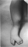
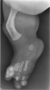
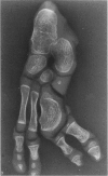
Detailed dissection of a malformed human foot was performed so that the skeletal and soft tissue anomalies in the foot could be compared and contrasted with those found in three previous specimens that we have dissected. The specimen described here consisted of a foot with a reduplicated hallux and two missing toes. Study of the bones revealed a wide medial metatarsal that articulated with a reduplicated hallux. There were two complete lateral toes with normal bones. Two toes and their metatarsals were missing with no remnants. The arterial pattern was similar to those seen previously by us. The dorsalis pedis artery was absent and the plantar arch was abnormal in that it did not terminate with a dorsal anastomosis. There was an extra lateral branch of the medial plantar artery. The digital arteries to the missing toes were also missing. The nerves of the foot were normal with the exception of an extra lateral branch from the medial plantar nerve. As with the arteries, the nerves to the missing toes were also missing. The muscles and tendons on the dorsal surface of the foot were all present but several of the tendons were inserted in abnormal locations in apparent response to the abnormal bone pattern. Most of the muscles and tendons of the plantar surface were present with the exception of the flexor hallucis longus and brevis muscles. Several of the remaining tendons apparently were influenced by the abnormal skeletal pattern and the missing muscles to become inserted on or near the replicated phalanges of the hallux. The anatomy of this specimen suggests that the teratogenic event occurred when specification of the limb bud mesoderm cells had progressed to the level of the distal tarsus and was located in the region that normally would have formed the metatarsals and phalanges of Toes 2 and 3. Further, we propose that the association between skeletal anomalies and arterial deficiencies has aetiological significance. We hypothesise that the abnormal arterial pattern put the limb at risk of teratogenic damage by reducing the number of collateral blood supply routes and that some event, such as extravasation of blood or embolisation, compromised the blood flow in the remaining blood vessels. These events could have resulted in both general shortening of the limb and the specific defects observed in this foot.
Full text
PDF


Images in this article
Selected References
These references are in PubMed. This may not be the complete list of references from this article.
- Gregosiewicz A., Wośko I., Okoński M. Girl with three legs. J Bone Joint Surg Br. 1989 Aug;71(4):708–709. doi: 10.1302/0301-620X.71B4.2768332. [DOI] [PubMed] [Google Scholar]
- Hamanishi C. Congenital short femur. Clinical, genetic and epidemiological comparison of the naturally occurring condition with that caused by thalidomide. J Bone Joint Surg Br. 1980 Aug;62(3):307–320. doi: 10.1302/0301-620X.62B3.7410462. [DOI] [PubMed] [Google Scholar]
- Hanley E. N., Jr, Stanitski C. L. Incomplete congenital duplication of a lower extremity. A case report. J Bone Joint Surg Am. 1980 Apr;62(3):479–481. [PubMed] [Google Scholar]
- Hootnick D. R., Levinsohn E. M., Crider R. J., Packard D. S., Jr Congenital arterial malformations associated with clubfoot. A report of two cases. Clin Orthop Relat Res. 1982 Jul;(167):160–163. [PubMed] [Google Scholar]
- Hootnick D. R., Levinsohn E. M., Randall P. A., Packard D. S., Jr Vascular dysgenesis associated with skeletal dysplasia of the lower limb. J Bone Joint Surg Am. 1980 Oct;62(7):1123–1129. [PubMed] [Google Scholar]
- Hootnick D. R., Packard D. S., Jr, Levinsohn E. M., Cady R. B. Soft tissue anomalies in a patient with congenital tibial aplasia and talo-calcaneal synchrondrosis. Teratology. 1987 Oct;36(2):153–162. doi: 10.1002/tera.1420360202. [DOI] [PubMed] [Google Scholar]
- Hootnick D. R., Packard D. S., Jr, Levinsohn E. M. Congenital tibial aplasia with preaxial polydactyly: soft tissue anatomy as a clue to teratogenesis. Teratology. 1983 Apr;27(2):169–179. doi: 10.1002/tera.1420270205. [DOI] [PubMed] [Google Scholar]
- Hootnick D. R., Packard D. S., Jr, Levinsohn E. M., Lebowitz M. R., Lubicky J. P. The anatomy of a congenitally short limb with clubfoot and ectrodactyly. Teratology. 1984 Apr;29(2):155–164. doi: 10.1002/tera.1420290202. [DOI] [PubMed] [Google Scholar]
- MILLEN J. W. TIMING OF HUMAN CONGENITAL MALFORMATIONS WITH A TIME-TABLE OF HUMAN DEVELOPMENT. Dev Med Child Neurol. 1963 Aug;5:343–350. doi: 10.1111/j.1469-8749.1963.tb05036.x. [DOI] [PubMed] [Google Scholar]
- Narang I. C., Mysorekar V. R., Mathur B. P. Diplopodia with double fibula and agenesis of tibia. A case report. J Bone Joint Surg Br. 1982;64(2):206–209. doi: 10.1302/0301-620X.64B2.7068742. [DOI] [PubMed] [Google Scholar]
- Sodre H., Bruschini S., Mestriner L. A., Miranda F., Jr, Levinsohn E. M., Packard D. S., Jr, Crider R. J., Jr, Schwartz R., Hootnick D. R. Arterial abnormalities in talipes equinovarus as assessed by angiography and the Doppler technique. J Pediatr Orthop. 1990 Jan-Feb;10(1):101–104. [PubMed] [Google Scholar]
- Summerbell D. The control of growth and the development of pattern across the anteroposterior axis of the chick limb bud. J Embryol Exp Morphol. 1981 Jun;63:161–180. [PubMed] [Google Scholar]
- Swanson G. J., Lewis J. The timetable of innervation and its control in the chick wing bud. J Embryol Exp Morphol. 1982 Oct;71:121–137. [PubMed] [Google Scholar]
- Williams L., Wientroub S., Getty C. J., Pincott J. R., Gordon I., Fixsen J. A. Tibial dysplasia. A study of the anatomy. J Bone Joint Surg Br. 1983 Mar;65(2):157–159. doi: 10.1302/0301-620X.65B2.6826620. [DOI] [PubMed] [Google Scholar]


