

Abstract
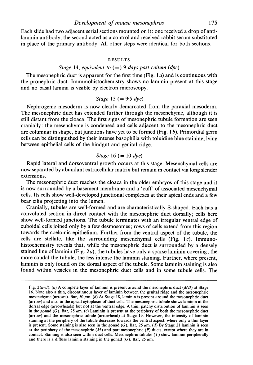
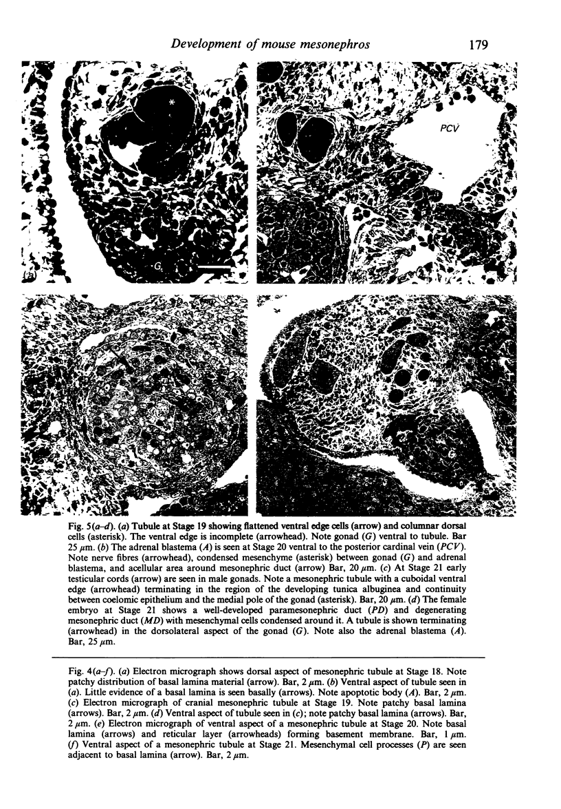
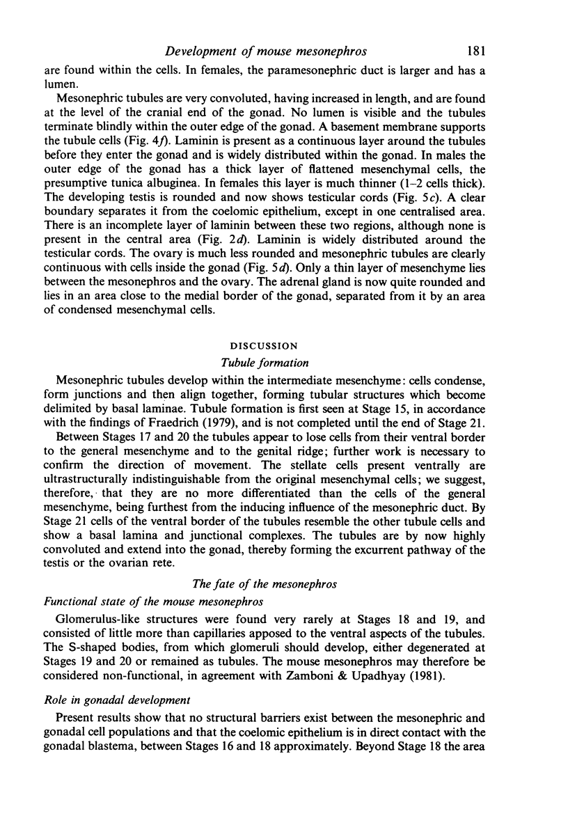
A study has been made of the development of mesonephric tubules in the mouse to investigate the possible transdifferentiation of tubule epithelial cell to gonadal somatic cells and/or adrenal cortical cells. Immunohistochemical localisation of laminin was carried out to study the development of basal laminae. Cells at the ventral aspect of mesonephric tubules did not show an epithelial phenotype during the period of somatic cell population of the gonadal blastema; a basal lamina appeared ventrally only after this period. Therefore, it is not necessary to postulate transdifferentiation of these cells.
Full text
PDF

Images in this article
Selected References
These references are in PubMed. This may not be the complete list of references from this article.
- Ekblom P., Alitalo K., Vaheri A., Timpl R., Saxén L. Induction of a basement membrane glycoprotein in embryonic kidney: possible role of laminin in morphogenesis. Proc Natl Acad Sci U S A. 1980 Jan;77(1):485–489. doi: 10.1073/pnas.77.1.485. [DOI] [PMC free article] [PubMed] [Google Scholar]
- Holtzer H., Strahs K., Biehl J., Somlyo A. P., Ishikawa H. Thick and thin filaments in postmitotic, mononucleated myoblasts. Science. 1975 May 30;188(4191):943–945. doi: 10.1126/science.1138363. [DOI] [PubMed] [Google Scholar]
- Klein G., Langegger M., Timpl R., Ekblom P. Role of laminin A chain in the development of epithelial cell polarity. Cell. 1988 Oct 21;55(2):331–341. doi: 10.1016/0092-8674(88)90056-6. [DOI] [PubMed] [Google Scholar]
- Motta P. M., Makabe S. Elimination of germ cells during differentiation of the human ovary: an electron microscopic study. Eur J Obstet Gynecol Reprod Biol. 1986 Sep;22(5-6):271–286. doi: 10.1016/0028-2243(86)90115-2. [DOI] [PubMed] [Google Scholar]
- OVERTON J. Studies of the mode of outgrowth of the amphibian pronephric duct. J Embryol Exp Morphol. 1959 Mar;7(1):86–93. [PubMed] [Google Scholar]
- Spiegelman M., Bennett D. A light- and electron-microscopic study of primordial germ cells in the early mouse embryo. J Embryol Exp Morphol. 1973 Aug;30(1):97–118. [PubMed] [Google Scholar]
- Upadhyay S., Zamboni L. Preliminary observations on the role of the mesonephros in the development of the adrenal cortex. Anat Rec. 1982 Jan;202(1):105–111. doi: 10.1002/ar.1092020112. [DOI] [PubMed] [Google Scholar]
- Wartenberg H. Development of the early human ovary and role of the mesonephros in the differentiation of the cortex. Anat Embryol (Berl) 1982;165(2):253–280. doi: 10.1007/BF00305481. [DOI] [PubMed] [Google Scholar]
- Wartenberg H. Human testicular development and the role of the mesonephros in the origin of a dual Sertoli cell system. Andrologia. 1978 Jan-Feb;10(1):1–21. doi: 10.1111/j.1439-0272.1978.tb01306.x. [DOI] [PubMed] [Google Scholar]
- Wartenberg H. Origin of gonadal blastemal cells in mammalian gonadogenesis. Arch Anat Microsc Morphol Exp. 1985;74(1):60–63. [PubMed] [Google Scholar]
- Zamboni L., Upadhyay S. Ephemeral, rudimentary glomerular structures in the mesonephros of the mouse. Anat Rec. 1981 Dec;201(4):641–644. doi: 10.1002/ar.1092010408. [DOI] [PubMed] [Google Scholar]