Abstract
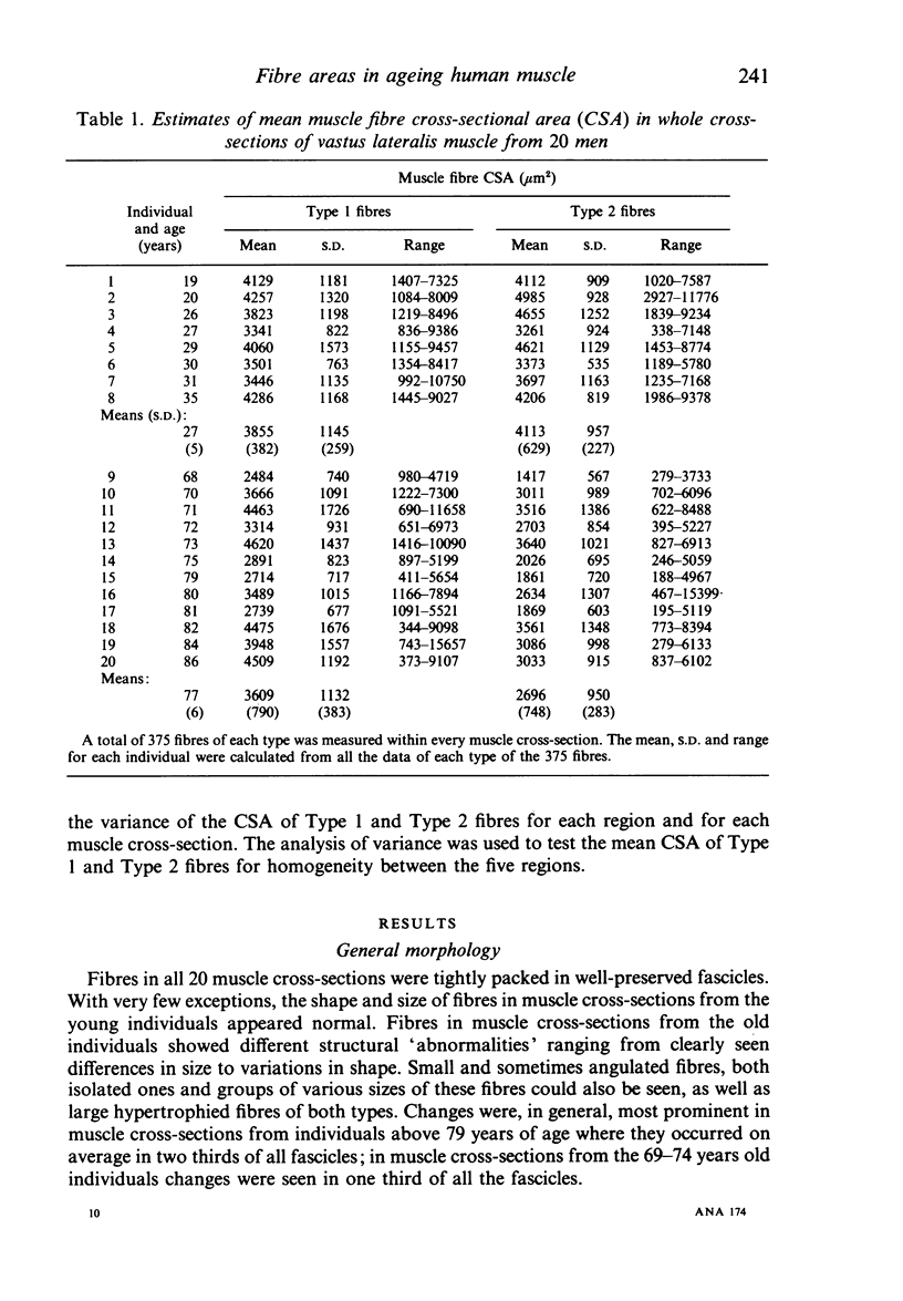
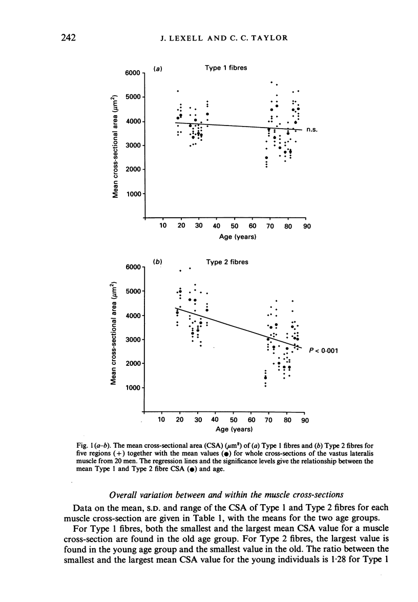
Cross-sections of whole vastus lateralis muscle from 20 men, 19 to 84 years of age, were prepared, and the cross-sectional area (microns2) of 375 type 1 and 375 Type 2 fibres was measured in five different regions throughout each muscle. In muscles from the old individuals, the mean CSA of Type 2 fibres was on average nearly 35% smaller (P less than 0.001) while the mean CSA of Type 1 fibres was on average just over 6% smaller (NS) than in muscles from the young individuals. There was a highly significant (P less than 0.001) variation in the mean CSA of both fibre types within all muscles. In the old muscles, there was no significant difference in mean fibre CSA between deep and superficial parts while in the young muscles the mean fibre CSA was significantly (P less than 0.05) larger in deep regions than superficially. The range of the fibre CSA was larger in the old muscles with an increased number of both hypotrophied and atrophied fibres as well as large, sometimes very large, fibres. The standard deviation of the fibre CSA of Type 2 fibres was significantly (P less than 0.001) larger than for Type 1 fibres in 60% of the regions of the old muscles compared to 12.5% of the regions of the young muscles, but the standard deviation for the whole muscles was more or less unaffected with increasing age. In the old age group, there were fewer muscles and regions with a correlation between the CSA of Type 1 and Type 2 fibres than in the young age group. In conclusion the age-related changes in the mean fibre CSA, and in the pattern of variation in fibre CSA throughout the muscle and in small sample regions, suggest a combination of a progressive denervation process and an altered physical activity level as the two major mechanisms underlying the effects of normal development and ageing on the human vastus lateralis muscle.
Full text
PDF
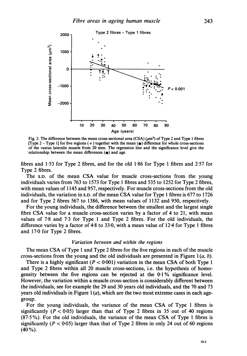
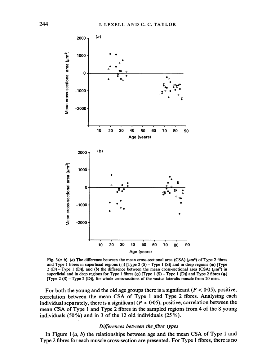
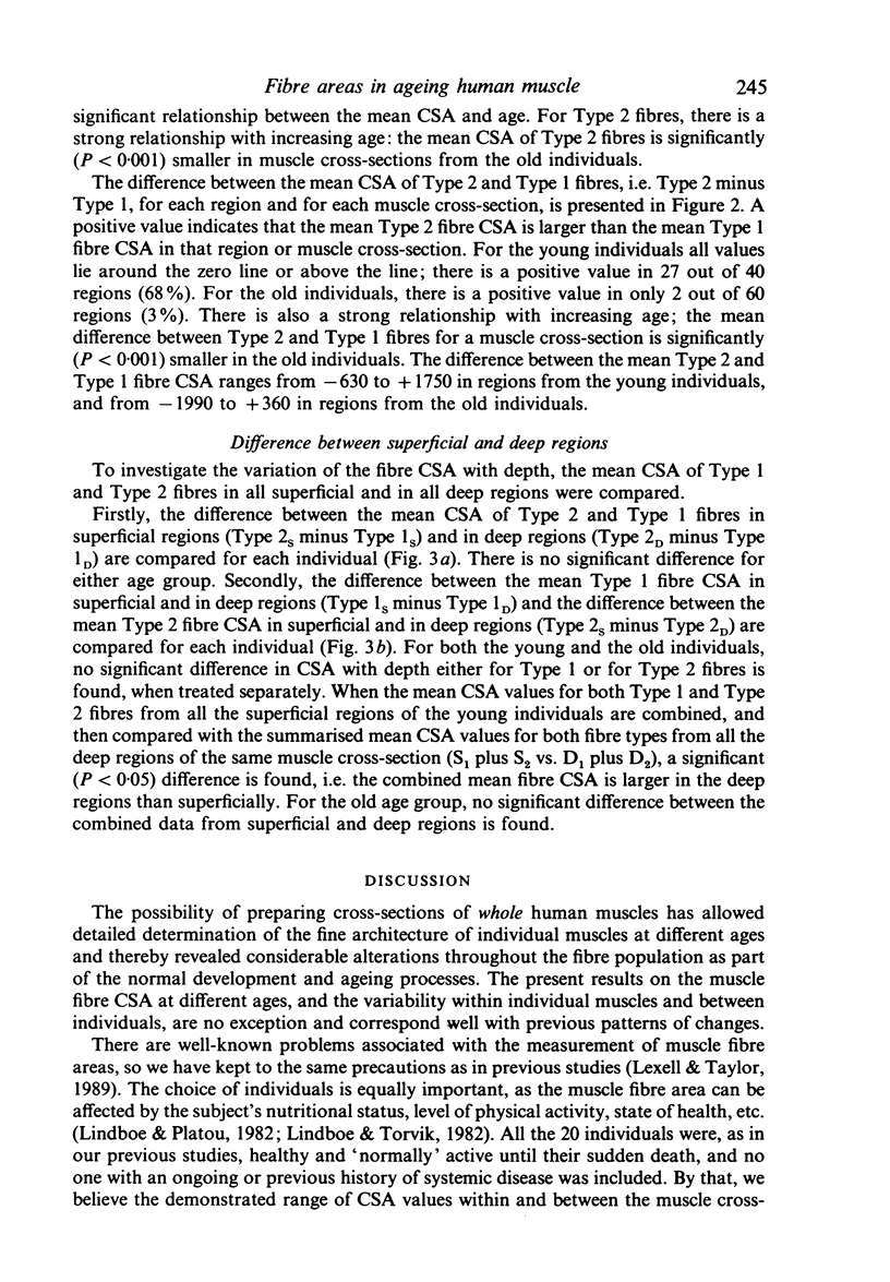
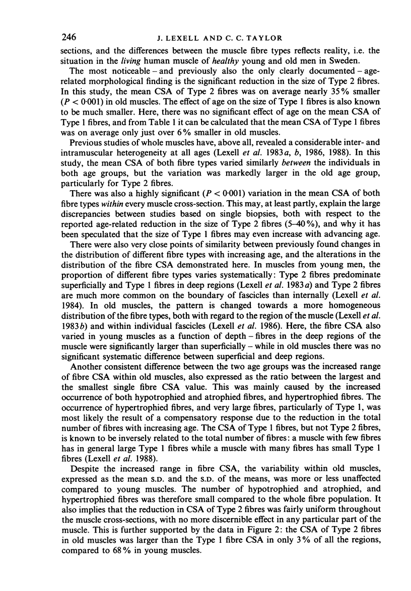
Selected References
These references are in PubMed. This may not be the complete list of references from this article.
- ALLEN T. H., ANDERSON E. C., LANGHAM W. H. Total body potassium and gross body composition in relation to age. J Gerontol. 1960 Oct;15:348–357. doi: 10.1093/geronj/15.4.348. [DOI] [PubMed] [Google Scholar]
- Aniansson A., Hedberg M., Henning G. B., Grimby G. Muscle morphology, enzymatic activity, and muscle strength in elderly men: a follow-up study. Muscle Nerve. 1986 Sep;9(7):585–591. doi: 10.1002/mus.880090702. [DOI] [PubMed] [Google Scholar]
- Boreham C. A., Watt P. W., Williams P. E., Merry B. J., Goldspink G., Goldspink D. F. Effects of ageing and chronic dietary restriction on the morphology of fast and slow muscles of the rat. J Anat. 1988 Apr;157:111–125. [PMC free article] [PubMed] [Google Scholar]
- Essén-Gustavsson B., Borges O. Histochemical and metabolic characteristics of human skeletal muscle in relation to age. Acta Physiol Scand. 1986 Jan;126(1):107–114. doi: 10.1111/j.1748-1716.1986.tb07793.x. [DOI] [PubMed] [Google Scholar]
- Grimby G., Danneskiold-Samsøe B., Hvid K., Saltin B. Morphology and enzymatic capacity in arm and leg muscles in 78-81 year old men and women. Acta Physiol Scand. 1982 May;115(1):125–134. doi: 10.1111/j.1748-1716.1982.tb07054.x. [DOI] [PubMed] [Google Scholar]
- Grimby G. Physical activity and effects of muscle training in the elderly. Ann Clin Res. 1988;20(1-2):62–66. [PubMed] [Google Scholar]
- Grimby G., Saltin B. The ageing muscle. Clin Physiol. 1983 Jun;3(3):209–218. doi: 10.1111/j.1475-097x.1983.tb00704.x. [DOI] [PubMed] [Google Scholar]
- Jennekens F. G., Tomlinson B. E., Walton J. N. Histochemical aspects of five limb muscles in old age. An autopsy study. J Neurol Sci. 1971 Nov;14(3):259–276. doi: 10.1016/0022-510x(71)90216-4. [DOI] [PubMed] [Google Scholar]
- Larsson L., Sjödin B., Karlsson J. Histochemical and biochemical changes in human skeletal muscle with age in sedentary males, age 22--65 years. Acta Physiol Scand. 1978 May;103(1):31–39. doi: 10.1111/j.1748-1716.1978.tb06187.x. [DOI] [PubMed] [Google Scholar]
- Lexell J., Downham D., Sjöström M. Distribution of different fibre types in human skeletal muscles. A statistical and computational study of the fibre type arrangement in m. vastus lateralis of young, healthy males. J Neurol Sci. 1984 Sep;65(3):353–365. doi: 10.1016/0022-510x(84)90098-4. [DOI] [PubMed] [Google Scholar]
- Lexell J., Downham D., Sjöström M. Distribution of different fibre types in human skeletal muscles. Fibre type arrangement in m. vastus lateralis from three groups of healthy men between 15 and 83 years. J Neurol Sci. 1986 Feb;72(2-3):211–222. doi: 10.1016/0022-510x(86)90009-2. [DOI] [PubMed] [Google Scholar]
- Lexell J., Henriksson-Larsén K., Sjöström M. Distribution of different fibre types in human skeletal muscles. 2. A study of cross-sections of whole m. vastus lateralis. Acta Physiol Scand. 1983 Jan;117(1):115–122. doi: 10.1111/j.1748-1716.1983.tb07185.x. [DOI] [PubMed] [Google Scholar]
- Lexell J., Henriksson-Larsén K., Winblad B., Sjöström M. Distribution of different fiber types in human skeletal muscles: effects of aging studied in whole muscle cross sections. Muscle Nerve. 1983 Oct;6(8):588–595. doi: 10.1002/mus.880060809. [DOI] [PubMed] [Google Scholar]
- Lexell J., Taylor C. C., Sjöström M. What is the cause of the ageing atrophy? Total number, size and proportion of different fiber types studied in whole vastus lateralis muscle from 15- to 83-year-old men. J Neurol Sci. 1988 Apr;84(2-3):275–294. doi: 10.1016/0022-510x(88)90132-3. [DOI] [PubMed] [Google Scholar]
- Lexell J., Taylor C. C. Variability in muscle fibre areas in whole human quadriceps muscle. How much and why? Acta Physiol Scand. 1989 Aug;136(4):561–568. doi: 10.1111/j.1748-1716.1989.tb08702.x. [DOI] [PubMed] [Google Scholar]
- Lindboe C. F., Platou C. S. Disuse atrophy of human skeletal muscle. An enzyme histochemical study. Acta Neuropathol. 1982;56(4):241–244. doi: 10.1007/BF00691253. [DOI] [PubMed] [Google Scholar]
- Lindboe C. F., Torvik A. The effects of ageing, cachexia and neoplasms on striated muscle. Quantitative histological and histochemical observations on an autopsy material. Acta Neuropathol. 1982;57(2-3):85–92. doi: 10.1007/BF00685374. [DOI] [PubMed] [Google Scholar]
- Nygaard E., Sanchez J. Intramuscular variation of fiber types in the brachial biceps and the lateral vastus muscles of elderly men: how representative is a small biopsy sample? Anat Rec. 1982 Aug;203(4):451–459. doi: 10.1002/ar.1092030404. [DOI] [PubMed] [Google Scholar]
- Poggi P., Marchetti C., Scelsi R. Automatic morphometric analysis of skeletal muscle fibers in the aging man. Anat Rec. 1987 Jan;217(1):30–34. doi: 10.1002/ar.1092170106. [DOI] [PubMed] [Google Scholar]
- Scelsi R., Marchetti C., Poggi P. Histochemical and ultrastructural aspects of m. vastus lateralis in sedentary old people (age 65--89 years). Acta Neuropathol. 1980;51(2):99–105. doi: 10.1007/BF00690450. [DOI] [PubMed] [Google Scholar]
- Sjöström M., Downham D. Y., Lexell J. Distribution of different fiber types in human skeletal muscles: why is there a difference within a fascicle? Muscle Nerve. 1986 Jan;9(1):30–36. doi: 10.1002/mus.880090105. [DOI] [PubMed] [Google Scholar]
- Tomlinson B. E., Walton J. N., Rebeiz J. J. The effects of ageing and of cachexia upon skeletal muscle. A histopathological study. J Neurol Sci. 1969 Sep-Oct;9(2):321–346. doi: 10.1016/0022-510x(69)90079-3. [DOI] [PubMed] [Google Scholar]
- Tomonaga M. Histochemical and ultrastructural changes in senile human skeletal muscle. J Am Geriatr Soc. 1977 Mar;25(3):125–131. doi: 10.1111/j.1532-5415.1977.tb00274.x. [DOI] [PubMed] [Google Scholar]
- Tzankoff S. P., Norris A. H. Effect of muscle mass decrease on age-related BMR changes. J Appl Physiol Respir Environ Exerc Physiol. 1977 Dec;43(6):1001–1006. doi: 10.1152/jappl.1977.43.6.1001. [DOI] [PubMed] [Google Scholar]


