

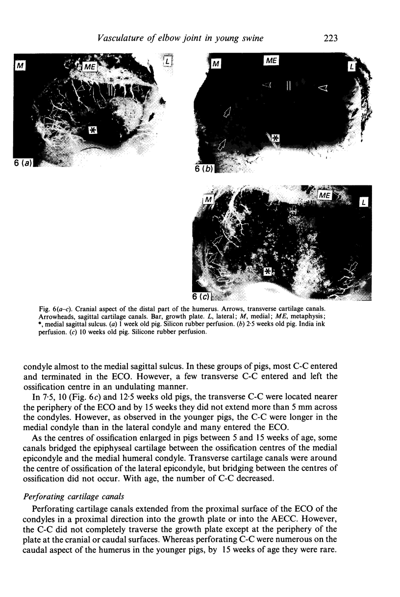
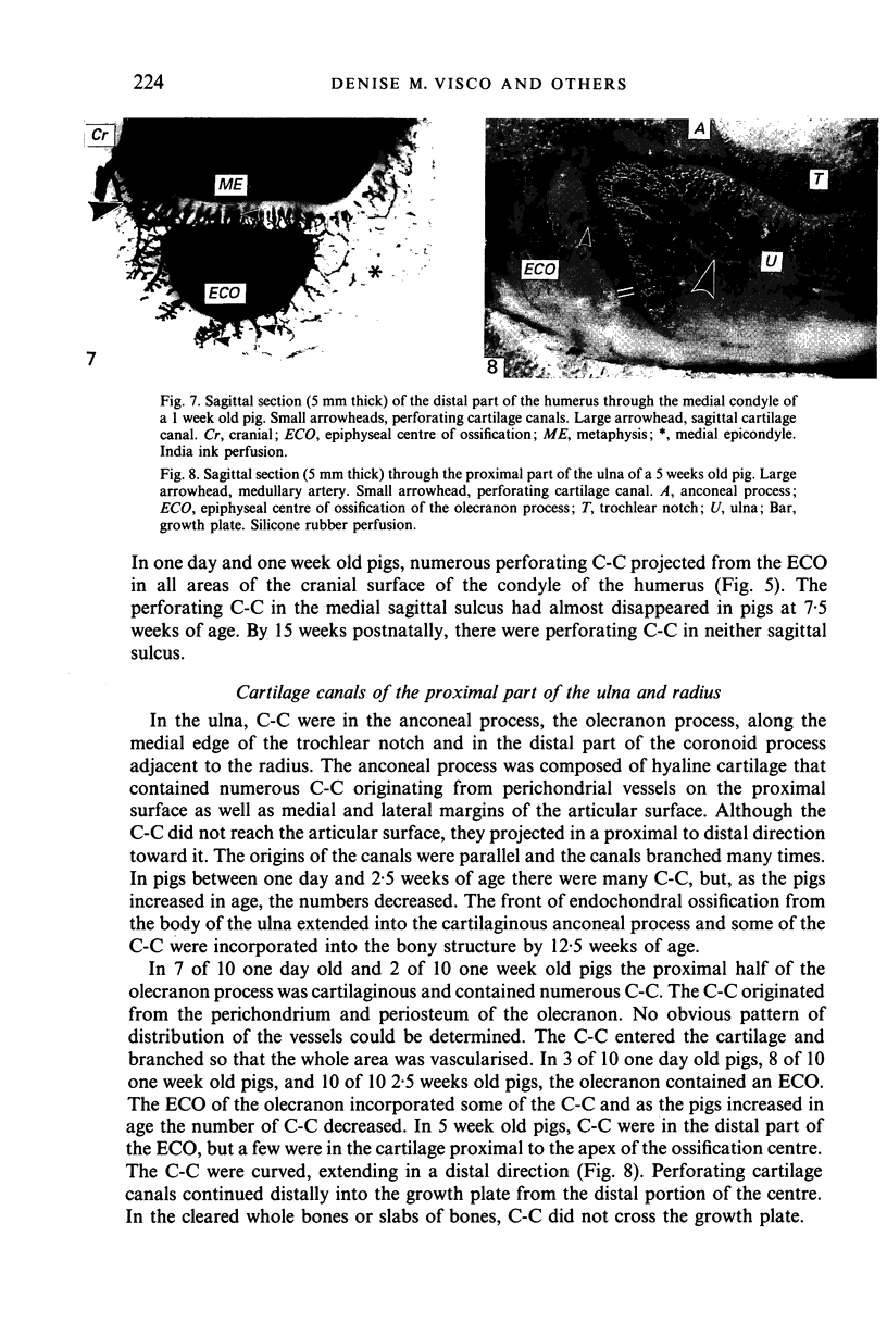
Abstract
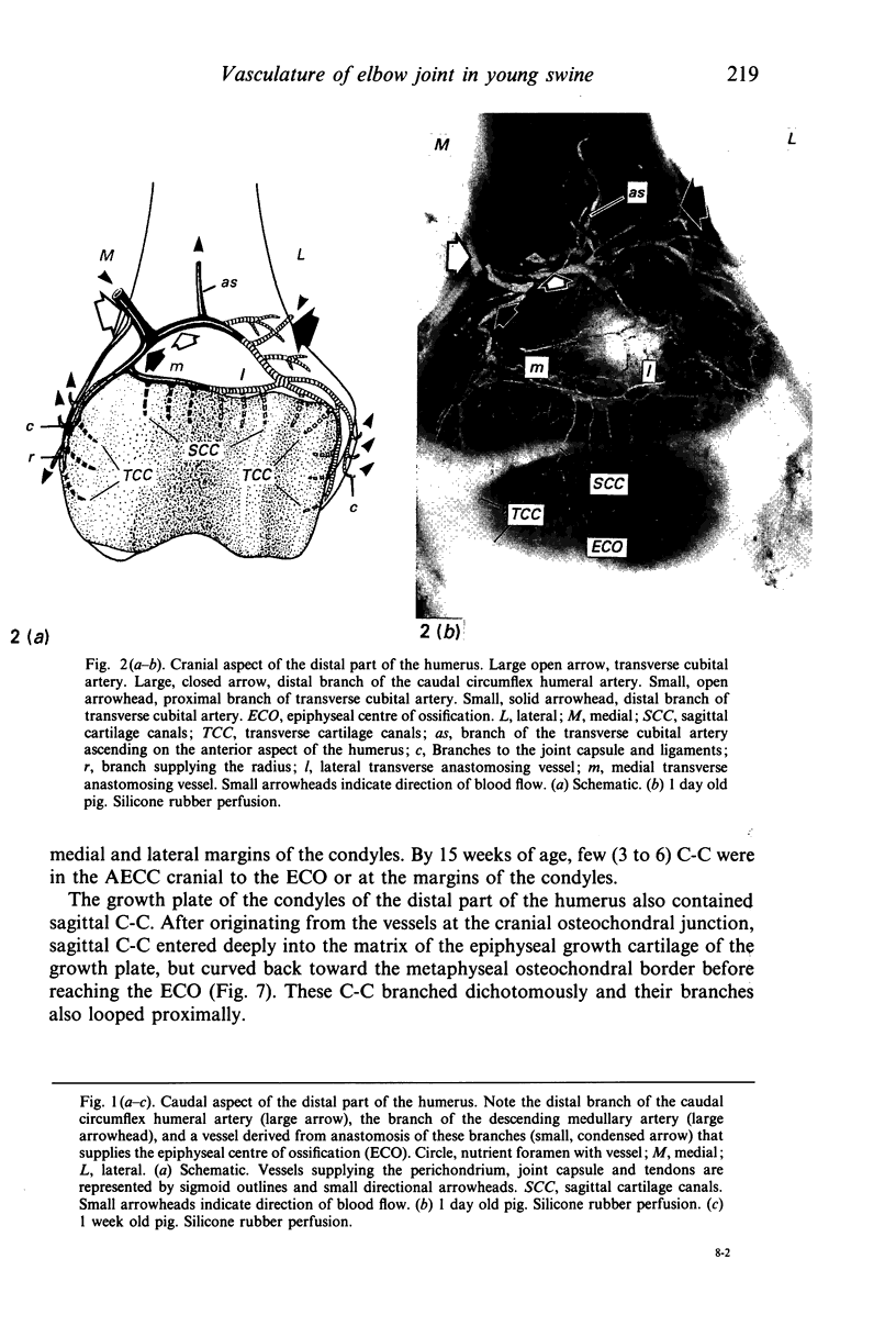
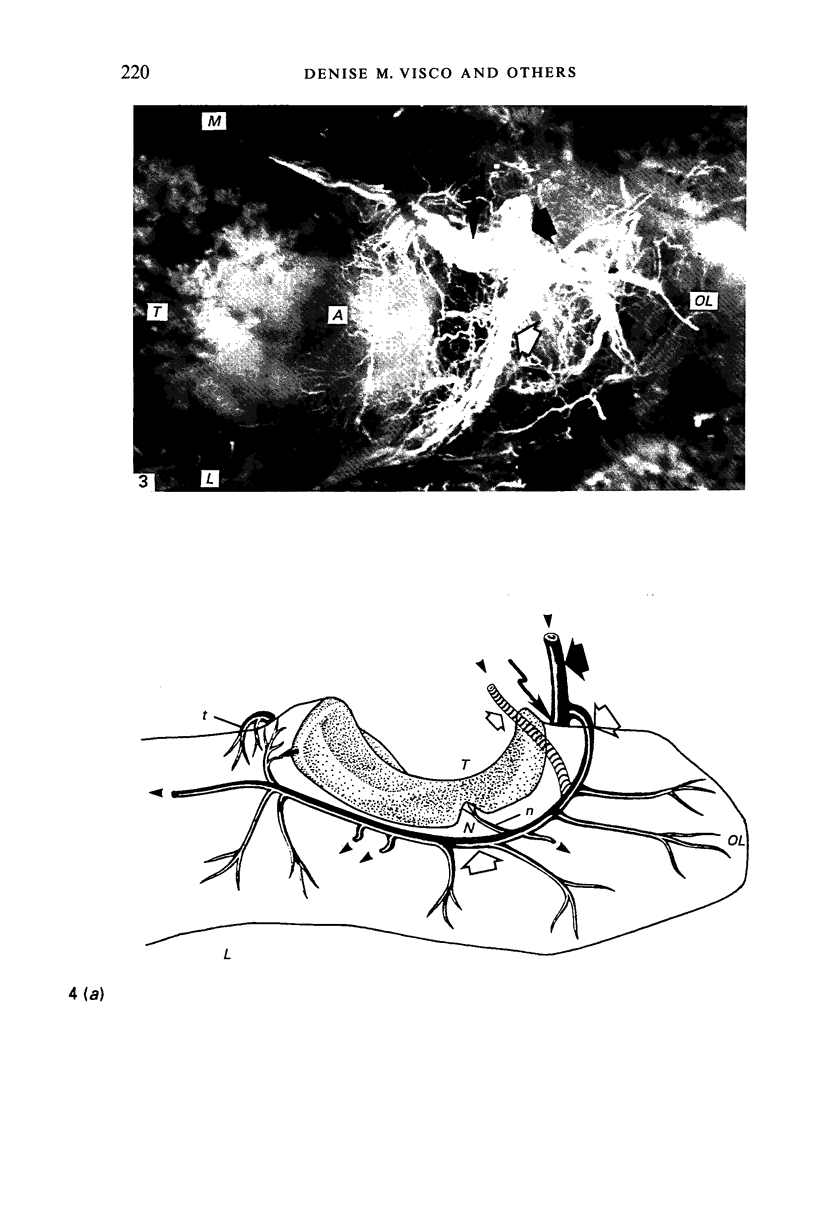
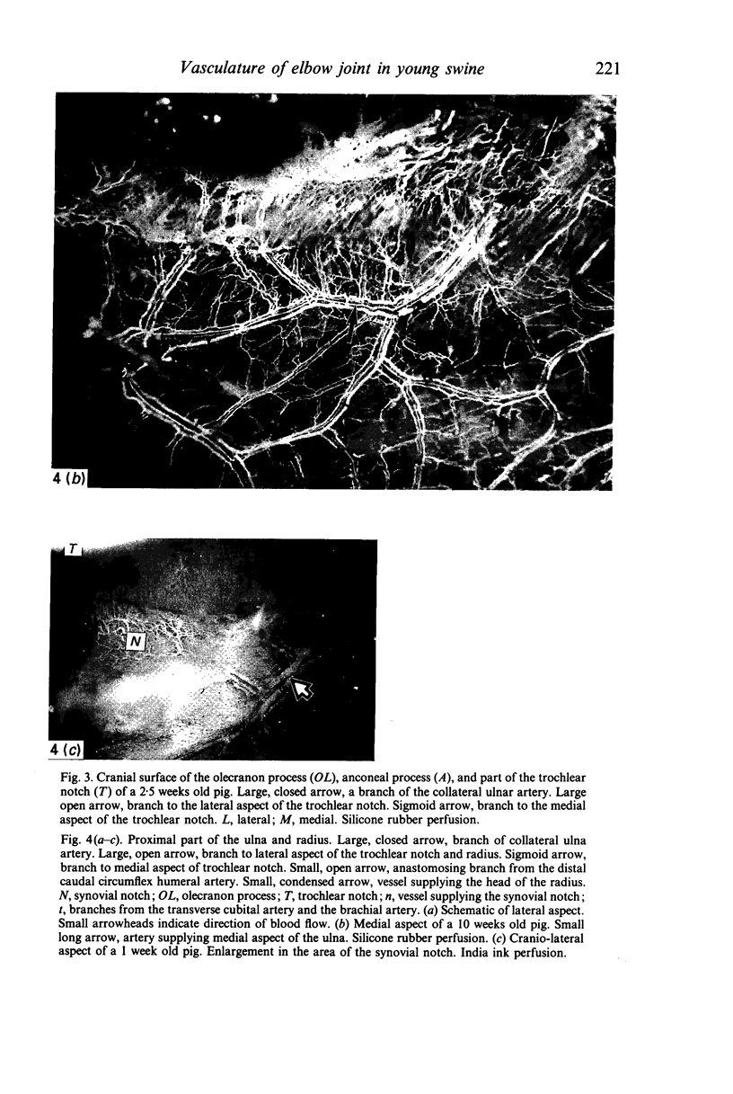
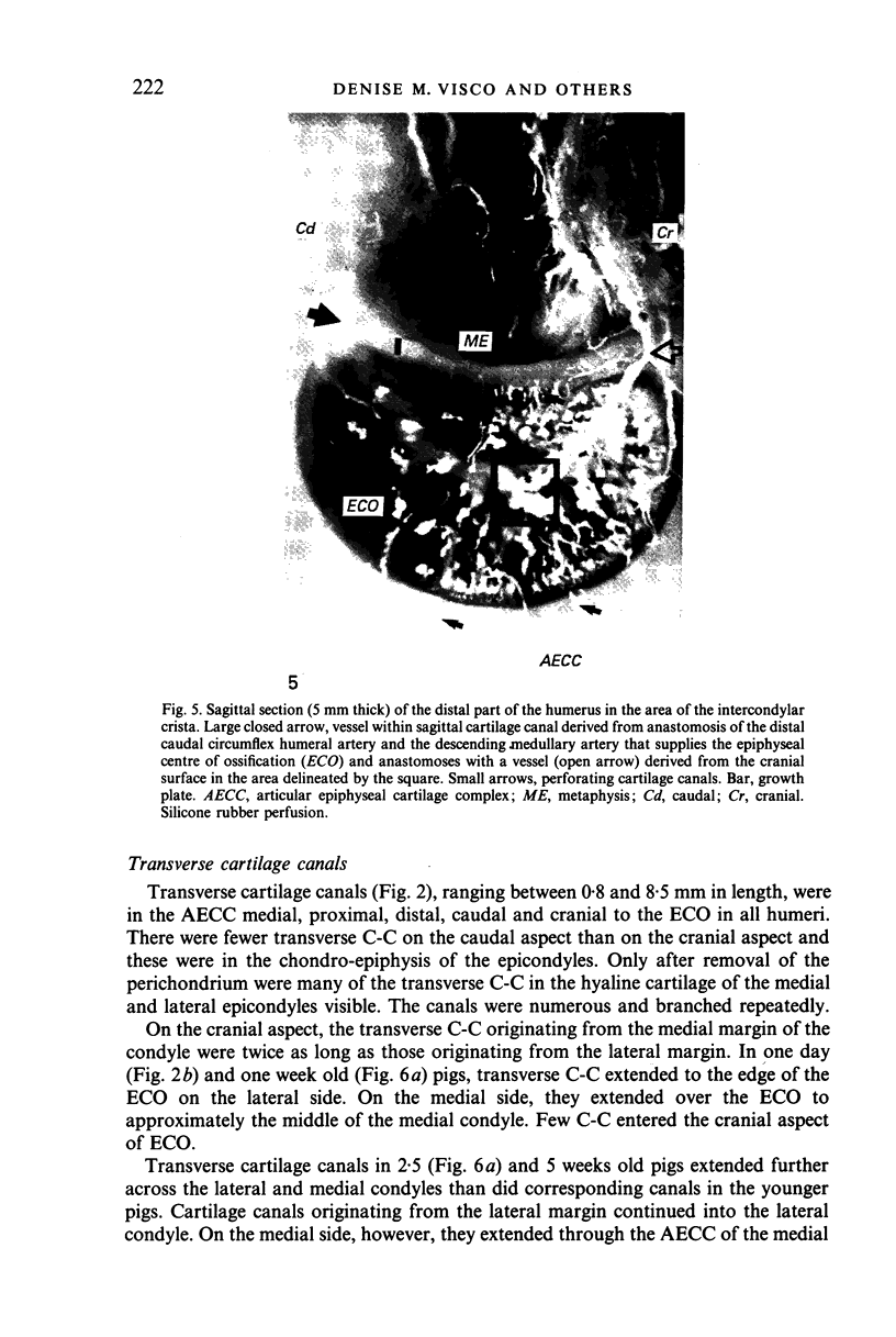
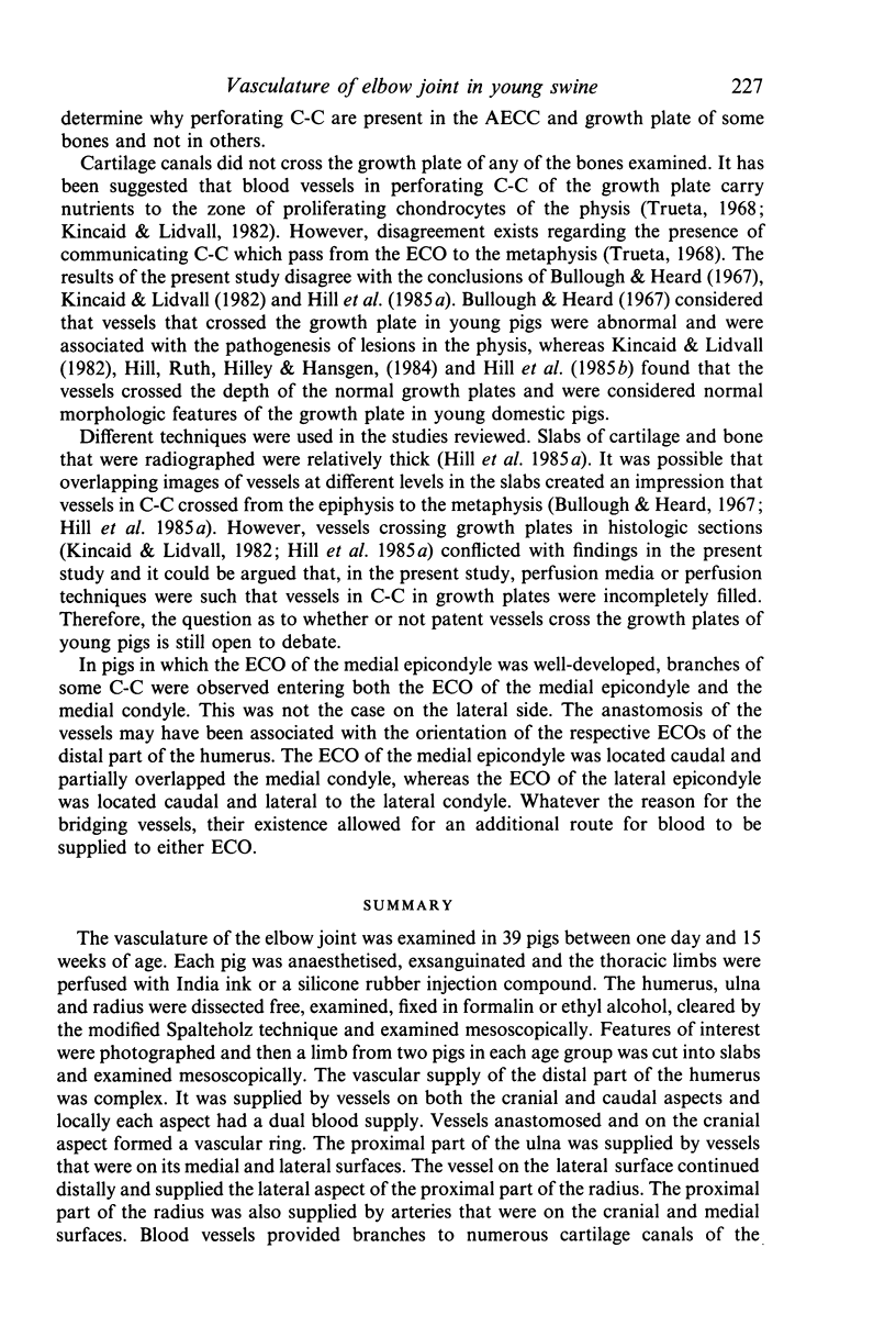
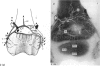
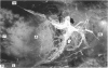
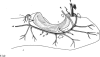
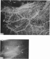
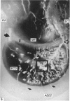
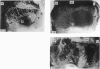
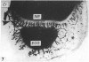
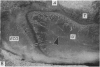
The vasculature of the elbow joint was examined in 39 pigs between one day and 15 weeks of age. Each pig was anaesthetised, exsanguinated and the thoracic limbs were perfused with India ink or a silicone rubber injection compound. The humerus, ulna and radius were dissected free, examined, fixed in formalin or ethyl alcohol, cleared by the modified Spalteholz technique and examined mesoscopically. Features of interest were photographed and then a limb from two pigs in each age group was cut into slabs and examined mesoscopically. The vascular supply of the distal part of the humerus was complex. It was supplied by vessels on both the cranial and caudal aspects and locally each aspect had a dual blood supply. Vessels anastomosed and on the cranial aspect formed a vascular ring. The proximal part of the ulna was supplied by vessels that were on its medial and lateral surfaces. The vessel on the lateral surface continued distally and supplied the lateral aspect of the proximal part of the radius. The proximal part of the radius was also supplied by arteries that were on the cranial and medial surfaces. Blood vessels provided branches to numerous cartilage canals of the articular-epiphyseal cartilage complexes, epiphyseal centres of ossification, and growth plates. The patterns of blood vessels in cartilage canals which were in sagittal or transverse planes were best exemplified by those in the distal part of the humerus. Perforating cartilage canals emerged from the epiphyseal centres of ossification. The pattern of cartilage canals was consistent in a general configuration, but individual variation did occur. Although cartilage canals were abundant in the youngest pigs, with increasing age the distribution of cartilage canals changed and the numbers of cartilage canals decreased.
Full text
PDF
Images in this article
Selected References
These references are in PubMed. This may not be the complete list of references from this article.
- Bullough P. G., Heard T. W. Pathological lesions associated with the "leg weakness" syndrome in pigs. Br Vet J. 1967 Jul;123(7):305–310. doi: 10.1016/s0007-1935(17)39907-4. [DOI] [PubMed] [Google Scholar]
- Cole A. A., Wezeman F. H. Perivascular cells in cartilage canals of the developing mouse epiphysis. Am J Anat. 1985 Oct;174(2):119–129. doi: 10.1002/aja.1001740203. [DOI] [PubMed] [Google Scholar]
- Firth E. C., Poulos P. W. Blood vessels in the developing growth plate of the equine distal radius and metacarpus. Res Vet Sci. 1982 Sep;33(2):159–166. [PubMed] [Google Scholar]
- HARALDSSON S. The vascular pattern of a growing and fullgrown human epiphysis. Acta Anat (Basel) 1962;48:156–167. doi: 10.1159/000141835. [DOI] [PubMed] [Google Scholar]
- Haines R. W. Cartilage Canals. J Anat. 1933 Oct;68(Pt 1):45–64. [PMC free article] [PubMed] [Google Scholar]
- Haines R. W. The pseudoepiphysis of the first metacarpal of man. J Anat. 1974 Feb;117(Pt 1):145–158. [PMC free article] [PubMed] [Google Scholar]
- Hill M. A., Ruth G. R., Bagent J. K., Torrison J. L., Leman A. D. Angiomicrographic investigation of the vessels associated with physes in young pigs. Res Vet Sci. 1985 Mar;38(2):151–159. [PubMed] [Google Scholar]
- Hill M. A., Ruth G. R., Hilley H. D., Hansgen D. C. Dyschondroplasias, including osteochondrosis, in boars between 25 and 169 days of age: histologic changes. Am J Vet Res. 1984 May;45(5):903–916. [PubMed] [Google Scholar]
- Hill M. A., Ruth G. R., Hilley H. D., Torrison J. L., Bagent J. K., Leman A. D. Dyschondroplasias of growth cartilages (osteochondrosis) in crossbred commercial pigs at one and 15 days of age: radiological, angiomicrographical and histological findings. Vet Rec. 1985 Jan 12;116(2):40–47. doi: 10.1136/vr.116.2.40. [DOI] [PubMed] [Google Scholar]
- Hill M. A., Ruth G. R., Van Sickle D. C., Hilley H. D., Torrison J. L., Bagent J. K., Leman A. D. Histochemical morphologic features of growth cartilages in long bones of pigs of various ages. Am J Vet Res. 1987 Oct;48(10):1477–1484. [PubMed] [Google Scholar]
- Hurrell D. J. The Vascularisation of Cartilage. J Anat. 1934 Oct;69(Pt 1):47–61. [PMC free article] [PubMed] [Google Scholar]
- Kincaid S. A., Lidvall E. R. Communicating cartilage canals of the physis of the distal part of the ulna of growing swine and their potential role in healing of metaphyseal dysplasia of osteochondrosis. Am J Vet Res. 1982 Jun;43(6):938–944. [PubMed] [Google Scholar]
- Kincaid S. A., Lidvall E. R. Observations on the postnatal morphogenesis of the porcine humeral condyle and the pathogenesis of osteochondrosis. Am J Vet Res. 1983 Nov;44(11):2095–2103. [PubMed] [Google Scholar]
- LEVENE C. THE PATTERNS OF CARTILAGE CANALS. J Anat. 1964 Oct;98:515–538. [PMC free article] [PubMed] [Google Scholar]
- Lutfi A. M. 35S-sulphate uptake by the growing tibia in the domestic fowl. J Anat. 1970 Nov;107(Pt 3):567–576. [PMC free article] [PubMed] [Google Scholar]
- Lutfi A. M. Mode of growth, fate and functions of cartilage canals. J Anat. 1970 Jan;106(Pt 1):135–145. [PMC free article] [PubMed] [Google Scholar]
- Reiland S. Morphology of osteochondrosis and sequelae in pigs. Acta Radiol Suppl. 1978;358:45–90. [PubMed] [Google Scholar]
- Rodríguez J. I., Delgado E., Paniagua R. Multivacuolated cells in human cartilage canals. Acta Anat (Basel) 1985;124(1-2):54–57. doi: 10.1159/000146096. [DOI] [PubMed] [Google Scholar]
- Sinha D. N., Varma H. C. Cartilage canal and chondroepiphysis of the lower end of the femur of human fetuses. Anat Anz. 1982;152(5):461–466. [PubMed] [Google Scholar]
- Stockwell R. A. The ultrastructure of cartilage canals and the surrounding cartilage in the sheep fetus. J Anat. 1971 Sep;109(Pt 3):397–410. [PMC free article] [PubMed] [Google Scholar]
- TRUETA J., HARRISON M. H. The normal vascular anatomy of the femoral head in adult man. J Bone Joint Surg Br. 1953 Aug;35-B(3):442–461. doi: 10.1302/0301-620X.35B3.442. [DOI] [PubMed] [Google Scholar]
- Wilsman N. J., Van Sickle D. C. Cartilage canals, their morphology and distribution. Anat Rec. 1972 May;173(1):79–93. doi: 10.1002/ar.1091730107. [DOI] [PubMed] [Google Scholar]
- Wilsman N. J., Van Sickle D. C. The relationship of cartilage canals to the initial osteogenesis of secondary centers of ossification. Anat Rec. 1970 Nov;168(3):381–391. doi: 10.1002/ar.1091680305. [DOI] [PubMed] [Google Scholar]