

Abstract
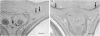
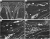
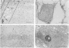
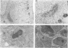
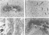
Membrane bone formation was examined in the developing chick frontal bone of Stage 32-36 embryos using alizarin red whole mounts, wax and resin histology, transmission electron microscopy and India ink injection techniques. Mineralised bone was seen at Stage 36, but was presaged by the appearance of twin mesenchymal condensations at Stage 35. The condensations appeared dorsolateral to the upper edge of the interorbital septum near its centre. The mesenchymal cells of the condensation were fusiform in shape and initially exhibited a close-packed, layered configuration which was avascular. Consequent differentiation of the central cells first into preosteoblasts and then into definitive osteoblasts, accompanied by secretion of bone matrix, coincided with blood vessel invasion of the frontal anlagen. It is suggested that these vascular changes associated with the onset of bone cell differentiation indicate that osteogenic cells may secrete an angiogenesis factor.
Full text
PDF
Images in this article
Selected References
These references are in PubMed. This may not be the complete list of references from this article.
- Archer C. W., Rooney P., Wolpert L. Cell shape and cartilage differentiation of early chick limb bud cells in culture. Cell Differ. 1982 Jun;11(4):245–251. doi: 10.1016/0045-6039(82)90072-0. [DOI] [PubMed] [Google Scholar]
- Bernard G. W., Pease D. C. An electron microscopic study of initial intramembranous osteogenesis. Am J Anat. 1969 Jul;125(3):271–290. doi: 10.1002/aja.1001250303. [DOI] [PubMed] [Google Scholar]
- Caplan A. I., Koutroupas S. The control of muscle and cartilage development in the chick limb: the role of differential vascularization. J Embryol Exp Morphol. 1973 Jun;29(3):571–583. [PubMed] [Google Scholar]
- Eisenstein R., Kuettner K. E., Neapolitan C., Soble L. W., Sorgente N. The resistance of certain tissues to invasion. III. Cartilage extracts inhibit the growth of fibroblasts and endothelial cells in culture. Am J Pathol. 1975 Nov;81(2):337–348. [PMC free article] [PubMed] [Google Scholar]
- Gould R. P., Selwood L., Day A., Wolpert L. The mechanism of cellular orientation during early cartilage formation in the chick limb and regenerating amphibian limb. Exp Cell Res. 1974 Feb;83(2):287–296. doi: 10.1016/0014-4827(74)90341-3. [DOI] [PubMed] [Google Scholar]
- Hallmann R., Feinberg R. N., Latker C. H., Sasse J., Risau W. Regression of blood vessels precedes cartilage differentiation during chick limb development. Differentiation. 1987;34(2):98–105. doi: 10.1111/j.1432-0436.1987.tb00055.x. [DOI] [PubMed] [Google Scholar]
- Marvaso V., Bernard G. W. Initial intramembraneous osteogenesis in vitro. Am J Anat. 1977 Aug;149(4):453–468. doi: 10.1002/aja.1001490403. [DOI] [PubMed] [Google Scholar]
- Pechak D. G., Kujawa M. J., Caplan A. I. Morphological and histochemical events during first bone formation in embryonic chick limbs. Bone. 1986;7(6):441–458. doi: 10.1016/8756-3282(86)90004-9. [DOI] [PubMed] [Google Scholar]
- Risau W. Developing brain produces an angiogenesis factor. Proc Natl Acad Sci U S A. 1986 Jun;83(11):3855–3859. doi: 10.1073/pnas.83.11.3855. [DOI] [PMC free article] [PubMed] [Google Scholar]
- Risau W., Ekblom P. Production of a heparin-binding angiogenesis factor by the embryonic kidney. J Cell Biol. 1986 Sep;103(3):1101–1107. doi: 10.1083/jcb.103.3.1101. [DOI] [PMC free article] [PubMed] [Google Scholar]
- Sorgente N., Kuettner K. E., Soble L. W., Eisenstein R. The resistance of certain tissues to invasion. II. Evidence for extractable factors in cartilage which inhibit invasion by vascularized mesenchyme. Lab Invest. 1975 Feb;32(2):217–222. [PubMed] [Google Scholar]
- Thorogood P. V., Hinchliffe J. R. An analysis of the condensation process during chondrogenesis in the embryonic chick hind limb. J Embryol Exp Morphol. 1975 Jun;33(3):581–606. [PubMed] [Google Scholar]
- Wilson D. J. Development of avascularity during cartilage differentiation in the embryonic limb. An exclusion model. Differentiation. 1986;30(3):183–187. doi: 10.1111/j.1432-0436.1986.tb00778.x. [DOI] [PubMed] [Google Scholar]