

Abstract
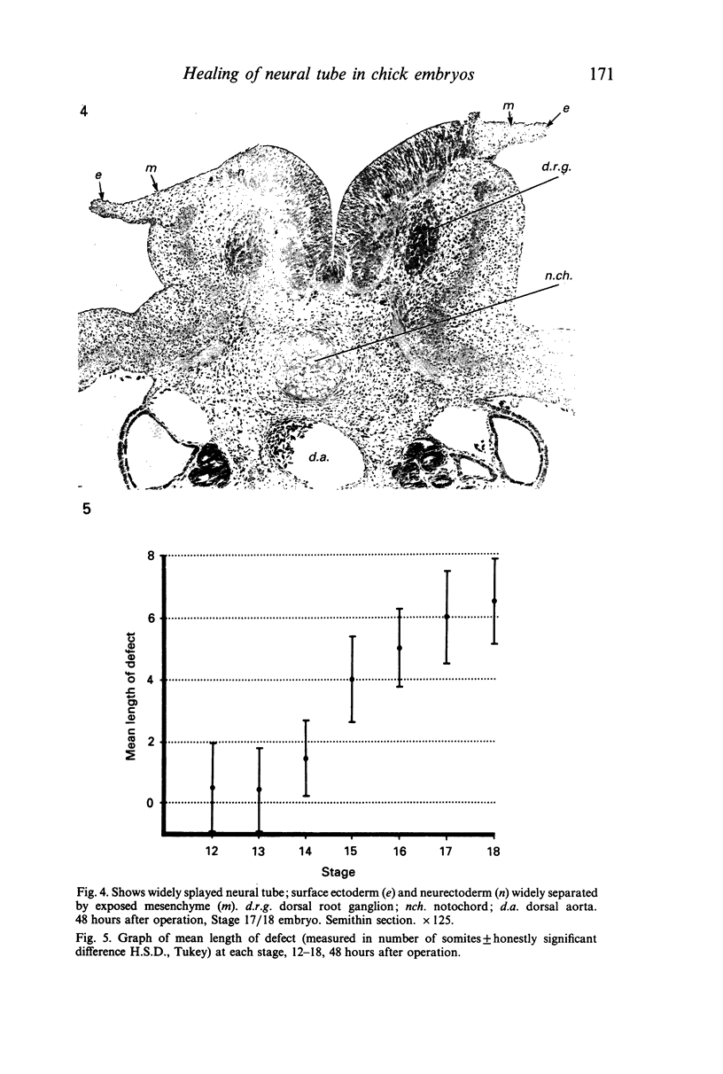
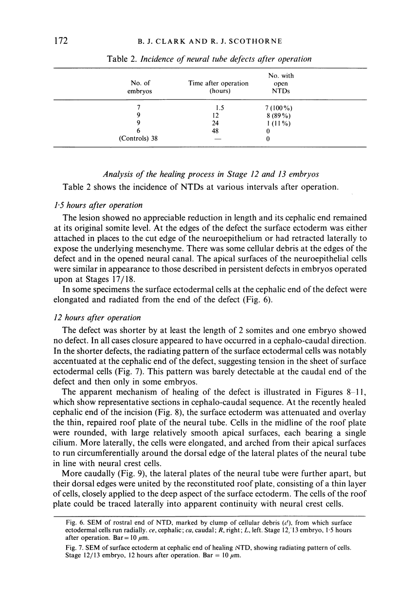
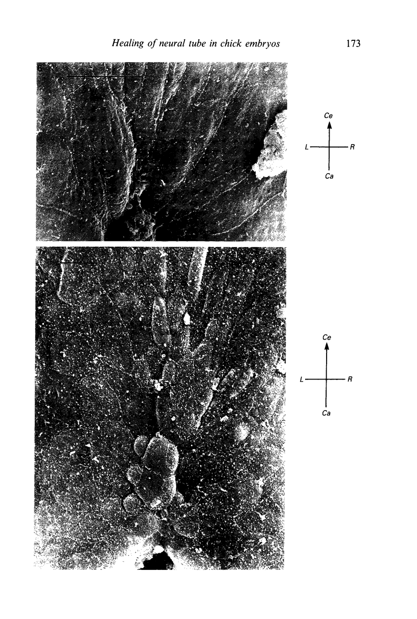
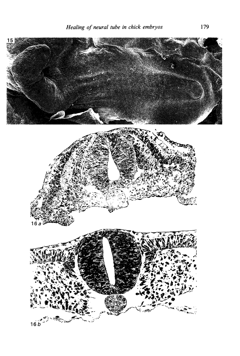
The effects of microsurgical reopening of the neural tube were examined in chick embryos of Stages 12-18. The roof plate of the thoracic neural tube was incised for a length equivalent to 7 somites. The site of incision was studied histologically and by SEM and TEM at intervals up to 48 hours. 48 hours after operation, persistent neural tube defects were more frequent and longer in embryos of more advanced stages at operation. Exposure of embryos to Streptomyces hyaluronidase, which inhibits neurulation in normal embryos, has no effect on the healing of the incised neural tube in the young embryos. Healing of the lesion in younger embryos appeared to occur in two stages: initially, by repair of the surface ectoderm, by a cephalo-caudal zipper-like mechanism, followed by a reconstitution of the roof plate by migration of neurectodermal cells on the deep surface of the ectoderm. Neural tubes of older embryos splay open more widely on incision of the roof plate, apparently making healing mechanically more difficult. This wider splaying may be related to the decline of forces which maintain occlusion of the neural canal in younger embryos.
Full text
PDF
Images in this article
Selected References
These references are in PubMed. This may not be the complete list of references from this article.
- Bancroft M., Bellairs R. The onset of differentiation in the epiblast of the chick blastoderm (SEM and TEM). Cell Tissue Res. 1974;155(3):399–418. doi: 10.1007/BF00222814. [DOI] [PubMed] [Google Scholar]
- DEKABAN A. S., BARTELMEZ G. W. COMPLETE DYSRAPHISM IN 14 SOMITE HUMAN EMBRYO. A CONTRIBUTION TO NORMAL AND ABNORMAL MORPHOGENESIS. Am J Anat. 1964 Jul;115:27–38. doi: 10.1002/aja.1001150104. [DOI] [PubMed] [Google Scholar]
- Desmond M. E. Description of the occlusion of the spinal cord lumen in early human embryos. Anat Rec. 1982 Sep;204(1):89–93. doi: 10.1002/ar.1092040112. [DOI] [PubMed] [Google Scholar]
- Desmond M. E., Schoenwolf G. C. Evaluation of the roles of intrinsic and extrinsic factors in occlusion of the spinal neurocoel during rapid brain enlargement in the chick embryo. J Embryol Exp Morphol. 1986 Sep;97:25–46. [PubMed] [Google Scholar]
- Desmond M. E., Schoenwolf G. C. Timing and positioning of occlusion of the spinal neurocele in the chick embryo. J Comp Neurol. 1985 May 22;235(4):479–487. doi: 10.1002/cne.902350406. [DOI] [PubMed] [Google Scholar]
- ECHLIN P. INTRA-CYTOPLASMIC MEMBRANOUS INCLUSIONS IN THE BLUE-GREEN ALGA, ANACYSTIS NIDULANS. Arch Mikrobiol. 1964 Oct 2;49:267–274. doi: 10.1007/BF00409749. [DOI] [PubMed] [Google Scholar]
- FORD E. H. An anencephalic human embryo of 33.5 millimetres. Acta Anat (Basel) 1956;28(1-2):149–155. doi: 10.1159/000141140. [DOI] [PubMed] [Google Scholar]
- GARDNER W. J. HYDRODYNAMIC MECHANISM OF SYRINGOMYELIA: ITS RELATIONSHIP TO MYELOCELE. J Neurol Neurosurg Psychiatry. 1965 Jun;28:247–259. doi: 10.1136/jnnp.28.3.247. [DOI] [PMC free article] [PubMed] [Google Scholar]
- GARDNER W. J. Rupture of the neural tube. Arch Neurol. 1961 Jan;4:1–7. doi: 10.1001/archneur.1961.00450070003001. [DOI] [PubMed] [Google Scholar]
- Gardner W. J. Embryologic origin of spinal malformations. Acta Radiol Diagn (Stockh) 1966;5:1013–1023. doi: 10.1177/02841851660050p245. [DOI] [PubMed] [Google Scholar]
- Gardner W. J. Hypothesis; overdistention of the neural tube may cause anomalies of non-neural organs. Teratology. 1980 Oct;22(2):229–238. doi: 10.1002/tera.1420220212. [DOI] [PubMed] [Google Scholar]
- Karfunkel P. The activity of microtubules and microfilaments in neurulation in the chick. J Exp Zool. 1972 Sep;181(3):289–301. doi: 10.1002/jez.1401810302. [DOI] [PubMed] [Google Scholar]
- Karfunkel P. The role of microtubules and microfilaments in neurulation in Xenopus. Dev Biol. 1971 May;25(1):30–56. doi: 10.1016/0012-1606(71)90018-2. [DOI] [PubMed] [Google Scholar]
- LEMIRE R. J., SHEPARD T. H., ALVORD E. C., Jr CAUDAL MYELOSCHISIS (LUMBO-SACRAL SPINA BIFIDA CYSTICA) IN A FIVE MILLIMETER (HORIZON XIV) HUMAN EMBRYO. Anat Rec. 1965 May;152:9–16. doi: 10.1002/ar.1091520103. [DOI] [PubMed] [Google Scholar]
- Lee H. Y., Kosciuk M. C., Nagele R. G., Roisen F. J. Studies on the mechanisms of neurulation in the chick: possible involvement of myosin in elevation of neural folds. J Exp Zool. 1983 Mar;225(3):449–457. doi: 10.1002/jez.1402250313. [DOI] [PubMed] [Google Scholar]
- Lee H. Y., Nagele R. G. Studies on the mechanisms of neurulation in the chick: interrelationship of contractile proteins, microfilaments, and the shape of neuroepithelial cells. J Exp Zool. 1985 Aug;235(2):205–215. doi: 10.1002/jez.1402350207. [DOI] [PubMed] [Google Scholar]
- Lee H., Nagele R. G. Neural tube defects caused by local anesthetics in early chick embryos. Teratology. 1985 Feb;31(1):119–127. doi: 10.1002/tera.1420310114. [DOI] [PubMed] [Google Scholar]
- Lee H., Nagele R. G., Pietrolungo J. F. Toxic and teratologic effects of caffeine on explanted early chick embryos. Teratology. 1982 Feb;25(1):19–25. doi: 10.1002/tera.1420250104. [DOI] [PubMed] [Google Scholar]
- Lemire R. J. Variations in development of the caudal neural tube in human embryos (Horizons XIV-XXI). Teratology. 1969 Nov;2(4):361–369. doi: 10.1002/tera.1420020410. [DOI] [PubMed] [Google Scholar]
- Marin-Padilla M. The closure of the neural tube in the golden hamster. Teratology. 1970 Feb;3(1):39–45. doi: 10.1002/tera.1420030109. [DOI] [PubMed] [Google Scholar]
- Morriss G. M., Solursh M. Regional differences in mesenchymal cell morphology and glycosaminoglycans in early neural-fold stage rat embryos. J Embryol Exp Morphol. 1978 Aug;46:37–52. [PubMed] [Google Scholar]
- O'Shea K. S., Kaufman M. H. Effect of acetaldehyde on the neuroepithelium of early mouse embryos. J Anat. 1981 Jan;132(Pt 1):107–118. [PMC free article] [PubMed] [Google Scholar]
- O'Shea K. S., Kaufman M. H. Neural tube closure defects following in vitro exposure of mouse embryos to xylocaine. J Exp Zool. 1980 Nov;214(2):235–238. doi: 10.1002/jez.1402140217. [DOI] [PubMed] [Google Scholar]
- Osaka K., Tanimura T., Hirayama A., Matsumoto S. Myelomeningocele before birth. J Neurosurg. 1978 Nov;49(5):711–724. doi: 10.3171/jns.1978.49.5.0711. [DOI] [PubMed] [Google Scholar]
- PATTEN B. M. Embryological stages in the establishing of myeloschisis with spina bifida. Am J Anat. 1953 Nov;93(3):365–395. doi: 10.1002/aja.1000930304. [DOI] [PubMed] [Google Scholar]
- PATTEN B. M. Overgrowth of the neural tube in young human embryos. Anat Rec. 1952 Aug;113(4):381–393. doi: 10.1002/ar.1091130402. [DOI] [PubMed] [Google Scholar]
- Padget D. H. Neuroschisis and human embryonic maldevelopment. New evidence on anencephaly, spina bifida and diverse mammalian defects. J Neuropathol Exp Neurol. 1970 Apr;29(2):192–216. [PubMed] [Google Scholar]
- Padget D. H. Spina bifida and embryonic neuroschisis--a causal relationship. Definition of the postnatal conformations involving a bifid spine. Johns Hopkins Med J. 1968 Nov;123(5):233–252. [PubMed] [Google Scholar]
- REYNOLDS E. S. The use of lead citrate at high pH as an electron-opaque stain in electron microscopy. J Cell Biol. 1963 Apr;17:208–212. doi: 10.1083/jcb.17.1.208. [DOI] [PMC free article] [PubMed] [Google Scholar]
- Rokos J., Knowles J. Experimental contribution to the pathogenesis of spina bifida. J Pathol. 1976 Jan;118(1):21–24. doi: 10.1002/path.1711180105. [DOI] [PubMed] [Google Scholar]
- Schoenwolf G. C., Desmond M. E. Timing and positioning of reopening of the occluded spinal neurocele in the chick embryo. J Comp Neurol. 1986 Apr 22;246(4):459–466. doi: 10.1002/cne.902460404. [DOI] [PubMed] [Google Scholar]
- Schoenwolf G. C., Fisher M. Analysis of the effects of Streptomyces hyaluronidase on formation of the neural tube. J Embryol Exp Morphol. 1983 Feb;73:1–15. [PubMed] [Google Scholar]
- Smedley M. J., Stanisstreet M. Scanning electron microscopy of wound healing in rat embryos. J Embryol Exp Morphol. 1984 Oct;83:109–117. [PubMed] [Google Scholar]
- Spratt N. T., Jr A Simple Method for Explanting and Cultivating Early Chick Embryos in Vitro. Science. 1947 Nov 7;106(2758):452–452. doi: 10.1126/science.106.2758.452. [DOI] [PubMed] [Google Scholar]
- Spurr A. R. A low-viscosity epoxy resin embedding medium for electron microscopy. J Ultrastruct Res. 1969 Jan;26(1):31–43. doi: 10.1016/s0022-5320(69)90033-1. [DOI] [PubMed] [Google Scholar]
- Stanisstreet M. Calcium and wound healing in Xenopus early embryos. J Embryol Exp Morphol. 1982 Feb;67:195–205. [PubMed] [Google Scholar]
- Stanisstreet M., Wakely J., England M. A. Scanning electron microscopy of wound healing in Xenopus and chicken embryos. J Embryol Exp Morphol. 1980 Oct;59:341–353. [PubMed] [Google Scholar]
- WARKANY J., WILSON J. G., GEIGER J. F. Myeloschisis and myelomeningocele produced experimentally in the rat. J Comp Neurol. 1958 Feb;109(1):35–64. doi: 10.1002/cne.901090103. [DOI] [PubMed] [Google Scholar]