Abstract
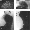
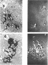
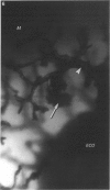
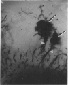
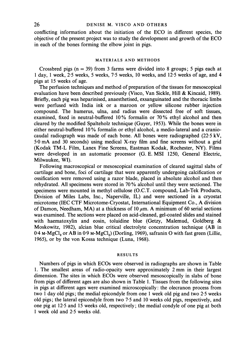
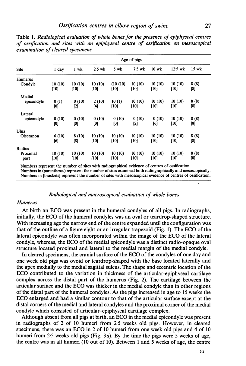
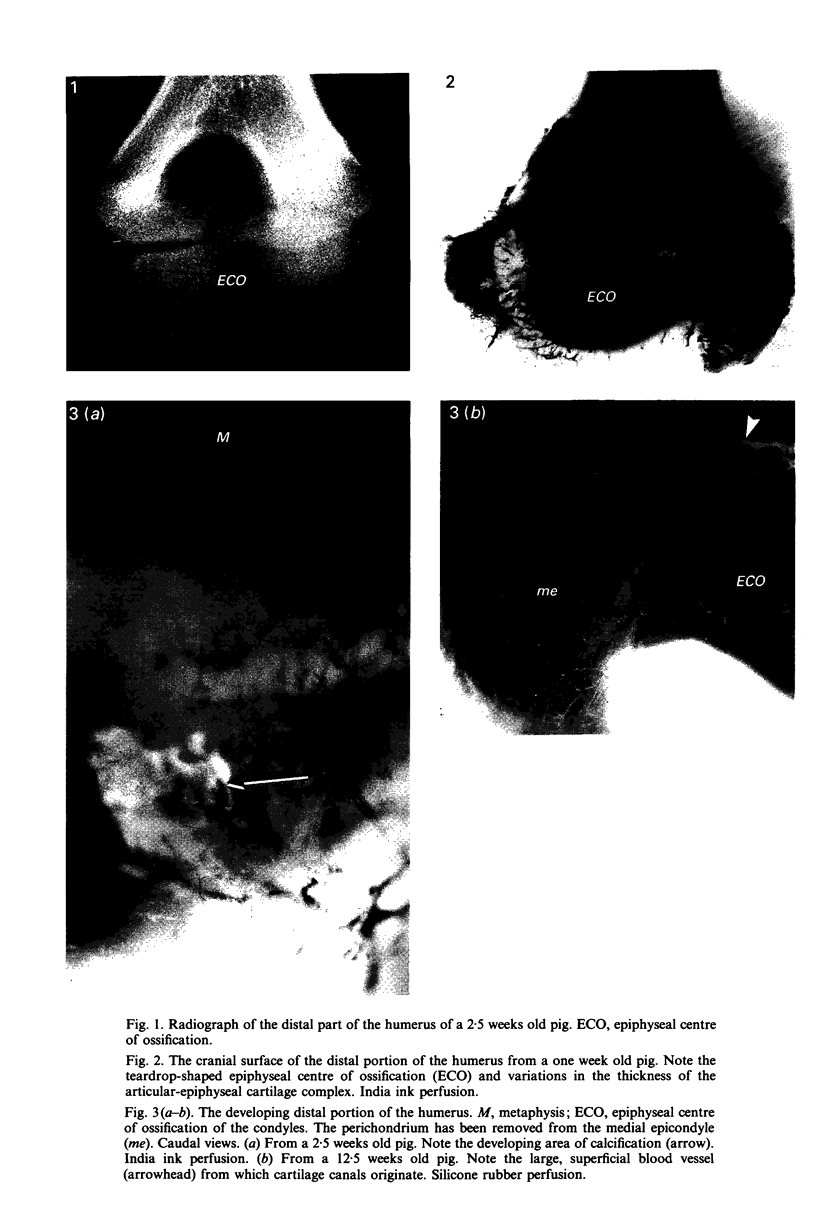
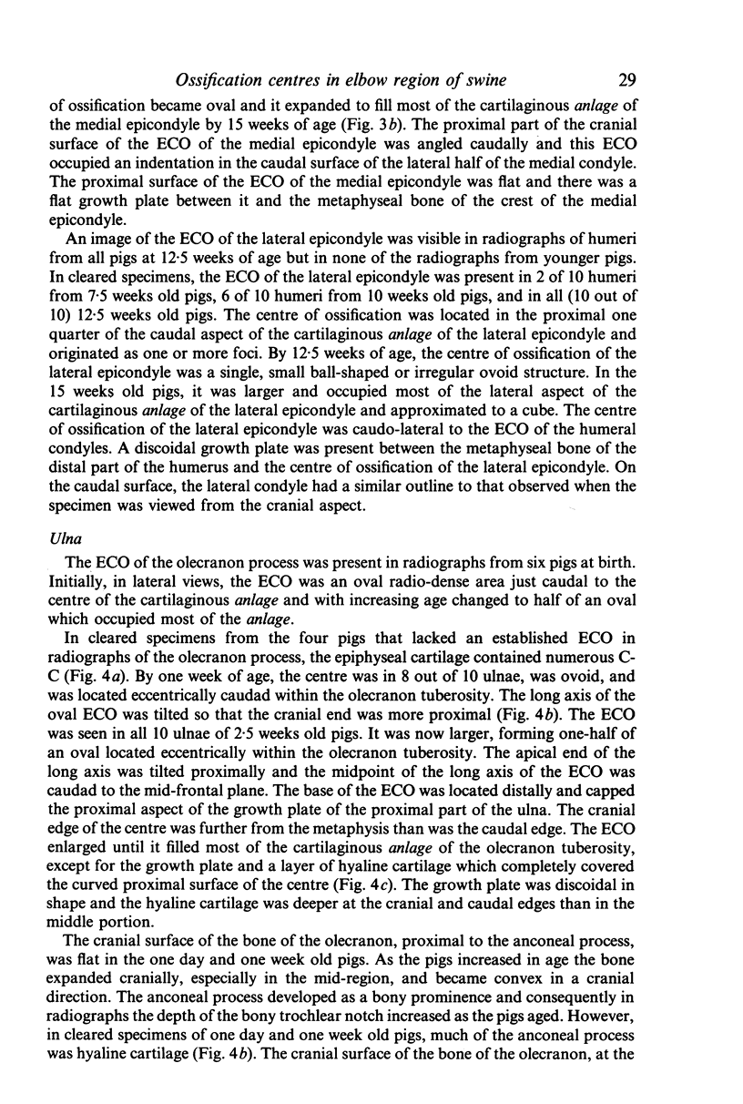
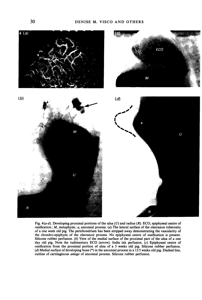
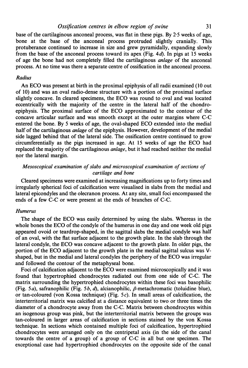
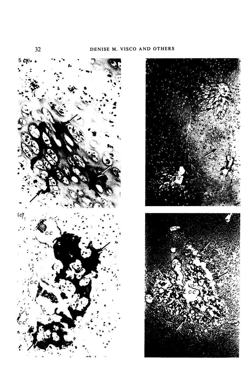
Epiphyseal centres of ossification in the bones forming the elbow joints of pigs between one day and 15 weeks of age were examined radiographically, macroscopically, mesoscopically and microscopically. Thoracic limbs from 39 pigs were perfused with India ink or silicone rubber injection compound and the bones were dissected free of soft tissues. The humerus, ulna and radius were fixed in formalin or ethyl alcohol and then cleared by the modified Spalteholz technique. Bones were radiographed, examined grossly, and then cut into slabs for mesoscopical evaluation. Foci considered to be calcifying within cartilaginous anlage were selected for microscopical examination. It was concluded that the epiphyseal centre of ossification develops at different times in different sites in the bones forming the elbow joint. Centres of ossification are initiated when foci of chondrocytes adjacent to one side of a cartilage canal undergo hypertrophy and the inter-territorial matrix becomes calcified. Osteogenesis then proceeds in the calcified focus, presumably with osteoprogenitor cells that originate within the cartilage canals. Subsequently, each epiphyseal centre of ossification enlarges by one of two methods. Firstly, the layer of cartilage adjacent to the centre undergoes endochondral ossification, thus allowing for the circumferential growth of the epiphyseal centre of ossification. Secondly, foci of calcification develop adjacent to the ends of cartilage canals near the epiphyseal centre of ossification and eventually the focus of calcification coalesces with the developing epiphyseal centre of ossification, thus establishing a new ossification front. Endochondral ossification continues at the periphery of the mass of bone. Mesoscopical examination is more useful than radiographical evaluation for identifying small foci of calcification which precede epiphyseal centres of ossification.
Full text
PDF
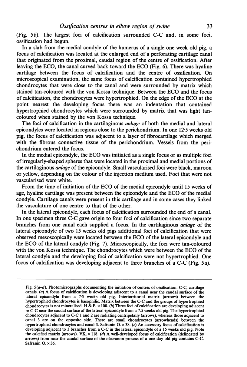
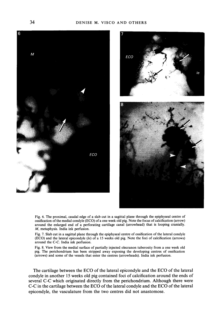
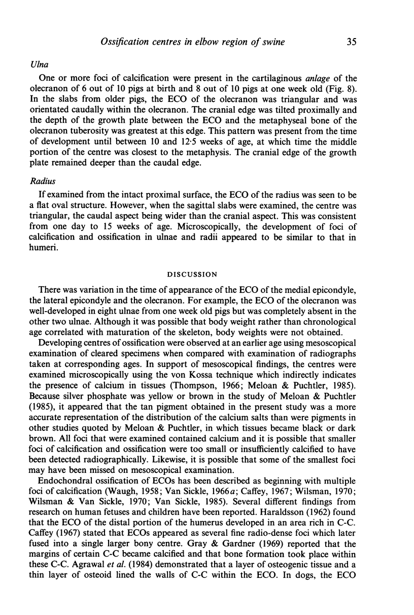
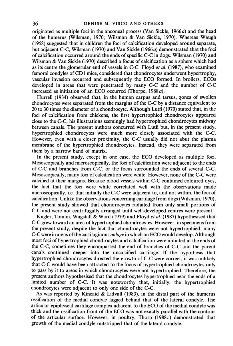
Images in this article
Selected References
These references are in PubMed. This may not be the complete list of references from this article.
- Agrawal P., Atre P. R., Kulkarni D. S. The role of cartilage canals in the ossification of the talus. Acta Anat (Basel) 1984;119(4):238–240. doi: 10.1159/000145891. [DOI] [PubMed] [Google Scholar]
- Brown M. P., MacCallum F. J. Anconeal process of ulna: separate centre of ossification in the horse. Br Vet J. 1974 Sep-Oct;130(5):434–439. doi: 10.1016/s0007-1935(17)35785-8. [DOI] [PubMed] [Google Scholar]
- Cole A. A., Wezeman F. H. Perivascular cells in cartilage canals of the developing mouse epiphysis. Am J Anat. 1985 Oct;174(2):119–129. doi: 10.1002/aja.1001740203. [DOI] [PubMed] [Google Scholar]
- Dorling J. "Critical electrolyte concentration" method in histochemistry. J Med Lab Technol. 1969 Apr;26(2):124–130. [PubMed] [Google Scholar]
- Floyd W. E., 3rd, Zaleske D. J., Schiller A. L., Trahan C., Mankin H. J. Vascular events associated with the appearance of the secondary center of ossification in the murine distal femoral epiphysis. J Bone Joint Surg Am. 1987 Feb;69(2):185–190. [PubMed] [Google Scholar]
- Frost H. M. A chondral modeling theory. Calcif Tissue Int. 1979 Nov 6;28(3):181–200. doi: 10.1007/BF02441236. [DOI] [PubMed] [Google Scholar]
- Gray D. J., Gardner E. The prenatal development of the human humerus. Am J Anat. 1969 Apr;124(4):431–445. doi: 10.1002/aja.1001240403. [DOI] [PubMed] [Google Scholar]
- HARALDSSON S. The vascular pattern of a growing and fullgrown human epiphysis. Acta Anat (Basel) 1962;48:156–167. doi: 10.1159/000141835. [DOI] [PubMed] [Google Scholar]
- Haines R. W. Cartilage Canals. J Anat. 1933 Oct;68(Pt 1):45–64. [PMC free article] [PubMed] [Google Scholar]
- Hurrell D. J. The Vascularisation of Cartilage. J Anat. 1934 Oct;69(Pt 1):47–61. [PMC free article] [PubMed] [Google Scholar]
- Kincaid S. A., Lidvall E. R. Observations on the postnatal morphogenesis of the porcine humeral condyle and the pathogenesis of osteochondrosis. Am J Vet Res. 1983 Nov;44(11):2095–2103. [PubMed] [Google Scholar]
- Kincaid S. A., Lidvall E. R. Postnatal morphogenesis of the anconeal process of the ulna of swine. Am J Vet Res. 1981 Dec;42(12):2158–2161. [PubMed] [Google Scholar]
- Kugler J. H., Tomlinson A., Wagstaff A., Ward S. M. The role of cartilage canals in the formation of secondary centres of ossification. J Anat. 1979 Oct;129(Pt 3):493–506. [PMC free article] [PubMed] [Google Scholar]
- LEVENE C. THE PATTERNS OF CARTILAGE CANALS. J Anat. 1964 Oct;98:515–538. [PMC free article] [PubMed] [Google Scholar]
- Lutfi A. M. Mode of growth, fate and functions of cartilage canals. J Anat. 1970 Jan;106(Pt 1):135–145. [PMC free article] [PubMed] [Google Scholar]
- Thorp B. H., Duff S. R. Effect of unilateral weight-bearing on pelvic limb development in broiler fowls: vascular studies. Res Vet Sci. 1988 Mar;44(2):164–174. [PubMed] [Google Scholar]
- Thorp B. H. Vascular pattern of the developing intertarsal joint in the domestic fowl. Res Vet Sci. 1988 Jan;44(1):100–111. [PubMed] [Google Scholar]
- Thorp B. H. Vascular pattern of the developing knee joint in the domestic fowl. Res Vet Sci. 1988 Jan;44(1):89–99. [PubMed] [Google Scholar]
- Visco D. M., Van Sickle D. C., Hill M. A., Kincaid S. A. The vascular supply of the chondro-epiphyses of the elbow joint in young swine. J Anat. 1989 Apr;163:215–229. [PMC free article] [PubMed] [Google Scholar]
- WAUGH W. The ossification and vascularisation of the tarsal navicular and their relation to Köhler's disease. J Bone Joint Surg Br. 1958 Nov;40-B(4):765–777. doi: 10.1302/0301-620X.40B4.765. [DOI] [PubMed] [Google Scholar]
- Wilsman N. J., Van Sickle D. C. The relationship of cartilage canals to the initial osteogenesis of secondary centers of ossification. Anat Rec. 1970 Nov;168(3):381–391. doi: 10.1002/ar.1091680305. [DOI] [PubMed] [Google Scholar]


