

Abstract
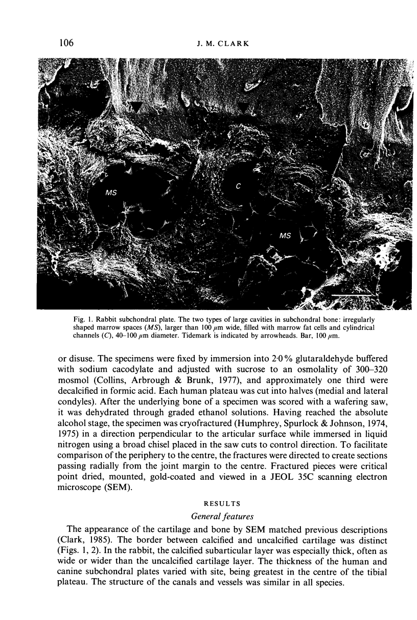
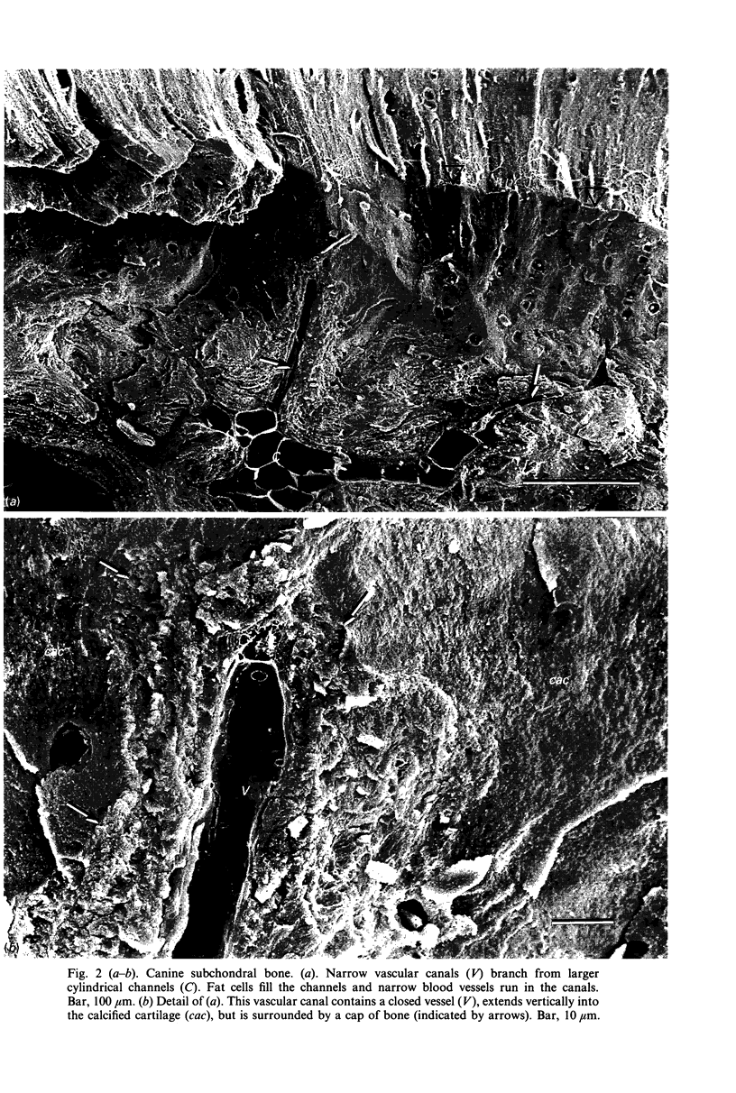
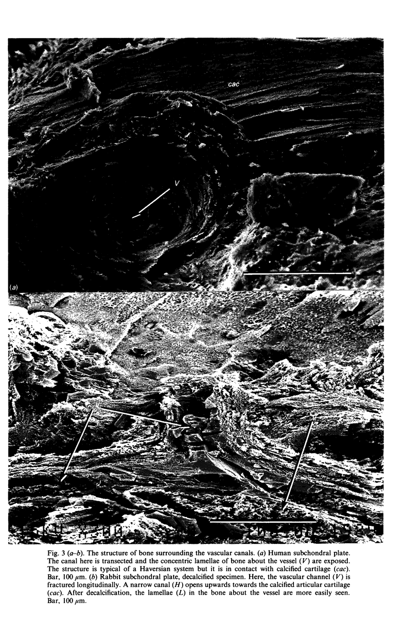
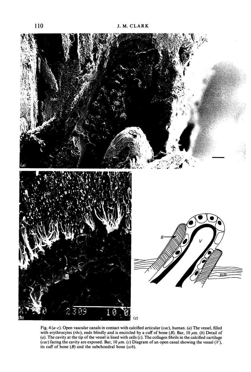
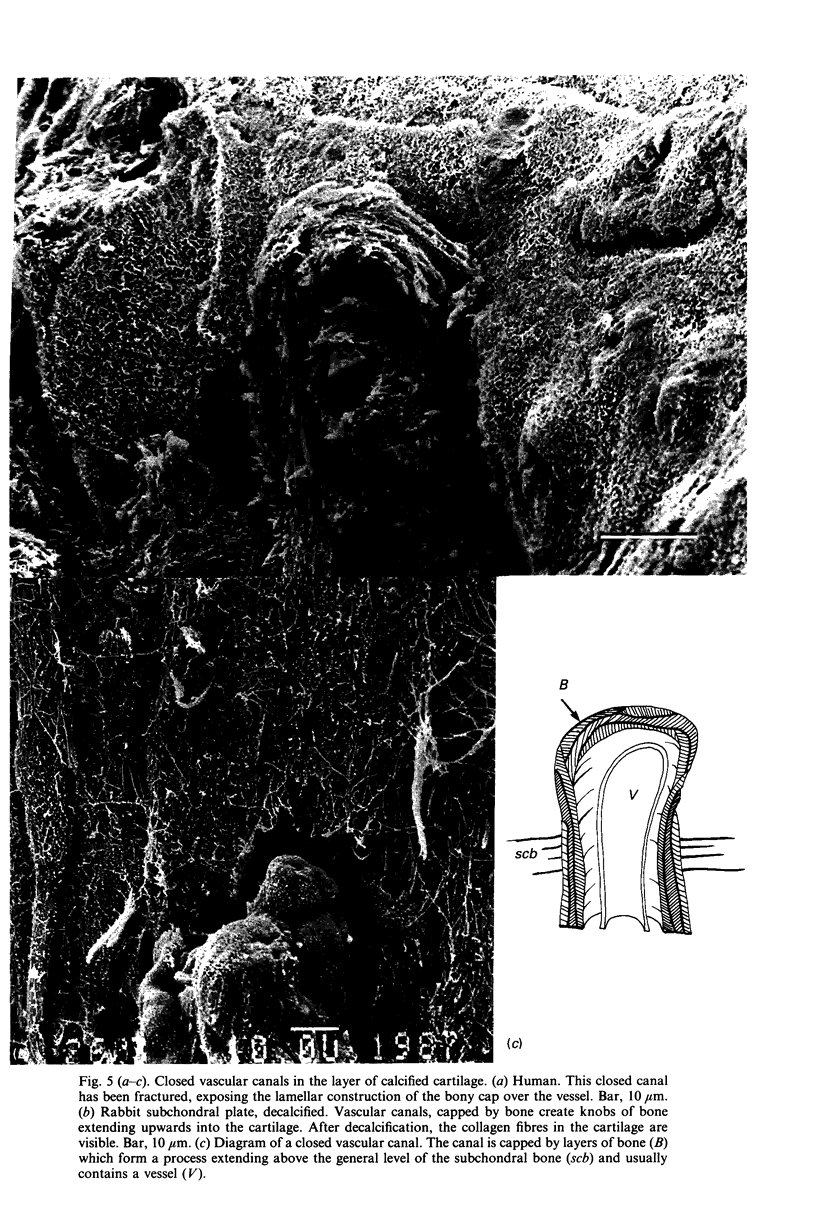
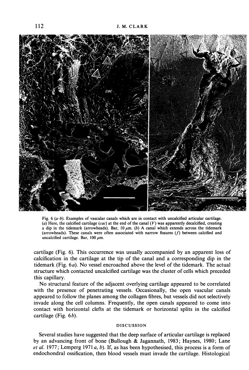
Human, rabbit and canine articular cartilage was prepared for SEM by fixation in isosmolal glutaraldehyde and freeze-fracture following dehydration. These techniques produced clear images of the bone, marrow and vessels in the subchondral region. Generally, cavities larger than 40 microns contained elements typical of marrow. Capillaries ran through the bone in cylindrical channels 20-40 microns wide. These channels were surrounded by concentric lamellae of bone and were in all respects Haversian canals within osteons. A minority of these channels opened into the calcified articular cartilage and there were preceded by cells which appeared to be cutting into the cartilage. Most vascular channels, however, were separated from the cartilage by a layer of bone. We conclude that the vessels within subchondral bone are present primarily to supply the bone through a network of mature osteons.
Full text
PDF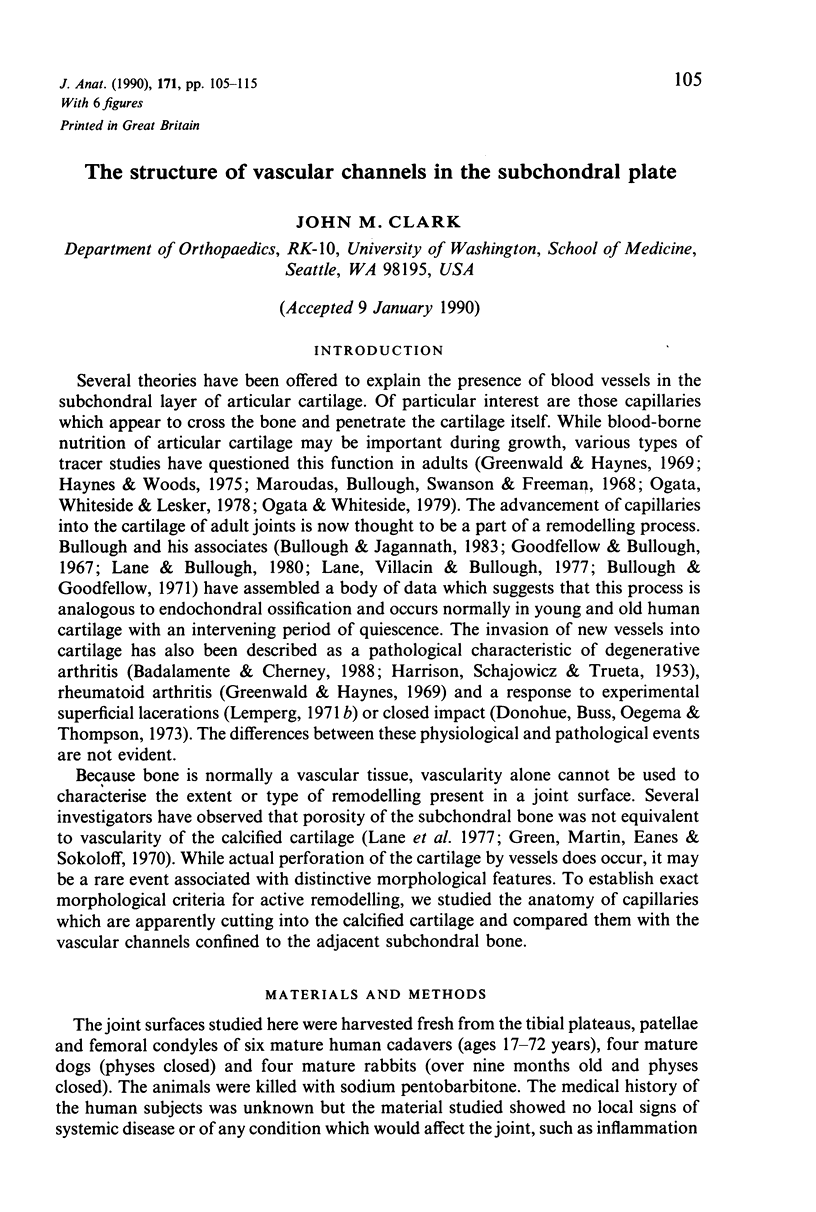

Images in this article


Selected References
These references are in PubMed. This may not be the complete list of references from this article.
- Bullough P. G., Jagannath A. The morphology of the calcification front in articular cartilage. Its significance in joint function. J Bone Joint Surg Br. 1983 Jan;65(1):72–78. doi: 10.1302/0301-620X.65B1.6337169. [DOI] [PubMed] [Google Scholar]
- Clark J. M. The organization of collagen in cryofractured rabbit articular cartilage: a scanning electron microscopic study. J Orthop Res. 1985;3(1):17–29. doi: 10.1002/jor.1100030102. [DOI] [PubMed] [Google Scholar]
- Collins V. P., Arborgh B., Brunk U. A comparison of the effects of three widely used glutaraldehyde fixatives on cellular volume and structure. A TEM, SEM, Volumetric and Cytochemical Study. Acta Pathol Microbiol Scand A. 1977 Mar;85A(2):157–168. doi: 10.1111/j.1699-0463.1977.tb00413.x. [DOI] [PubMed] [Google Scholar]
- Donohue J. M., Buss D., Oegema T. R., Jr, Thompson R. C., Jr The effects of indirect blunt trauma on adult canine articular cartilage. J Bone Joint Surg Am. 1983 Sep;65(7):948–957. [PubMed] [Google Scholar]
- Duncan H., Jundt J., Riddle J. M., Pitchford W., Christopherson T. The tibial subchondral plate. A scanning electron microscopic study. J Bone Joint Surg Am. 1987 Oct;69(8):1212–1220. [PubMed] [Google Scholar]
- Fisher D. E., Bickel W. H., Holley K. E. Histologic demonstration of fat emboli in aseptic necrosis associated with hypercortisonism. Mayo Clin Proc. 1969 Apr;44(4):252–259. [PubMed] [Google Scholar]
- Goodfellow J. W., Bullough P. G. The pattern of ageing of the articular cartilage of the elbow joint. J Bone Joint Surg Br. 1967 Feb;49(1):175–181. [PubMed] [Google Scholar]
- Green W. T., Jr, Martin G. N., Eanes E. D., Sokoloff L. Microradiographic study of the calcified layer of articular cartilage. Arch Pathol. 1970 Aug;90(2):151–158. [PubMed] [Google Scholar]
- Greenwald A. S., Haynes D. W. A pathway for nutrients from the medullary cavity to the articular cartilage of the human femoral head. J Bone Joint Surg Br. 1969 Nov;51(4):747–753. [PubMed] [Google Scholar]
- HARRISON M. H., SCHAJOWICZ F., TRUETA J. Osteoarthritis of the hip: a study of the nature and evolution of the disease. J Bone Joint Surg Br. 1953 Nov;35-B(4):598–626. doi: 10.1302/0301-620X.35B4.598. [DOI] [PubMed] [Google Scholar]
- Havelka S. Evidence of vascularization of osteochondral junctions by a new approach. Acta Univ Carol Med (Praha) 1986;32(5-6):491–498. [PubMed] [Google Scholar]
- Humphreys W. J., Spurlock B. O., Johnson J. S. Transmission electron microscopy of tissue prepared for scanning electron microscopy by ethanol-cryofracturing. Stain Technol. 1975 Mar;50(2):119–125. doi: 10.3109/10520297509117045. [DOI] [PubMed] [Google Scholar]
- Inoue H., Kodama T., Fujita T. Scanning electron microscopy of normal and rheumatoid articular cartilages. Arch Histol Jpn. 1969 Aug;30(5):425–435. doi: 10.1679/aohc1950.30.425. [DOI] [PubMed] [Google Scholar]
- Kawai K., Tamaki A., Hirohata K. Steroid-induced accumulation of lipid in the osteocytes of the rabbit femoral head. A histochemical and electron microscopic study. J Bone Joint Surg Am. 1985 Jun;67(5):755–763. [PubMed] [Google Scholar]
- Lane L. B., Bullough P. G. Age-related changes in the thickness of the calcified zone and the number of tidemarks in adult human articular cartilage. J Bone Joint Surg Br. 1980 Aug;62(3):372–375. doi: 10.1302/0301-620X.62B3.7410471. [DOI] [PubMed] [Google Scholar]
- Lane L. B., Villacin A., Bullough P. G. The vascularity and remodelling of subchondrial bone and calcified cartilage in adult human femoral and humeral heads. An age- and stress-related phenomenon. J Bone Joint Surg Br. 1977 Aug;59(3):272–278. doi: 10.1302/0301-620X.59B3.893504. [DOI] [PubMed] [Google Scholar]
- Lemperg R. The subchondral bone plate of the femoral head in adult rabbits. I. Spontaneus remodelling studied by microradiography and tetracycline labelling. Virchows Arch A Pathol Pathol Anat. 1971;352(1):1–13. doi: 10.1007/BF00549758. [DOI] [PubMed] [Google Scholar]
- Lemperg R. The subchondral bone plate of the femoral head in adult rabbits. II. Changes induced by intracartilaginous defects studied by microradiography and tetracycline labelling. Virchows Arch A Pathol Pathol Anat. 1971;352(1):14–25. doi: 10.1007/BF00549759. [DOI] [PubMed] [Google Scholar]
- Lester K. S., Ash M. M., Jr, Lillie J. H. Condylar cartilage: a scanning electron microscope study of anorganic mammalian condyles. J Prosthet Dent. 1981 Apr;45(4):422–429. doi: 10.1016/0022-3913(81)90105-0. [DOI] [PubMed] [Google Scholar]
- Maroudas A., Bullough P., Swanson S. A., Freeman M. A. The permeability of articular cartilage. J Bone Joint Surg Br. 1968 Feb;50(1):166–177. [PubMed] [Google Scholar]
- Meachim G., Bentley G. Horizontal splitting in patellar articular cartilage. Arthritis Rheum. 1978 Jul-Aug;21(6):669–674. doi: 10.1002/art.1780210610. [DOI] [PubMed] [Google Scholar]
- Ogata K., Whiteside L. A. Barrier to material transfer at the bone-cartilage interface: measurement with hydrogen gas in vivo. Clin Orthop Relat Res. 1979 Nov-Dec;(145):273–276. [PubMed] [Google Scholar]
- Ogata K., Whiteside L. A., Lesker P. A. Subchondral route for nutrition to articular cartilage in the rabbit. Measurement of diffusion with hydrogen gas in vivo. J Bone Joint Surg Am. 1978 Oct;60(7):905–910. [PubMed] [Google Scholar]