Abstract
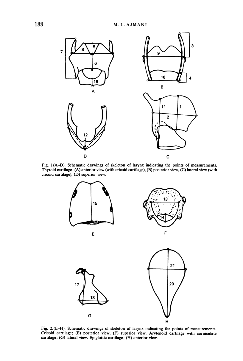
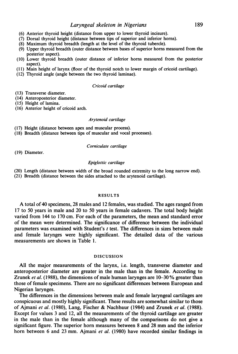
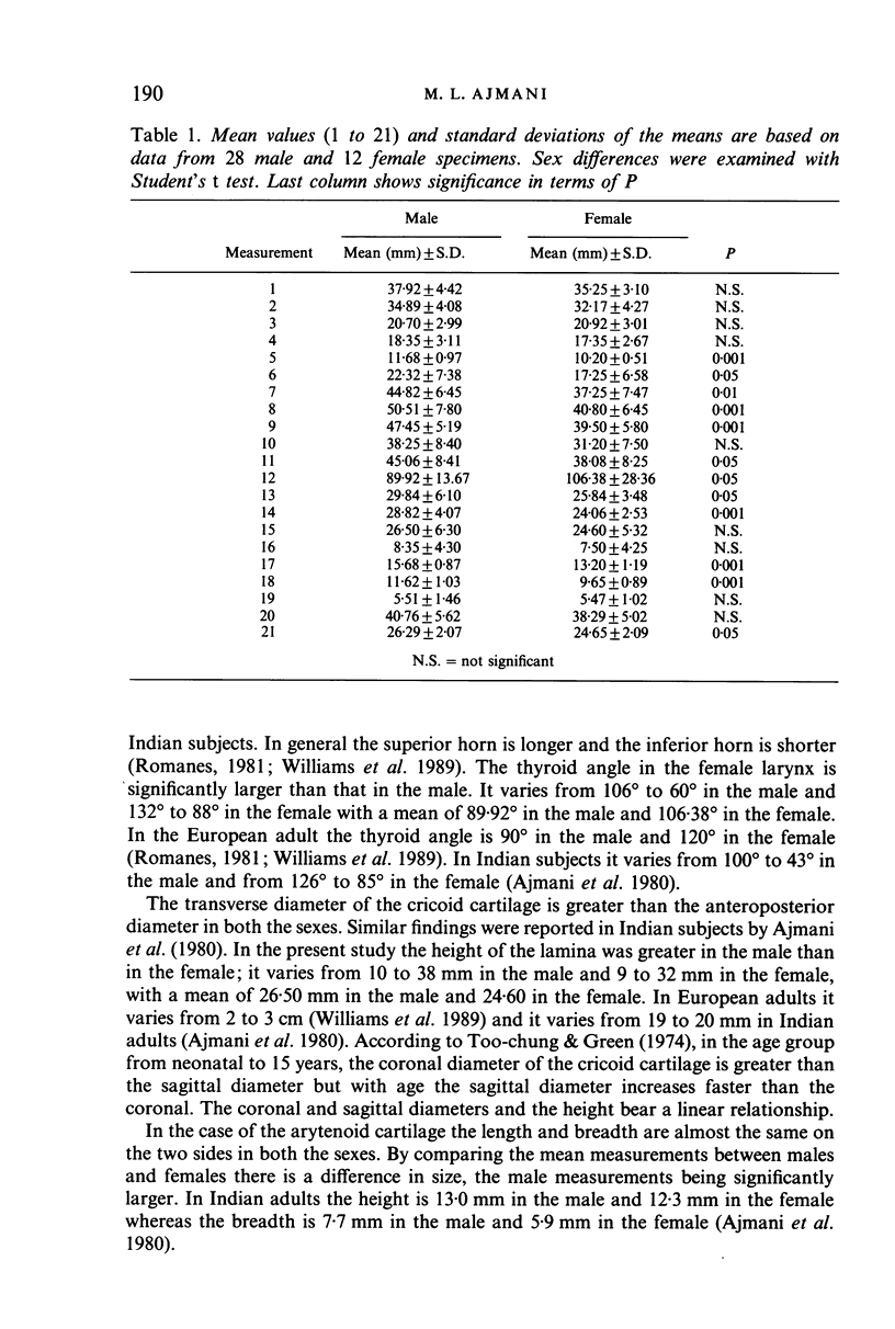
Laryngeal cartilages were studied in 40 dissection room specimens of adult age groups ranging from 17 to 50 years in both the sexes. Various dimensions of the laryngeal skeleton were measured and statistical analysis of the data for male and female were evaluated separately. Conspicuous and highly significant differences of the dimensions between male and female laryngeal cartilages were observed. The incidence of the cuneiform cartilage and cartilago triticea was greater in the female than in the male.
Full text
PDF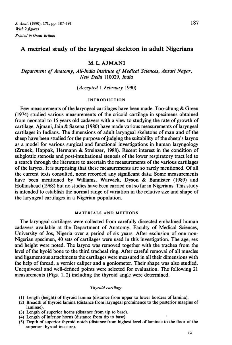
Selected References
These references are in PubMed. This may not be the complete list of references from this article.
- Ajmani M. L., Jain S. P., Saxena S. K. A metrical study of laryngeal cartilages and their ossification. Anat Anz. 1980;148(1):42–48. [PubMed] [Google Scholar]
- Lang J., Fischer K., Nachbaur S. Uber Masse, Form und Formvarianten der Cartilagines thyreoidea et cricoidea. Gegenbaurs Morphol Jahrb. 1984;130(5):639–657. [PubMed] [Google Scholar]
- Negus V. E. Function of the Cartilages of Santorini. J Anat. 1929 Jul;63(Pt 4):430–433. [PMC free article] [PubMed] [Google Scholar]
- Too-Chung M. A., Green J. R. The rate of growth of the cricoid cartilage. J Laryngol Otol. 1974 Jan;88(1):65–70. doi: 10.1017/s0022215100078348. [DOI] [PubMed] [Google Scholar]
- Zrunek M., Happak W., Hermann M., Streinzer W. Comparative anatomy of human and sheep laryngeal skeleton. Acta Otolaryngol. 1988 Jan-Feb;105(1-2):155–162. doi: 10.3109/00016488809119460. [DOI] [PubMed] [Google Scholar]


