Abstract
In human somatic cells, homologous recombination is a rare event. To facilitate the targeted modification of the genome for research and gene therapy applications, efforts should be directed toward understanding the molecular mechanisms of homologous recombination in human cells. Although human genes homologous to members of the RAD52 epistasis group in yeast have been identified, no genes have been demonstrated to play a role in homologous recombination in human cells. Here, we report that RAD54B plays a critical role in targeted integration in human cells. Inactivation of RAD54B in a colon cancer cell line resulted in severe reduction of targeted integration frequency. Sensitivity to DNA-damaging agents and sister-chromatid exchange were not affected in RAD54B-deficient cells. Parts of these phenotypes were similar to those of Saccharomyces cerevisiae tid1/rdh54 mutants, suggesting that RAD54B may be a human homolog of TID1/RDH54. In yeast, TID1/RDH54 acts in the recombinational repair pathway via roles partially overlapping those of RAD54. Our findings provide the first genetic evidence that the mitotic recombination pathway is functionally conserved from yeast to humans.
Keywords: homologous recombination/human cells/mitosis/sister-chromatid exchange/targeted integration
Introduction
Gene targeting experiments in the chicken B-cell line DT40 and mouse embryonic stem (ES) cells have contributed to our understanding of the molecular mechanisms of homologous recombination in higher eukaryotes (Morrison and Takeda, 2000; Khanna and Jackson, 2001; Thompson and Schild, 2001). The reduced frequency of targeted integration and hypersensitivity to ionizing radiation in RAD54 null cells have indicated that RAD54 function is conserved from Saccharomyces cerevisiae to mice (Bezzubova et al., 1997; Essers et al., 1997). In contrast, RAD51 and RAD52 functions are not conserved throughout evolution. RAD51 in higher eukaryotes is essential for cell viability, while yeast RAD51 is not (Lim and Hasty, 1996; Tsuzuki et al., 1996). RAD52 does not affect radiosensitivity in higher eukaryotes, while inactivation of RAD52 in yeast results in hypersensitivity to ionizing radiation (Rijkers et al., 1998; Yamaguchi-Iwai et al., 1998). Thus, knockout experiments in DT40 and mouse ES cells have revealed that the functions of recombination genes are not always conserved from S.cerevisiae to mice, although their structures are well conserved throughout evolution, with the exception of RAD52, in which the homology is concentrated in the N-terminal part.
Despite the successful genetic manipulation of genomes of interest in DT40 and mouse ES cells, such manipulation has rarely been achieved in human cells (Yáñez and Porter, 1998; Sedivy and Dutriaux, 1999). The major obstacle to this is the low frequency of homologous recombination in humans. To improve this frequency, it is essential to understand the mechanisms of homologous recombination in human cells. It is possible that functions of recombination genes are not always conserved among higher eukaryotes. For this reason, gene targeting of these genes in human cells is indispensable.
Human RAD54B was isolated by screening a testis cDNA library with an expressed sequence-tagged (EST) probe homologous to S.cerevisiae RAD54 (Hiramoto et al., 1999). The gene encodes a protein containing ATPase domains that are highly conserved in members of the SWI2/SNF2 superfamily, including RAD54. The N-terminal half of RAD54B shares significant similarity with a yeast recombination gene TID1/RDH54, but not with RAD54, suggesting that RAD54B may be a mammalian homolog of TID1/RDH54. Consistent with its putative role in recombination, Rad54B forms a protein complex with Rad51, Rad54 and Brca1 (Tanaka et al., 2000). Rad51 is recruited to sites of DNA damage, indicating that this complex plays a role in recombinational repair of DNA damage (Tashiro et al., 2000).
To clarify the role of RAD54B in homologous recombination in humans, we generated RAD54B-deficient colon cancer cell lines by sequential gene targeting. The frequency of targeted integration in these cells was dramatically reduced compared with that in RAD54B-expressing cells. Unlike chicken and mouse mutants of other recombination genes, RAD54B mutants did not show hypersensitivity to DNA-damaging agents (Luo et al., 1999; Yamaguchi-Iwai et al., 1999; Deans et al., 2000; Takata et al., 2000, 2001). Sister-chromatid exchange (SCE), which was suppressed in RAD54-deficient DT40 and mouse ES cells (Sonoda et al., 1999; Dronkert et al., 2000), was not affected in RAD54B mutants. These phenotypes partially overlapped those of S.cerevisiae tid1/rdh54 mutants (Klein, 1997; Shinohara et al., 1997), suggesting that RAD54B may be a human homolog of TID1/RDH54. Thus, this system should contribute to our understanding of recombination in humans.
Results
Targeting of the RAD54B locus in HCT116 cells
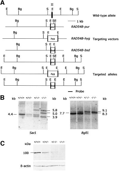
Puromycin-resistant colonies generated with the RAD54B-pur vector were screened for targeted integration by Southern blot analysis. Insertion of the pur gene into the locus was expected to give 3.9 kb SacI and 8.3 kb BglII fragments, in addition to 4.4 kb SacI and 7.7 kb BglII wild-type fragments (Figure 1A). Two of 25 puromycin-resistant clones showed the targeted bands (Figure 1B). The RAD54B-hyg vector was introduced into one of these clones, RAD54B+/+/–p37. Hygromycin-resistant clones were screened for targeted integration. Insertion of the hyg gene into the locus was expected to give 5.8 kb SacI and 9.1 kb BglII fragments (Figure 1A). Nine of 61 hygromycin-resistant RAD54B+/+/–p37 subclones showed the targeting bands without removing the pur gene (Figure 1B). After double knockout, the wild-type band was still present, suggesting that HCT116 cells harbor at least three alleles of RAD54B. The RAD54B-bsd vector was then introduced into one of these clones, RAD54B+/–/–h9. Blasticidin-resistant clones were screened for targeted integration. Insertion of the bsd gene into the locus was expected to give 5.0 kb SacI and 8.3 kb BglII fragments (Figure 1A). Two of 78 blasticidin-resistant RAD54B+/–/–h9 subclones showed the targeted band without removing the pur and hyg genes (Figure 1B). After triple knockout, the wild-type band disappeared. Western blot analysis revealed that the levels of Rad54B protein correlated with targeting events (Figure 1C).
Fig. 1. Gene targeting at the human RAD54B locus. (A) Schematic representation of the RAD54B locus, the targeting vectors and the targeted locus. Relevant EcoRI (E), SacI (S) and BglII (Bg) restriction sites, and the position of the probe used for Southern blot analysis are shown. Exon II is indicated by a numbered solid box. (B) Southern blot analysis of DNA from HCT116 cells digested with SacI or BglII. Both blots were hybridized with the probe indicated in (A). (C) Western blot analysis of whole-cell extracts from HCT116 cells. The blot was incubated with a 1:200 dilution of anti-Rad54B antiserum.
Reduced frequencies of targeted integration in RAD54B-deficient cells
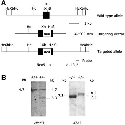
To examine the role of RAD54B in targeted integration in HCT116 cells, frequencies of targeted integration at the XRCC2 and RAD51L2/RAD51C loci were analyzed. The XRCC2-neo vector was constructed from isogenic DNA. Targeting at the locus was expected to give 3.3 kb HincII and 8.2 kb XbaI fragments, in addition to 4.7 kb HincII and 7.3 kb XbaI wild-type fragments (Figure 2). The frequency of targeted integration in wild-type HCT116 cells was 6.4%. In single- and double-knockout cells, the frequencies were 7.5 and 8.4%, respectively. No significant difference in the frequency of targeted integration was observed between these cell lines. For measurement of the frequency in triple-knockout cells, two independent cell lines, RAD54B–/–/–b125 and RAD54B–/–/–b140, were examined. The frequencies of targeted integration at the XRCC2 locus in these RAD54B-deficient cells were 0.6 and 0%, respectively, values that were reduced >10-fold compared with those for the wild-type cells (Table I). The difference in targeted integration between the wild-type and RAD54B-deficient cells was statistically significant. To prove that the phenotype of RAD54B-deficient cells was caused by the RAD54B mutation, a complementation experiment was performed. RAD54B–/–/–b125 cells were electroporated with a mammalian expression vector expressing the wild-type RAD54B cDNA and the drug-resistant marker zeocin. Cell lines that stably expressed RAD54B were identified from phleomycin-resistant colonies (data not shown). The frequency of targeted integration in the transformed line was 15.0%, a value that was stimulated 25-fold compared with that for RAD54B–/–/–b125. Expression of the wild-type RAD54B cDNA corrected the targeted integration at the XRCC2 locus to a level that was not significantly different from that of the wild-type cells (p = 0.95) but was significantly different from that of RAD54B–/–/–b125 cells (p <0.01) (Table I).
Fig. 2. Gene targeting at the human XRCC2 locus. (A) Schematic representation of the XRCC2 locus, the targeting vector and the targeted locus. Relevant XhoI (Xh), SacI (S), HincII (Hc) and XbaI (Xb) restriction sites, and the position of the probe used for Southern blot analysis are shown. The positions of the primers used for PCR are indicated by arrows. Exon III is indicated by a numbered solid box. (B) Southern blot analysis of DNA from HCT116 cells digested with HincII or XbaI. Both blots were hybridized with the probe indicated in (A).
Table I. Frequency of targeted integration in HCT116 cells.
| HCT116 cells | Targeting construct | |||
|---|---|---|---|---|
| |
XRCC2-neo (%)a |
p value relative to parent |
RAD51L2-neo (%)a |
p value relative to parent |
| RAD54B+/+/+ | 6.4 (6/94) | – | 0.083 (6/7197) | – |
| RAD54B+/+/–p37 | 7.5 (7/93) | 0.49 | 0.101 (6/5948) | 0.48 |
| RAD54B+/–/–h9 | 8.4 (14/166) | 0.37 | 0.108 (7/6509) | 0.43 |
| RAD54B–/–/–b125 | 0.6 (1/173) | 8.5 × 10⁻³ | 0.010 (1/10382) | 2.1 × 10⁻² |
| RAD54B–/–/–b140 | 0 (0/120) | 6.6 × 10⁻³ | 0 (0/7398) | 1.4 × 10⁻² |
| RAD54B–/–/–b125 + RAD54B-zeo | 15.0 (3/20) | 0.95 | ND | ND |
aThe frequency of targeted integration is shown as a percentage of correctly targeted clones relative to the total number of drug-resistant clones analyzed; absolute numbers are given in parentheses. The difference in frequency between RAD54B–/–/–b125 and RAD54B–/–/–b125 + RAD54B-zeo for XRCC2-neo was statistically significant (p = 3.6 × 10⁻³). ND, not determined. p values were calculated using Fisher’s exact test.
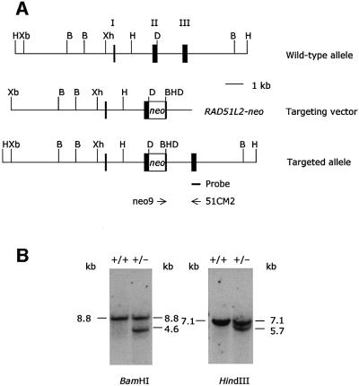
In contrast to XRCC2, targeted integration at the RAD51L2/RAD51C locus with the RAD51L2-neo vector was extremely low, partly because the vector was constructed from non-isogenic DNA. Targeting at the RAD51L2/RAD51C locus was expected to give 4.6 kb BamHI and 5.7 kb HindIII fragments, in addition to 8.8 kb BamHI and 7.1 kb wild-type HindIII fragments (Figure 3). The frequency in the wild type was 0.083%. In single- and double-knockout cells, the frequencies were 0.101 and 0.108%, respectively. No significant difference in the frequency of targeted integration was observed between these cell lines. In contrast, the frequencies were 0.010 and 0% in RAD54B-deficient cells, values that were reduced >8-fold compared with those for the wild-type cells (Table I). The difference in targeted integration between the wild-type and RAD54B-deficient cells was statistically significant.
Fig. 3. Gene targeting at the human RAD51L2/RAD51C locus. (A) Schematic representation of the RAD51L2/RAD51C locus, the targeting vector and the targeted locus. Relevant XhoI (Xh), XbaI (Xb), DraIII (D), BamHI (B) and HindIII (H) restriction sites, and the position of the probe used for Southern blot analysis are shown. The positions of the primers used for PCR are indicated by arrows. Exons I, II and III are indicated by numbered solid boxes. (B) Southern blot analysis of DNA from HCT116 cells digested with BamHI or HindIII. Both blots were hybridized with the probe indicated in (A).
No effect of RAD54B on cell growth, cell survival or SCE
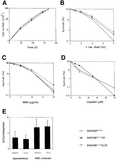
The growth of RAD54B-deficient cells was similar to that of wild-type and RAD54B+/–/– cells (Figure 4A). Sensitivity to DNA-damaging agents was monitored by the ability to form colonies after irradiation, treatment with methyl methanesulfate (MMS) or cisplatin. Cell survival assays revealed that the sensitivity of RAD54B-deficient cells to these agents was similar to that of wild-type and RAD54B+/–/– cells (Figure 4B–D). Compared with wild-type cells, levels of spontaneous and mitomycin C (MMC)-induced SCE were not changed in RAD54B-deficient cells (Figure 4E). No significant difference in karyotypes was observed among the original cell line, and the single-, double- and triple-knockout lines (data not shown).
Fig. 4. Proliferative characteristics, sensitivity to DNA-damaging agents and levels of SCE of wild-type and targeted HCT116 cells. (A) Growth curves. (B) Sensitivity to ionizing radiation. (C) Sensitivity to MMS. (D) Sensitivity to cisplatin. All measurements in growth curves and cell survival were performed in triplicate. Results of representative experiments are shown here. Consistent results were obtained between different sets of experiments. (E) Levels of SCE. The RAD54B-deficient cell line used in this study was RAD54B–/–/–b125. One hundred cells were analyzed in each preparation. The mean number of SCEs per metaphase and the standard deviation are shown.
Discussion
The present work demonstrates that RAD54B plays a critical role in targeted integration in human cells without affecting cell growth, cell survival to DNA-damaging agents or SCE. Since RAD54B shares structural similarity with S.cerevisiae TID1/RDH54 not only in ATPase domains but also in the N-terminal region, it is probable that RAD54B is a human homolog of TID1/RDH54. The properties of the rdh54 single mutants and the rad54 tid1/rdh54 double mutants in mitosis and meiosis have already been characterized (Klein, 1997; Shinohara et al., 1997). The yeast mutants did not show hypersensitivity to MMS. The TID1/RDH54 gene was required for interchromosomal recombination but not for intrachromosomal gene conversion in mitosis. This phenotype was observed in the tid1/rdh54 single mutant (Klein, 1997), whereas elsewhere it was reported that the tid1/rdh54 single mutant did not show deficiency in mitotic recombination (Shinohara et al., 1997). Interchromosomal recombination at the HIS4 locus was reduced in the rad54 tid1/rdh54 double mutant but not in the tid1/rdh54 single mutant, suggesting that TID1/RDH54 and RAD54 act in the recombinational repair pathway with partially overlapping roles. In meiosis, the tid/rdh54 mutant exhibited significant defects in sporulation, spore viability and recombination.
Although few comparative experiments have been performed between RAD54B-deficient human cells and the tid1/rdh54 mutants, there are a couple of similarities between these mutants. First, in contrast to RAD54 mutants in S.cerevisiae (Kanaar et al., 1996), chickens (Bezzubova et al., 1997) and mice (Essers et al., 1997), neither of these mutants showed hypersensitivity to MMS. Secondly, both mutants were defective in recombination, although the respective experiments were not comparable. The effect of TID1/RDH54 on targeted integration, which is reduced in RAD54 mutants in mice (Essers et al., 1997), chickens (Bezzubova et al., 1997), Schizosaccharomyces pombe (Muris et al., 1997) and S.cerevisiae (Arbel et al., 1999) as well as in RAD54B mutants, has not been reported. Interchromosomal recombination has been studied in tid/rdh54 mutants. Although the relationship between targeted integration and interchromosomal recombination has not been established, they may share some similarity, since both use homologous sequences that are not present on the same chromosome. Since the roles of RAD54B in meiosis and the overlapping roles of RAD54B and RAD54 in humans remain to be demonstrated, we are not able to conclude that RAD54B is the functional homolog of the yeast TID1/RDH54 gene. However, our findings indicate that some, if not all, roles in the recombinational repair pathway in mitosis might be conserved between RAD54B and TID1/RDH54.
In the mitotic cell cycle of yeast, RAD54 is essential for repair by the sister chromatid, whereas TID1/RDH54 is not required for this pathway (Arbel et al., 1999). The role for RAD54 in sister-chromatid-based repair may be conserved from yeast to higher eukaryotes. RAD54-deficient DT40 cells have been shown to be extremely sensitive to ionizing radiation in G2 as well as G1, while wild-type DT40 cells were resistant to irradiation in G2 (Takata et al., 1998). The sister chromatids serve as the main template for recombination in G2. Therefore, hypersensitivity to irradiation of the RAD54 mutant in G2 implies that RAD54 is required for DNA repair mediated by sister chromatids. Consistent with this idea, DNA damage-induced SCE was reduced in RAD54-deficient DT40 and mouse ES cells (Sonoda et al., 1999; Dronkert et al., 2000). These phenotypes of RAD54 mutants were not observed in either human RAD54B-deficient cells or yeast tid1/rdh54 mutants, supporting the idea that RAD54 acts in homologous recombination in a pathway distinct from that of RAD54B. However, given that both mutations in mammals show synergic defects in recombination, RAD54B and RAD54 may act in the recombinational pathway with partially overlapping roles.
Rad54B forms a protein complex with Rad51, Rad54 and Brca1 (Tanaka et al., 2000). In RAD54-deficient ES cells, Rad51 focus formation has been shown to be blocked (Tan et al., 1999). In analogy with RAD54, inactivation of RAD54B may result in the reduction of Rad51 focus formation. Another possible scenario for the effect of Rad54B on the protein complex is that Rad54B may affect the colocalization of Rad51 and other proteins that play a role in recombination. In the meiotic cell cycle of yeast tid1/rdh54 mutants, the colocalization of Rad51 and Dmc1 has been shown to be reduced, although Rad51 formed foci (Shinohara et al., 2000). DMC1 is only expressed in meiosis (Bishop et al., 1992). There may be other proteins that interact with Rad51 and Rad54B in the mitotic recombination pathway.
RAD54B plays a unique role in homologous recombination because it does not affect the sensitivity to DNA-damaging agents. With the exception of RAD52, other recombination genes, including RAD51, RAD54, MRE11 and RAD51 paralogs, do affect the sensitivity to such agents. To improve targeted integration in the human genome, it will be important to modify the functions of genes that play a central role in targeted integration without affecting other cellular properties. RAD54B is an excellent candidate for such modification. Targeted integration events were hardly detectable in RAD54B-deficient cells in the present study. In RAD52-deficient ES cells, no such drastic reduction of targeted integration has been reported (Rijkers et al., 1998). Thus, this system provides new insight into genetic manipulation of the human genome.
Materials and methods
Construction of RAD54B targeting vectors
Promoterless targeting vectors were designed to insert drug resistance genes in exon 2 in-frame. The right homology arm was isolated as a 1.2 kb EcoRI fragment from the EMBL3 SP6/T7 human genomic library (Clontech) and inserted into pBluescript SK. A 3′ 130 bp fragment with the EcoRI site was deleted with a combination of exonuclease III and S1 nuclease digestion. The left homology arm was isolated as a 6.4 kb EcoRI fragment from the same library and inserted into the unique EcoRI site of the vector containing the right arm. The 5′ EcoRI site was removed by treatment with exonuclease III and S1 nuclease. A puromycin resistance cassette was amplified from pKO SelectPuro (Lexicon Genetics) with puro-1 (5′-GCGAATTCCATGACCGAGTACAAG-3′) and BGHpA-2 (5′-GCGAATTCGCCTGCTATTGTCTTCC-3′) and inserted into the EcoRI site of the targeting vector. A hygromycin resistance cassette was generated from pcDNA3.1/Hygro (Invitrogen) by PCR. Since the coding sequence for this cassette contains an EcoRI site, PCR was performed with mismatch primers converting the EcoRI to a HindIII site without loss of hygromycin resistance. Upstream of the EcoRI site was amplified with hyg1 (5′-GCCGAATTCGATGAAAAAGCCTGA-3′) and hyg2 (5′-CGGAATTCAAGCTTCCAATGTCAAGC-3′) and inserted into the EcoRI site of the vector containing both arms. Downstream of the EcoRI site was amplified with hyg3 (5′-ATAAGCTTCAGCGAGAGCCTG-3′) and hyg4 (5′-CGAAGCTTTCATTAGGCACC-3′) and inserted into the HindIII site converted from the EcoRI site by hyg2. A blasticidin resistance cassette was amplified from pCMV/Bsd (Invitrogen) with BSD-1 (5′-GCGAATTCCATGGCCAAGCCTTTG-3′) and BSD-2 (5′-CCGGGAATTCAGACATG-3′) and inserted into the EcoRI of the targeting vector. The sequences of all PCR products were confirmed.
Generation of RAD54B-deficient cells
HCT116 cells derived from human colon carcinoma (ATCC CCL-247) were cultured in McCoy’s 5A medium with 10% fetal calf serum. Targeting vectors linearized with KpnI were introduced into 2 × 10⁷ cells using a Bio-Rad Gene Pulsar at 1200 V and 50 µF. For selection of targeted cells, hygromycin, puromycin and blasticidin were added at 200, 1.25 and 20 µg/ml, respectively. After 14 days, colonies were isolated and expanded. Genomic DNA from individual colonies was digested with SacI or BglII and subjected to Southern blot analysis. The blots were hybridized with an external probe 3′ of the targeting constructs. Western blot analysis was performed as described previously (Tanaka et al., 2000).
Expression of the RAD54B cDNA
The human RAD54B cDNA (DDBJ/EMBL/GenBank accession No. AF112481) was cloned into the pcDNA3.1/Zeo(+) mammalian expression vector (Invitrogen) and checked by sequencing. The vector was transfected into RAD54B-deficient cells by electroporation and selected with 240 µg/ml phleomycin (Sigma).
Construction of the XRCC2 targeting vector
A promoterless targeting vector was designed to insert a neomycin resistance cassette at the XhoI site in exon 3 in-frame. The 1.6 kb right homology arm was amplified from isogenic DNA of HCT116 cells with E3-1 (5′-GTAGTCACCCATCTCTCT-3′) and I3-1 (5′-TCTTTGCCTGTGCCTATG-3′) and inserted into pCR2.1 (Invitrogen) by the TA cloning method. A neomycin resistance cassette was amplified from pMC1neo-polyA (Stratagene) with S-X-neo (5′-ATGAGCTCGAGCATGGGATCGGCCATT-3′) and neo8-S (5′-TGAGCTCAAGCTTGGCTGCAGGT-3′) and replaced with a 244 bp exonic fragment flanked with SacI in the right arm vector. The 2.7 kb left homology arm was amplified from the isogenic DNA with I2-1 (5′-CTCCTGGGTTCAACTGAT-3′) and E3-2 (5′-TCTTCTGATGAGCTCGAG-3′) and inserted into pCR2.1. A 56 bp XhoI fragment was replaced with a 2.3 kb XhoI fragment released from the right arm vector containing both the neomycin resistance cassette and the right arm.
Measurement of targeted integration frequencies at the XRCC2 locus
The XRCC2-neo vector was introduced into 2 × 10⁷ wild-type or RAD54B mutant HCT116 cells by electroporation. After 24 h, G418 was added to a final concentration of 400 µg/ml. After selection, DNA was extracted from individual colonies. Targeted integration events were screened by PCR with neo9 (5′-ATCGCCTTCTTGACGAGT-3′) and I3-2 (5′-TTGGCGTCTTCGTCATGA-3′). Southern blot analysis was also used to confirm targeted integration. Genomic DNA was digested with HincII or XbaI and the blots were hybridized with an external probe.
Construction of the RAD51L2/RAD51C targeting vector
A promoterless targeting vector was designed to insert a neomycin resistance cassette at the DraIII site in exon 2 in-frame. A 4.4 kb XhoI fragment containing exons 1 and 2 was isolated from the EMBL3 SP6/T7 human genomic library and inserted into pUC19. A neomycin resistance cassette was amplified from pMC1neo-polyA (Stratagene) with neodras (5′-CGCACCAGGTGCCAATATGGGATCGGCCA-3′) and neodraas (5′-GGCACCTGGTGAAGCTTGGCTGCAGGTC-3′) and inserted at the DraIII site in exon 2. A 5.1 kb XbaI–XhoI fragment containing the promoter region upstream of exon 1 was isolated from the same library and inserted into pBluescript SK. A 5.3 kb XhoI fragment with the neomycin resistance cassette released from the pUC19 vector was inserted into the XhoI site of the pBluescript vector with the upstream region.
Measurement of targeted integration frequencies at the RAD51L2/RAD51C locus
The RAD51L2-neo targeting vector was introduced into 1.5–3.7 × 10⁸ wild-type or RAD54B mutant HCT116 cells by electroporation. After 24 h, G418 was added to a final concentration of 400 µg/ml. After selection, DNA was extracted from pools of 20–100 colonies. Targeted integration events were screened by PCR with neo9 and 51CM2 (5′-TGCACATCTACTGCCAAC-3′). To test for PCR conditions, a control vector that included the annealing site for 51CM2 was constructed and introduced into HCT116 cells. PCR conditions were optimized to detect a single targeted integration event in a pool of 100 negative G418-resistant cells. To confirm the targeting event by Southern blot analysis, single positive clones were obtained from positive pools of G418-resistant colonies by limiting dilution. Their genomic DNA was subjected to Southern blot analysis; the blots were hybridized with a probe outside the targeting vector.
Analysis of cell growth
HCT116 cells were plated at a density of 104 cells per 60 mm dish and cultured. On the days indicated, cells were counted. All measurements were performed in triplicate.
Cell survival assay
HCT116 cells were plated at a density of 103 cells per 60 mm dish and irradiated with a 60Co source or treated with MMS (Sigma). To measure sensitivity to cisplatin (Nihon-Kayaku), cells were incubated in the presence of cisplatin for 1 h and washed twice. The cells were grown for 7 days and, after fixing and staining, colonies were counted. All measurements were performed in triplicate.
Analysis of SCEs
Cells were cultured in 16 µM 5-bromodeoxyuridine (BUdR) for 36 h and pulsed with 0.1 µg/ml colcemid for the last 1 h. To examine MMC-induced SCEs, cells were incubated in the presence of 0.8 µg/ml MMC for 8 h. Harvested cells were treated with 75 mM KCl:1% sodium citrate (4:1) for 20 min and fixed with methanol:acetic acid (3:1). Cells were fixed on slides and incubated with 12.8 µg/ml Hoechst 33258 for 20 min. The slides were irradiated with ultraviolet radiation (λ = 352 nm) for 1 h at 60°C and stained with 4% Giemsa solution. The cell lines were coded to prevent bias in the analysis.
Acknowledgments
Acknowledgements
We thank M.Ohtaki for statistical analysis. This work was supported by Grants-in-Aid from the Ministry of Education, Science, Sports and Culture of Japan.
References
- Arbel A., Zenvirth,D. and Simchen,G. (1999) Sister chromatid-based DNA repair is mediated by RAD54, not by DMC1 or TID1. EMBO J., 18, 2648–2658. [DOI] [PMC free article] [PubMed] [Google Scholar]
- Bezzubova O., Silbergleit,A., Yamaguchi-Iwai,Y., Takeda,S. and Buerstedde,J.-M. (1997) Reduced X-ray resistance and homologous recombination frequencies in a RAD54–/– mutant of the chicken DT40 cell line. Cell, 89, 185–193. [DOI] [PubMed] [Google Scholar]
- Bishop D.K., Park,D., Xu,L. and Kleckner,N. (1992) DMC1: a meiosis-specific yeast homolog of E. coli recA required for recombination, synaptonemal complex formation, and cell cycle progression. Cell, 69, 439–456. [DOI] [PubMed] [Google Scholar]
- Deans B., Griffin,C.S., Maconochie,M. and Thacker,J. (2000) Xrcc2 is required for genetic stability, embryonic neurogenesis and viability in mice. EMBO J., 19, 6675–6685. [DOI] [PMC free article] [PubMed] [Google Scholar]
- Dronkert M.L.G., Beverloo,H.B., Johnson,R.D., Hoeijmakers,J.H.J., Jasin,M. and Kanaar,R. (2000) Mouse RAD54 affects DNA double-strand break repair and sister chromatid exchange. Mol. Cell. Biol., 20, 3147–3156. [DOI] [PMC free article] [PubMed] [Google Scholar]
- Essers J., Hendriks,R.W., Swagemakers,S.M.A., Troelstra,C., de Wit,J., Bootsma,D., Hoeijmakers,J.H.J. and Kanaar,R. (1997) Disruption of mouse RAD54 reduces ionizing radiation resistance and homologous recombination. Cell, 89, 195–204. [DOI] [PubMed] [Google Scholar]
- Hiramoto T. et al. (1999) Mutations of a novel human RAD54 homologue, RAD54B, in primary cancer. Oncogene, 18, 3422–3426. [DOI] [PubMed] [Google Scholar]
- Kanaar R. et al. (1996) Human and mouse homologs of the Saccharomyces cerevisiae RAD54 DNA repair gene: evidence for functional conservation. Curr. Biol., 6, 828–838. [DOI] [PubMed] [Google Scholar]
- Khanna K.K. and Jackson,S.P. (2001) DNA double-strand breaks: signaling, repair and the cancer connection. Nature Genet., 27, 247–254. [DOI] [PubMed] [Google Scholar]
- Klein H.L. (1997) RDH54, a RAD54 homologue in Saccharomyces cerevisiae, is required for mitotic diploid-specific recombination and repair and for meiosis. Genetics, 147, 1533–1543. [DOI] [PMC free article] [PubMed] [Google Scholar]
- Lim D.-S. and Hasty,P. (1996) A mutation in mouse rad51 results in an early embryonic lethal that is suppressed by a mutation in p53. Mol. Cell. Biol., 16, 7133–7143. [DOI] [PMC free article] [PubMed] [Google Scholar]
- Luo G., Yao,M.S., Bender,C.F., Mills,M., Bladl,A.R., Bradley,A. and Petrini,J.H.J. (1999) Disruption of mRad50 causes embryonic stem cell lethality, abnormal embryonic development, and sensitivity to ionizing radiation. Proc. Natl Acad. Sci. USA, 96, 7376–7381. [DOI] [PMC free article] [PubMed] [Google Scholar]
- Morrison C. and Takeda,S. (2000) Genetic analysis of homologous DNA recombination in vertebrate somatic cells. Int. J. Biochem. Cell Biol., 32, 817–831. [DOI] [PubMed] [Google Scholar]
- Muris D.F.R., Vreeken,K., Schmidt,H., Ostermann,K., Clever,B., Lohman,P.H.M. and Pastink,A. (1997) Homologous recombination in the fission yeast Schizosaccharomyces pombe: different requirements for the rhp51+, rhp54+ and rad22+ genes. Curr. Genet., 31, 248–254. [DOI] [PubMed] [Google Scholar]
- Rijkers T., van den Ouweland,J., Morolli,B., Rolink,A.G., Baarends, W.M., van Sloun,P.P.H., Lohman,P.H.M. and Pastink,A. (1998) Targeted inactivation of mouse RAD52 reduces homologous recombination but not resistance to ionizing radiation. Mol. Cell. Biol., 18, 6423–6429. [DOI] [PMC free article] [PubMed] [Google Scholar]
- Sedivy J.M. and Dutriaux,A. (1999) Gene targeting and somatic cell genetics: a rebirth or a coming of age? Trends Genet., 15, 88–90.10203800 [Google Scholar]
- Shinohara M., Shita-Yamaguchi,E., Buerstedde,J.-M., Shinagawa,H., Ogawa,H. and Shinohara,A. (1997) Characterization of the roles of the Saccharomyces cerevisiae RAD54 gene and a homologue of RAD54, RDH54/TID1, in mitosis and meiosis. Genetics, 147, 1545–1556. [DOI] [PMC free article] [PubMed] [Google Scholar]
- Shinohara M., Gasior,S.L., Bishop,D. and Shinohara,A. (2000) Tid1/Rdh54 promotes colocalization of Rad51 and Dmc1 during meiotic recombination. Proc. Natl Acad. Sci. USA, 97, 10814–10819. [DOI] [PMC free article] [PubMed] [Google Scholar]
- Sonoda E., Sasaki,M.S., Morrison,C., Yamaguchi-Iwai,Y., Takata,M. and Takeda,S. (1999) Sister chromatid exchanges are mediated by homologous recombination in vertebrate cells. Mol. Cell. Biol., 19, 5166–5169. [DOI] [PMC free article] [PubMed] [Google Scholar]
- Takata M. et al. (1998) Homologous recombination and non-homologous end-joining pathways of DNA double-strand break repair have overlapping roles in the maintenance of chromosomal integrity in vertebrate cells. EMBO J., 17, 5497–5508. [DOI] [PMC free article] [PubMed] [Google Scholar]
- Takata M. et al. (2000) The Rad51 paralog Rad51B promotes homologous recombinational repair. Mol. Cell. Biol., 20, 6476–6482. [DOI] [PMC free article] [PubMed] [Google Scholar]
- Takata M., Sasaki,M.S., Tachiiri,S., Fukushima,T., Sonoda,E., Shild,D., Thompson,L.H. and Takeda,S. (2001) Chromosome instability and defective recombinational repair in knockout mutants of the five Rad51 paralogs. Mol. Cell. Biol., 21, 2858–2866. [DOI] [PMC free article] [PubMed] [Google Scholar]
- Tan T.L.R., Essers,J., Citterio,E., Swagemakers,S.M.A., de Wit,J., Benson,F.E., Hoeijmakers,J.H.J. and Kanaar,R. (1999) Mouse Rad54 affects DNA conformation and DNA-damage-induced Rad51 foci formation. Curr. Biol., 9, 325–328. [DOI] [PubMed] [Google Scholar]
- Tanaka K., Hiramoto,T., Fukuda,T. and Miyagawa,K. (2000) A novel human Rad54 homologue, Rad54B, associates with Rad51. J. Biol. Chem., 275, 26316–26321. [DOI] [PubMed] [Google Scholar]
- Tashiro S., Walter,J., Shinohara,A., Kamada,N. and Cremer,T. (2000) Rad51 accumulation at sites of DNA damage and in postreplicative chromatin. J. Cell Biol., 150, 283–291. [DOI] [PMC free article] [PubMed] [Google Scholar]
- Thompson L.H. and Schild,D. (2001) Homologous recombinational repair of DNA ensures mammalian chromosome stability. Mutat. Res., 477, 131–153. [DOI] [PubMed] [Google Scholar]
- Tsuzuki T., Fujii,Y., Sakumi,K., Tominaga,Y., Nakao,K., Sekiguchi,M., Matsushiro,A., Yoshimura,Y. and Morita,T. (1996) Targeted disruption of the Rad51 gene leads to lethality in embryonic mice. Proc. Natl Acad. Sci. USA, 93, 6236–6240. [DOI] [PMC free article] [PubMed] [Google Scholar]
- Yamaguchi-Iwai Y., Sonoda,E., Buerstedde,J.-M., Bezzubova,O., Morrison,C., Takata,M., Shinohara,A. and Takeda,S. (1998) Homolo gous recombination, but not DNA repair, is reduced in vertebrate cells deficient in RAD52. Mol. Cell. Biol., 18, 6430–6435. [DOI] [PMC free article] [PubMed] [Google Scholar]
- Yamaguchi-Iwai Y. et al. (1999) Mre11 is essential for the maintenance of chromosomal DNA in vertebrate cells. EMBO J., 18, 6619–6629. [DOI] [PMC free article] [PubMed] [Google Scholar]
- Yáñez R.J. and Porter,A.C.G. (1998) Therapeutic gene targeting. Gene Ther., 5, 149–159. [DOI] [PubMed] [Google Scholar]


