Abstract
Since the discovery of RNA recombination in polioviruses, there has been a general belief that this mechanism operates only in positive-sense RNA viruses. Recently, studying wild-type Tula hantavirus, we observed a mosaic-like structure of the S RNA segment that was consistent with generation by recombination between viruses from two genetic lineages. Here we show transfection-mediated rescue of Tula virus carrying recombinant S RNA segment. Independent attempts yielded S RNA molecules of similar structure; the majority of them carried a break point located close to one of the break points suggested for natural recombinants. Recombinant virus purified from the original variant was able to grow to the same titers in cell culture and showed the same characteristic immunofluorescence pattern when stained for the nucleocapsid protein. While competent, the recombinant virus appeared to be slightly less competitive than the wild type. Sequence analysis of the S cDNA clones obtained from the purified recombinant virus confirmed that all S RNA molecules were of recombinant origin. This provides the first example of a negative-sense RNA virus constructed using homologous recombination.
Keywords: hantavirus/negative-sense RNA virus/recombination
Introduction
Recombination in RNA viruses is a special mechanism of genetic exchange. It involves insertion of one nucleotide sequence, ‘the donor’, into another sequence, ‘the acceptor’, resulting in a new combination of genetic information from the two sources (for a review see Worobey and Holmes, 1999). The consensus is that a copy-choice (template-switch) model (Cooper et al., 1974) agrees with the vast majority of experimental data on recombination in RNA viruses. However, other mechanisms such as transesterification (Chetverin et al., 1997) and ‘primer alignment and extension’ (Pierangeli et al., 1999) have also been described. Under the copy-choice model, it is assumed that the viral RNA polymerase, together with the attached nascent RNA strand, ‘jumps’ from one viral RNA template to another, thus generating hybrid RNAs.
Homologous recombination (HRec; i.e. recombination between homologous parental RNAs through crossovers at homologous sites) has been established for positive-sense RNA viruses since its first description in the early 1960s (Hirst, 1962; Ledinko, 1963). In negative-sense RNA viruses (which include important human pathogens such as influenza, Ebola and hantaviruses), HRec is not generally believed to occur. These viruses have been thought to generate genetic diversity, needed for successful evolution, via mutation and, for those with segmented genomes, reassortment. Recently, in a collaborative study, we observed a mosaic-like structure of the small (S) RNA segment and the encoded nucleocapsid (N) protein of Tula hantavirus (TULV), which most probably had been generated by HRec between viruses from two distinct genetic lineages (Sibold et al., 1999). This prompted our attempts to reproduce the phenomenon experimentally and thus show that HRec can indeed operate in a negative-sense RNA virus.
Hantaviruses constitute a genus in the Bunyaviridae family (Elliott et al., 2000). They have a tripartite RNA genome of negative polarity, which, besides the S segment encoding the N protein, includes the medium (M) segment encoding two glycoproteins (G1 and G2), and the large (L) segment encoding an RNA-dependent RNA polymerase. Some hantaviruses are important human pathogens that cause hemorrhagic fever with renal syndrome (fatality rate up to 12%) and hantavirus pulmonary syndrome (fatality rate up to 40%), which are prime examples of emerging virus infections (for reviews see Nichol et al., 1996; Plyusnin et al., 1996; Schmaljohn and Hjelle, 1997). Other hantaviruses (including TULV) are apathogenic for humans and therefore may be used safely as laboratory models. Hantaviruses are carried by their natural hosts, primarily rodents, in which they cause a life-long persistent, non-pathogenic infection. The detection of natural reassortants (Henderson et al., 1995; Li et al., 1995) suggests that simultaneous infection of a host with two genetically distinct variants of the same hantavirus species can occur. Obviously, this also creates an opportunity for recombination, as proposed in the above-mentioned study of the wild-type TULV (Sibold et al., 1999). We now show that transfection-mediated HRec in vitro resulted in the rescue of functionally competent Tula hantavirus with the recombinant S (recS) RNA segment.
Results
Transfection-mediated generation of recS RNA molecules
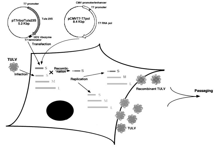
To model the recombination in TULV, two distinct types of its S RNA segment were delivered into Vero E6 cells by means of infection and transfection (Figure 1). For infection, we used the virus isolate Tula/Moravia/Ma5302V/94 originating from the European common vole (Microtus arvalis, the natural host of TULV) trapped in Moravia in 1994 (Vapalahti et al., 1996). The plasmid for transfection contained a cDNA copy of the S segment from the wild-type strain Tula/Tula/Ma23/87 recovered from lung tissue of the common vole trapped in Tula (Russia), the very place where the virus had first been discovered (Plyusnin et al., 1994). In the cDNA clone, the first 34 nucleotides (nt) were deleted. As the terminal sequences of hantaviral genomic RNAs are thought to contain a recognition site for the viral polymerase (Plyusnin et al., 1996), the cDNA-encoded, truncated S RNA molecule would not be capable of replication. The appearance of recombinant RNA (recRNA) in Vero E6 cells containing the two types of TULV S RNA (originating from infected virus and from cDNA-containing plasmid and referred to below as ‘V-type’ and ‘P-type’, respectively) was monitored by RT–PCR.
Fig. 1. Generation of Tula virus carrying recS RNA. Vero E6 cells were infected with TULV and, after 7 days, transfected with the plasmids pT7ribo/Tula23 and pCMV/T7-T7pol. It is assumed that the transiently expressed T7 polymerase produces the truncated S RNA of TULV (depicted as a dark gray bar) in the same cells where the normal type of the TULV S RNA, together with the M and L segments (depicted as light gray bars), was generated during infection. As the terminal sequences of hantaviral RNAs are thought to contain a recognition site for the viral polymerase (Plyusnin et al., 1996), the cDNA-encoded truncated S RNA molecule would not be capable of replication. Thus, in the cells, the two types of TULV S RNA co-existed, which allowed them to recombine during viral replication. As a result, TULV with two types of S RNA, normal and recombinant, was produced.
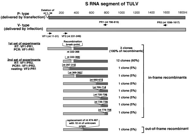
Nucleotide S segment sequences of the two TULV strains differed at 286 of the total of 1853 positions, with some substitutions clustered. This allowed the design of oligonucleotide primers for the RT–PCR, which, in theory, would selectively hybridize to only one of the two sequences. Primers for the RT–PCR, VF1 and PR1 (Figure 2) were designed to reverse-transcribe and amplify recS RNA (minus sense), which had acquired the V-type 3′-part and the P-type 5′-part. As it was not known where the recombination point(s), if any, would be located along the S RNA, in the first set of experiments we attempted to amplify and clone the left half of the S RNA molecule (nt 11–815). Three out of the 39 cDNA clones that were obtained carried identical in-frame recS RNA sequences, with the recombination break point located somewhere between nt 332 and 368 (better precision could not be achieved as sequences of the two S RNA types are identical in this region). Thus, recS RNA molecules were generated in our system of infection–supertransfection with a frequency that allowed us to detect them by RT–PCR. Control experiments with a mechanical mixture of RNA purified from cells, which had been either infected or transfected, yielded no recS RNA. This proved that the recombinant molecules described above were genuine products of the RNA-dependent RNA polymerase of TULV and not artefacts produced by the reverse transcriptase, which is well known to generate recombinant genomes during replication of retroviruses (Robertson et al., 1995).
Fig. 2. Amplification and sequencing of the TULV recS RNA. The two types of the S RNA segment of TULV are depicted as light gray and dark gray bars. Numeration of nucleotides is given for the positive-sense S RNA. Primers used for RT–PCR are depicted by arrows; numbers in parentheses correspond to the primer annealing sites (for details, see Materials and methods).
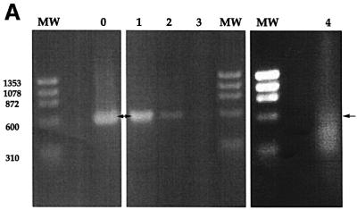
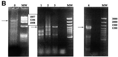
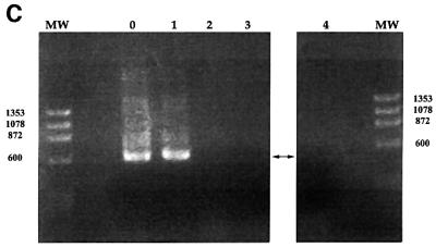
The rest of the 39 cDNA clones carried either only V-type (30 clones) or P-type (six clones) TULV sequences, all resulting from unexpected annealing of one of the primers to a wrong sequence. This showed that the detection system we used was neither very efficient nor highly specific. Therefore, the strategy used in the RT–PCR was corrected, aiming to detect recS RNA molecules carrying break points in the region located between nt 200 and 800. In addition, an attempt was made to monitor the presence of such molecules during sequential passages of the virus. RT with primers VF1 and PR2 (Figure 2), followed by nested PCR with primers VF1– PR2/VF2–PR1, was performed on RNA purified from infected–supertransfected cells and from four subsequent passages. This yielded a band of the expected size (585 bp) corresponding to the V-type 3′-part of the sequence fused to the P-type 5′-part (Figure 3A). The intensity of this band decreased with passaging, in contrast to the intensity of the corresponding band amplified with V-specific primers from the original Vero E6 cell-adapted TULV that was stable through all the passages (Figure 3B). To check for the presence of original plasmid pTUL23-T7ribo used for transfection, control nested PCR was performed using two pairs of primers specific for the vector part of the plasmid; after the first passage, the plasmid was not detectable (Figure 3C). Note that the nested PCR test was able to detect one molecule of the plasmid DNA (data not shown). This ruled out PCR artefacts, i.e. the possibility that the recS RNA sequences had been generated not by genuine recombination but by PCR fusion.
Fig. 3. Monitoring of TULV recS RNA during sequential passages of the virus. (A) Recombinant PCR amplicons (585 bp, indicated by arrows) obtained in RT-nested PCR with primers VR1–PR2/VF2–PR1 on RNA from infected and supertransfected Vero E6 cells (0) and on following passages (1, 2, 3 and 4). (B and C) Controls: PCR amplicons produced on TULV RNA with V-specific primers (1241 bp), and on plasmid DNA with P-specific primers (599 bp), respectively. MW, molecular weight markers.
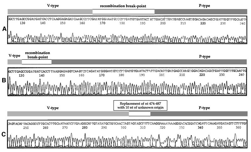
The recombinant V–P-type PCR amplicons from the second passage were cloned, and sequencing of 20 individual cDNA clones showed the presence of recombination break points (Figure 2). Twelve clones carried recS sequences identical to those obtained in the first set of experiments, with a break point located between nt 332 and 368 (the sequence of one of these clones is shown in Figure 4A). Furthermore, a third independent attempt to generate recS RNA molecules using transfection followed by infection, again, resulted in the production of recombinants of the same structure (data not shown). These observations show the region between nt 332 and 368 to be a ‘hot-spot’ for recombination. Each of the remaining eight clones from the second set of experiments carried a unique recS sequence with a break point located outside this region (Figure 4B and C). One of these clones had a replacement of 14 nt, located between positions 474 and 487, with 10 nt of unknown origin; this is the only ‘out-of-frame’ recS RNA sequence (as well as the only product of non-homologous recombination) that was observed (Figure 4C).
Fig. 4. Sequence analysis of selected recS RNA clones (second passage). (A) One of the 12 ‘major’ in-frame recombinants with the break point located between nt 332 and 368. The mapping is based on the following sequence differences: the V-type sequence contains CCTTC at position 327–331 while the P-type sequence contains TCTCA, and the V-type sequence contains ‘C’ at position 369 while the P-type sequence contains ‘T’ at this position. (B) One of the seven ‘minor’ in-frame recombinants with the break point located between nt 298 and 311. The mapping is based on the following sequence differences: the V-type sequence contains GCTTGAG at position 291–297 while the P-type sequence contains CCTTGAA, and the V-type sequence contains ‘C’ at position 312 while the P-type sequence contains ‘T’. (C) Out-of-frame recombinant. The V-type sequence contains TAG at position 471–473 while the P-type sequence contains CCG, and the V-type sequence contains ‘G’ at position 501 while the P-type sequence contains ‘A’. The sequence AGGGAGGCAAACAA at position 474–487 is replaced with the sequence TAGTAGCTTT of unknown origin (nt 490–500 are identical in both V- and P-type sequences). The virus- and plasmid-derived sequences are depicted as light gray and dark gray bars, respectively.
Our search for other break points within the regions 11–231 and 815–1617 in the long PCR amplicons, which were produced after the first 30 rounds of PCR, revealed none. Thus, all ‘in-frame’ recS RNA molecules appeared to have only one break point, in contrast to the ‘out-of-frame’ recS RNA that contained two of them.
Isolation of pure recombinant Tula virus
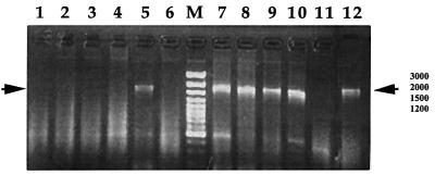
As recS RNA molecules gradually disappeared from the genetic swarm of the original Vero E6 cell-adapted variant on sequential passages, an attempt to separate viral particles with the recS segment from the original virus was made. Six-well plates of monolayer culture of Vero E6 cells were infected with diluted supernatant from passage 2 (∼1–2 focus-forming units per well). The presence of original and recS RNA types was monitored using RT–PCR with virus- or recombinant-specific primers, respectively. Results presented in Figure 5 show the presence of the recS RNA in one of the wells, indicating that the aliquot of the diluted supernatant delivered to this well contained the recombinant virus. The other five wells on the same plate received aliquots containing the original virus, thus confirming that it dominated in the mixed virus population. The recombinant virus from well 5 (designated ‘recTULV5’) was purified further through infection of fresh cells with a 1:104 dilution of the supernatant. Re-checking the offspring using recombinant-specific, virus-specific and universal RT–PCR confirmed that recTULV5 contained recS RNA type only. Analysis of 14 cDNA clones that were generated from the S segment of recTULV5 with primers VF1 and PR2 showed that each of them carried a single recombinant break point. In 13 cDNA clones, the break point was located between nt 332 and 368, and, in one cDNA clone, between nt 261 and 279. Thus, recS RNA molecules of the first type represented the vast majority of the genetic swarm of recTULV5. This correlated well with the dominance of this type of molecule in the originally generated population of recS RNA (Figure 2).
Fig. 5. Selection of recombinant Tula hantavirus from original variants. Lanes 1–6 show recombinant PCR amplicons (1607 bp) obtained in RT–PCR with primers VF1–PLR2 from six individual wells of Vero E6 cells infected with diluted (1:1000) supernatant from passage 2. Lanes 7–12 show virus-type PCR amplicons (1672 bp) obtained in RT–PCR with primers VF1–VR1 from the same six wells. M, molecular weight marker.
recTULV5 was also able to grow steadily in the Vero E6 cell culture after repeated passages, forming regular foci and at approximately the same titers as the original TULV (2–5 × 10³ focus-forming units per ml of supernatant). Gradual accumulation of cells with typical granular fluorescence pattern of the N protein was detected (Figure 6A), similarly to infection with the original TULV isolate (Lundkvist et al., 1996; Vapalahti et al., 1996). Both Coomassie Blue staining (data not shown) and immunoblotting of the N protein from recTULV5 (Figure 6B) showed a characteristic band of ∼48 kDa indistinguishable from that of the original TULV. These features of recTULV5 allowed consideration of this virus as functionally competent and stable.
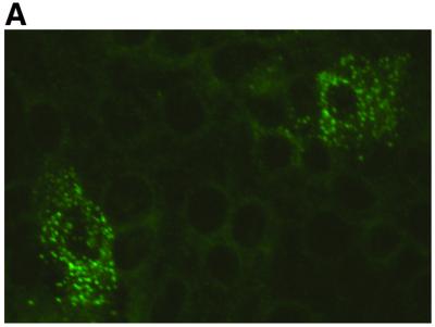
Fig. 6. (A) Immunofluorescence pattern of Vero E6 cells infected with recombinant Tula virus. Vero E6 cells infected for 3 days with recTULV5 stained with rabbit hantavirus N-specific antiserum showing typical granular distribution of the nucleocapsid protein. (B) Immunoblotting of the recombinant N protein (lane 3). For comparison, the N protein of the original Tula virus (lane 2) and Puumala virus (lane 1) are given. MW, molecular weight marker.
Discussion
Transfection-mediated homologous recombination yields functionally competent Tula virus carrying a recS RNA segment
In Vero E6 cells that were first infected with TULV and then transfected with two plasmids producing truncated S RNA molecules, we have been observing almost exclusively ‘in-frame’ recS RNA molecules, and most of those contained a single break point located between nt 332 and 368. Notably, this region is very close to one of the putative break points in the S segment of wild-type TULV strains from eastern Slovakia that was considered to be a product of HRec occurring in nature (Sibold et al., 1999). Moreover, the suspected ‘parents’ of the recombinant Slovakian TULV strains are thought to belong to the Moravian and Russian genetic lineages, represented in our experiments by the TULV isolate and the wild-type TULV strain used as a source for the truncated cDNA clone, plasmid pT7ribo/Tula23. It is tempting to speculate that the observed recombination ‘hot-spot’ is located within a ‘special’ region of the S RNA, e.g. one having particularly strong secondary structure. However, our first attempts to calculate the secondary structure did not reveal any peculiarities of the region in question. Perhaps, like in other RNA viruses, N protein or other chaperones promote the formation of a special secondary structure of the S RNA, thus enhancing recombination at a particular spot (Negroni and Buc, 2000).
The fact that all ‘in-frame’ recS RNA molecules appeared to have only one break point suggests that they resulted from single template-switching events. The only ‘out-of-frame’ recombinant looks like a product of two consecutive ‘jumps’ of the viral RNA polymerase: first from the viral S RNA template to another, non-viral RNA molecule, and back again. The nature of this non-viral RNA remains obscure due to the short length of the sequence introduced into the recS RNA.
The preservation of the recS RNA molecules on serial passages showed that viral particles which possessed them had replicated as part of the genetic swarm of the original Vero E6 cell-adapted variant, i.e. they were functionally competent. These recS RNA molecules (as well as the corresponding virus particles) should also be considered relatively stable as they are able to survive at least 1 month of passaging. Their replication, however, was not a complete success, as the amount of corresponding PCR amplicons decreased continuously from passage 1 to 4. Thus, virus particles carrying recS RNA seemed to be less competitive than those having the wild-type S segment. This is not totally unexpected; passaging through cell culture creates an opportunity to outcompete variants with lower fitness, i.e. in this case, the recombinants.
When separated from original variant, however, the virus-carried recS RNA segment was able to grow steadily in sequential passages (four so far), yielding titers similar to those of the original, non-recombinant virus. These observations confirmed the stability and functional competence of the recombinant Tula hantavirus, which represents the first case of a negative-strand virus engineered using HRec.
HRec: universal mechanism operating in evolution of RNA viruses?
The data presented in this paper demonstrate that HRec, which had been suggested based on genetic analyses of naturally circulating TULV strains, can be reproduced experimentally in vitro. TULV thus provides the first example of a negative-sense RNA virus for which HRec has been shown to occur. So far, it has been a general belief that the negative-sense RNA viruses do not recombine because their RNA of both genome and anti-genome polarity is tightly packaged into nucleocapsids, thus not allowing for polymerase ‘jumps’. Such a view, however, totally ignored the existence of defective interfering RNA viruses with mosaic-like structure in their genome (Lazzarini et al., 1981; Nayak et al., 1985) and reports on non-homologous recombination, i.e. recombination between unrelated RNA sequences (Khatchikian et al., 1989; Bergmann et al., 1992; Orlich et al., 1994), which demonstrated the potential of a negative-sense RNA virus polymerase for a template switch. Our observations on Tula virus indeed confirm such a potential.
Together with the well-established knowledge on HRec in the evolution of positive-sense RNA viruses and the more recent findings on double-stranded RNA viruses (Suzuki et al., 1998), our observations suggest that HRec can operate in all three groups of RNA viruses. Thus, HRec seems to be a common mechanism of generating and spreading advantageous genetic combinations in viruses with an RNA genome, which represent the most populated domain in the virus kingdom. How widely HRec occurs in negative-sense RNA viruses remains to be seen. One could speculate that viruses with a high level of interference, short reproduction time and/or strong cytopathic capacity (like vesicular stomatitis virus, a classical example of a negative-sense virus) would have relatively small chances to recombine, simply due to the extremely low frequency of double infection, which is an obligatory prerequisite for recombination. On the other hand, persistent, less cytopathic and interfering viruses (like bunyaviruses, including hantaviruses) might have reasonable chances to recombine. However, for viruses with a segmented genome, those chances are expected to be smaller than (or even uncomparable with) the chances for reassortment.
It seems certain that HRec contributes to the success of several human RNA viral pathogens. The most prominent examples are human immunodeficiency virus (Robertson et al., 1997), human enteroviruses (Santti et al., 1999) and dengue virus (Worobey et al., 1999). The evolutionary impact of the discovered phenomenon for the negative-sense RNA viruses, as well as theoretical complications connected with their use as vaccine vectors, remain to be evaluated.
Materials and methods
Generation of Tula hantavirus carrying recS RNA
A monolayer culture of Vero E6 cells (5 × 10⁶ cells) was infected with 104 focus-forming units of TULV, strain Moravia/Ma5302V/94 (Vapalahti et al., 1996) and, after 7 days, transfected with plasmids pT7ribo/Tula23 and pCMV/T7-T7pol (Brisson et al., 1999) using LipofectAMINE reagent (Gibco BRL, Life Technologies) according to the manufacturer’s recommendations. The plasmid pT7ribo/Tula23 carries a truncated (nt 1–34 deleted) cDNA for the S RNA segment of TULV, strain Tula/Tula/Ma23/87 (Plyusnin et al., 1994) inserted into pT7ribo (Dunn et al., 1995) under the T7 promoter; the downstream-located hepatitis δ virus antigenome ribozyme sequence allowed the precise cutting of the 3′ terminus of the RNA transcript. The plasmid pCMV/T7-T7pol carries the gene encoding the T7 RNA polymerase under both cytomegalovirus (CMV) and T7 promoters. Cells were used for RNA extraction and the supernatant (∼20 ml) was collected and added to fresh Vero E6 cells. Second and third passages were performed in the same way. From the fourth passage onwards, 2 ml of the supernatant from the previous passage were used to infect fresh cells.
Isolation of pure recombinant Tula virus
Six-well plates of a monolayer culture of Vero E6 cells (5 × 10⁵ cells per well) were infected with diluted supernatant from passage 2 (∼1–2 focus-forming units per well). After 10 days, the supernatant (∼2 ml) was collected and used for infection of fresh Vero E6 cells using a dilution of 1:104. Further passages of the recombinant virus were performed under standard conditions, i.e. with undiluted supernatant.
RT–PCR
RNA was purified using the TriPure isolation reagent (Boehringer Mannheim) according to the manufacturer’s recommendations. RT was performed with the Moloney murine leukemia virus reverse transcriptase (New England Biolabs) according to the manufacturer’s recommendations. For PCR, AmpliTaq DNA polymerase (Perkin Elmer, Roche Molecular Systems) was used. In the first set of experiments, RT and first PCR were performed with primers VF1 (5′-CTCCTTGAAAAG CTACTACTA-3′, nt 11–31) and PR1 (5′-TCTTCTTCTTTCCC AGGAGC-3′, nt 796–815). In the second set, RT and first PCR were performed with primers VF1 and PR2 (5′-CAATAAACAAAGA CTTAACTTA-3′, nt 1596–1617), and nested PCR with primers VF2 (5′-GCAGCTTGCAGATCTGGTG-3′, nt 231–149) and PR1 (Figure 2). The PCR amplicons were separated on agarose gels, purified with the QIAquick kit (Qiagen GmbH) and cloned into the pGEM-T vector (Promega). Sequencing was performed using the ABI PRISM Dye Terminator sequencing kit [Perkin Elmer Applied Biosystems Division (PE/ABI)] according to the manufacturer’s instructions, and the reactions were run on an ABI 373 A sequencer (PE/ABI).
To monitor the presence of TULV RNA when a mixture of original and recombinant viruses was passaged, RT and first PCR were performed with primers VF1 and VR1 (5′-CAATAATGAACTTACACTAGTG-3′, nt 1661–1682), and second PCR (semi-nested) with primers VF3 (5′-CTTTGCACTTGCAATAATCTTG-3′, nt 420–442) and VR1; the authenticity of the PCR amplicons was confirmed by direct sequencing. To monitor the presence of plasmid DNA on passages, first PCR was performed with primers PLVecF1 (5′-TCAACAGCGGTAAGATCC TTG-3′, nt 1741–1762) and PLVecR1 (5′-GAGCGGTATCAG CTCACTCAA-3′, nt 3325–3346), and nested PCR with primers PLVecF2 (5′-CTGCGGCCAACTTACTTCTGA-3′, nt 1988–2008) and PLVecR2 (5′-GAACGAAAACTCACGTTAAGGGA-3′, nt 2564– 2586).
During purification of the recombinant Tula virus from the original variant, three types of RT–PCR were used. To detect S RNA of the original TULV, RT and PCR were performed with primers VF1 and VR1. To detect S RNA of the recombinant TULV, RT and PCR were performed with primers VF1 and PR2. To detect simultaneously both original and recombinant viruses, RT and PCR were performed with universal primers UF1 (5′-CCATGAACAGCAAATTGTCATTG-3′, nt 81–103) and UR1 (5′-GAACACTC(G/A)TTACTACGAATGG-3′, nt 820–840). The authenticity of the PCR amplicons was confirmed by direct sequencing.
Immunofluorescence assay
Vero E6 cells were grown to confluency on coverslips and infected with recTULV5 for 3 days before acetone fixation and staining with rabbit polyclonal antiserum raised against recombinant Puumala virus N-gluta thione S-transferase fusion protein as described before (Vapalahti et al., 1995).
Immunoblotting
Immunoblotting was performed essentially as described previously (Plyusnin et al., 1994; Vapalahti et al., 1996). Recombinant Tula virus was purified by centrifugation through a sucrose cushion (20%). Proteins were separated by using standard 10% SDS–PAGE and transferred to Immobilon filters (Millipore S.A., Malsheim, France). Filters were pre-absorbed with 1% non-fat dry milk in phosphate-buffered saline, then incubated at room temperature with 1/200 dilution of polyclonal protein-specific rabbit antisera for 2 h, followed by anti-rabbit peroxidase conjugates (Dakopatts) for 1 h at 37°C. Specific antibody binding was detected with diaminobenzidine substrate (Sigma, St Louis, MO).
Acknowledgments
Acknowledgements
The authors thank E.Dunn for the plasmid pT7ribo and M.Brisson for the plasmid pCMV/T7-T7pol. Åke Lundkvist is acknowledged for his help with the focus titration protocol. The constructive criticism and helpful comments of Petteri Arstila, John Brunstein, Timo Hyypiä and Erkki Ruoslahti are greatly appreciated. This work was supported by research grants from the Academy of Finland, EU grant QLK2-CT-1999-0119 and the Sigrid Jusélius Foundation.
References
- Bergmann M., Garcia-Sastre,A. and Palese,P. (1992) Transfection-mediated recombination of influenza A virus. J. Virol., 66, 7576–7580. [DOI] [PMC free article] [PubMed] [Google Scholar]
- Brisson M., He,Y., Li,S., Yand,J.P. and Huang,L. (1999) A novel T7 RNA polymerase autogene for efficient cytoplasmic expression of target genes. Gene Ther., 6, 263–270. [DOI] [PubMed] [Google Scholar]
- Chetverin A.B., Chetverina,H.V., Demidenko,A.A. and Ugarov,V.I. (1997) Nonhomologous RNA recombination in a cell-free system: evidence for a transesterification mechanism guided by secondary structure. Cell, 88, 503–513. [DOI] [PMC free article] [PubMed] [Google Scholar]
- Cooper P.D., Steiner-Pryor,A.S., Scotti,P.D. and Delong,D. (1974) On the nature of poliovirus genetic recombinants. J. Gen. Virol., 23, 41–49. [DOI] [PubMed] [Google Scholar]
- Dunn E.F., Pritlove,D.C., Jin,H. and Elliott,R.M. (1995) Transcription of a recombinant bunyavirus RNA template by transiently expressed bunyavirus proteins. Virology, 211, 133–143. [DOI] [PubMed] [Google Scholar]
- Elliott R.M., Bouloy,M., Calisher,C.H., Goldbach,R., Moyer,J.T., Nichol,S.T., Pettersson,R., Plyusnin,A. and Schmaljohn,C.S. (2000) Family Bunyaviridae. In van Regenmortel,M.H.V. et al. (eds), Virus Taxonomy. VIIth Report of the International Committee on Taxonomy of Viruses. Academic Press, San Diego, CA, pp. 599–621.
- Henderson W.W., Monroe,M.C., St. Jeor,S.C., Thayer,W.P., Rowe,J.E., Peters,C.J. and Nichol,S.T. (1995) Naturally occurring Sin Nombre virus genetic reassortants. Virology, 214, 602–610. [DOI] [PubMed] [Google Scholar]
- Hirst G.K. (1962) Genetic recombination with Newcastle disease virus, polioviruses and influenza virus. Cold Spring Harb. Symp. Quant. Biol., 27, 303–309. [DOI] [PubMed] [Google Scholar]
- Khatchikian D., Orlich,M. and Rott,R. (1989) Increased viral pathogenicity after insertion of a 28S ribosomal RNA sequence into the hemagglutinin gene of influenza virus. Nature, 340, 156–157. [DOI] [PubMed] [Google Scholar]
- Lazzarini R.A., Keene,J.D. and Schubert,M. (1981) The origins of defective interfering particles of the negative-strand RNA viruses. Cell, 26, 145–154. [DOI] [PubMed] [Google Scholar]
- Ledinko N. (1963) Genetic recombination with poliovirus type 1: studies of crosses between a normal horse serum-resistant mutant and several guanidine-resistant mutants of the same strain. Virology, 20, 107–119. [DOI] [PubMed] [Google Scholar]
- Li D., Schmaljohn,A.L., Anderson,K. and Schmaljohn,C.S. (1995) Complete nucleotide sequences of the M and S segments of two hantavirus isolates from California: evidence for reassortment in nature among viruses related to hantavirus pulmonary syndrome. Virology, 206, 973–983. [DOI] [PubMed] [Google Scholar]
- Lundkvist Å., Vapalahti,O., Plyusnin,A., Brus Sjölander,K., Niklasson,B. and Vaheri,A. (1996) Characterization of Tula hantavirus antigenic determinants defined by monoclonal antibodies raised against baculovirus-expressed nucleocapsid protein. Virus Res., 45, 29–44. [DOI] [PubMed] [Google Scholar]
- Nayak D.P., Chambers,T.M. and Akkina,R.K. (1985) Defective interfering RNAs of influenza viruses: origin, structure, expression and interference. Curr. Top. Microbiol. Immunol., 114, 103–151. [DOI] [PubMed] [Google Scholar]
- Negroni M. and Buc,H. (2000) Copy-choice recombination by reverse transcriptases: reshuffling of genetic markers mediated by RNA chaperones. Proc. Natl Acad. Sci. USA, 97, 6385–6390. [DOI] [PMC free article] [PubMed] [Google Scholar]
- Nichol S.T., Ksiazek,T.G., Rollin,P.E. and Peters,C.J. (1996) Hantavirus pulmonary syndrome and newly described hantaviruses in the United States. In Elliott,R.M. (ed.), The Bunyaviridae. Plenum Press, New York, NY, pp. 269–280.
- Orlich M., Gottwald,H. and Rott,R. (1994) Nonhomologous recombination between the hemagglutinin gene and the nucleoprotein gene of an influenza virus. Virology, 204, 462–465. [DOI] [PubMed] [Google Scholar]
- Pierangeli A., Bucci,M., Forzan,M., Paganotti,P., Equestre,M. and Perez Bercoff,R. (1999) ‘Primer alignment-and-extension’: a novel mechanism of viral RNA recombination responsible for the rescue of inactivated poliovirus cDNA clones. J. Gen. Virol., 80, 1889–1897. [DOI] [PubMed] [Google Scholar]
- Plyusnin A. et al. (1994) Tula virus: a newly detected hantavirus carried by European common voles. J. Virol., 68, 7833–7839. [DOI] [PMC free article] [PubMed] [Google Scholar]
- Plyusnin A., Vapalahti,O. and Vaheri,A. (1996) Hantaviruses: genome structure, expression and evolution. J. Gen. Virol., 77, 2677–2687. [DOI] [PubMed] [Google Scholar]
- Robertson D.L., Hahn,B.H. and Sharp,P.M. (1995) Recombination in AIDS viruses. J. Mol. Evol., 40, 249–259. [DOI] [PubMed] [Google Scholar]
- Robertson D., Gao,F., Hahn,B. and Sharp,P. (1997) Intersubtype recombinant HIV-1 sequences. In Korber,B., Hahn,B. and Foley,B. (eds), The Human Retroviruses and AIDS. Los Alamos National Laboratory, Los Alamos, NM, pp. 25–30.
- Santti J., Hyypiä,T., Kinnunen,L. and Salminen,M. (1999) Evidence of recombination among enteroviruses. J. Virol., 73, 8741–8749. [DOI] [PMC free article] [PubMed] [Google Scholar]
- Schmaljohn C.S. and Hjelle,B. (1997) Hantaviruses: a global disease problem. Emerg. Infect. Dis., 3, 95–104. [DOI] [PMC free article] [PubMed] [Google Scholar]
- Sibold C., Meisel,H., Krüger,D.H., Labuda,M., Lysy,J., Kozuch,O., Pejcoch,M., Vaheri,A. and Plyusnin,A. (1999) Recombination in Tula hantavirus evolution: analysis of genetic lineages from Slovakia. J. Virol., 73, 667–675. [DOI] [PMC free article] [PubMed] [Google Scholar]
- Suzuki Y., Gojobori,T. and Nakagomi,O. (1998) Intragenic recombin ation in rotaviruses. FEBS Lett., 427, 183–187. [DOI] [PubMed] [Google Scholar]
- Vapalahti O. et al. (1995) Human B-cell epitopes of Puumala virus nucleocapsid protein, the major antigen in early serological response. J. Med. Virol., 46, 293–303. [DOI] [PubMed] [Google Scholar]
- Vapalahti O. et al. (1996) Isolation and characterization of Tula virus: a distinct serotype in genus Hantavirus, family Bunyaviridae. J. Gen. Virol., 77, 3063–3067. [DOI] [PubMed] [Google Scholar]
- Worobey M. and Holmes,E.C. (1999) Evolutionary aspects of recombination in RNA viruses. J. Gen. Virol., 80, 2535–2543. [DOI] [PubMed] [Google Scholar]
- Worobey M., Rambaut,A. and Holmes,E.C. (1999) Widespread serotype recombination in natural populations of dengue virus. Proc. Natl Acad. Sci. USA, 96, 7352–7357. [DOI] [PMC free article] [PubMed] [Google Scholar]


