

Abstract
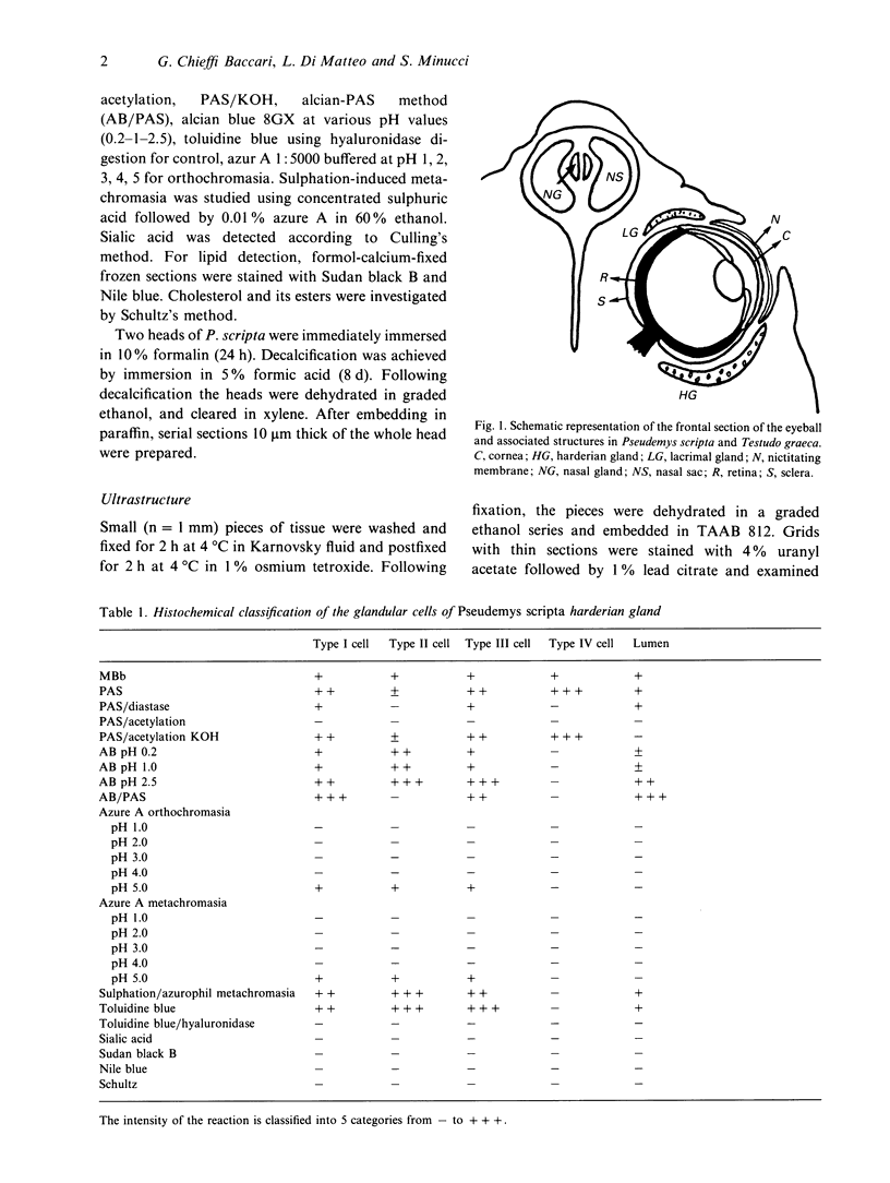
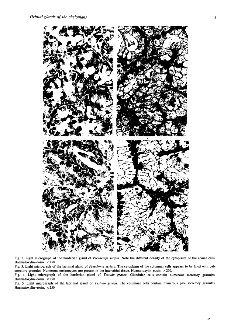
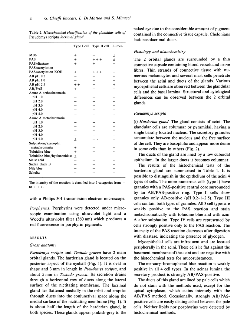
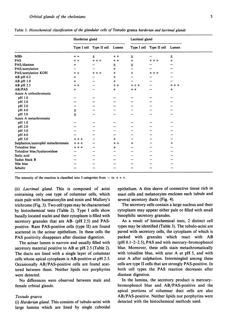
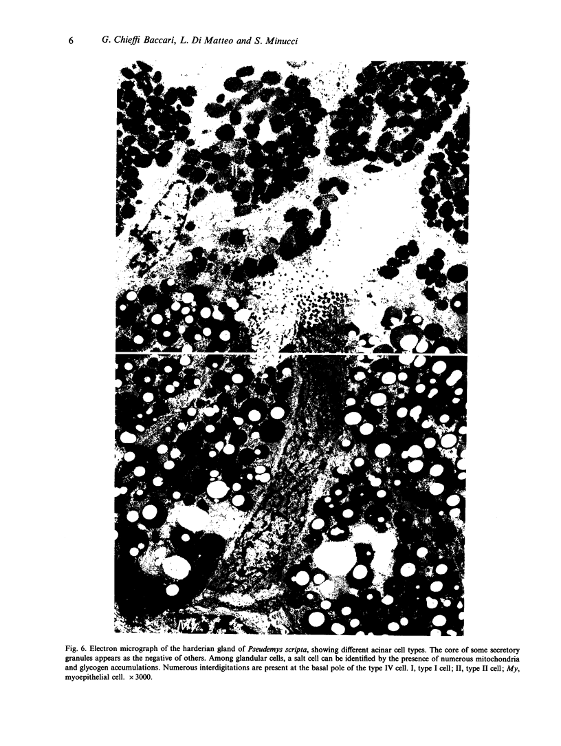
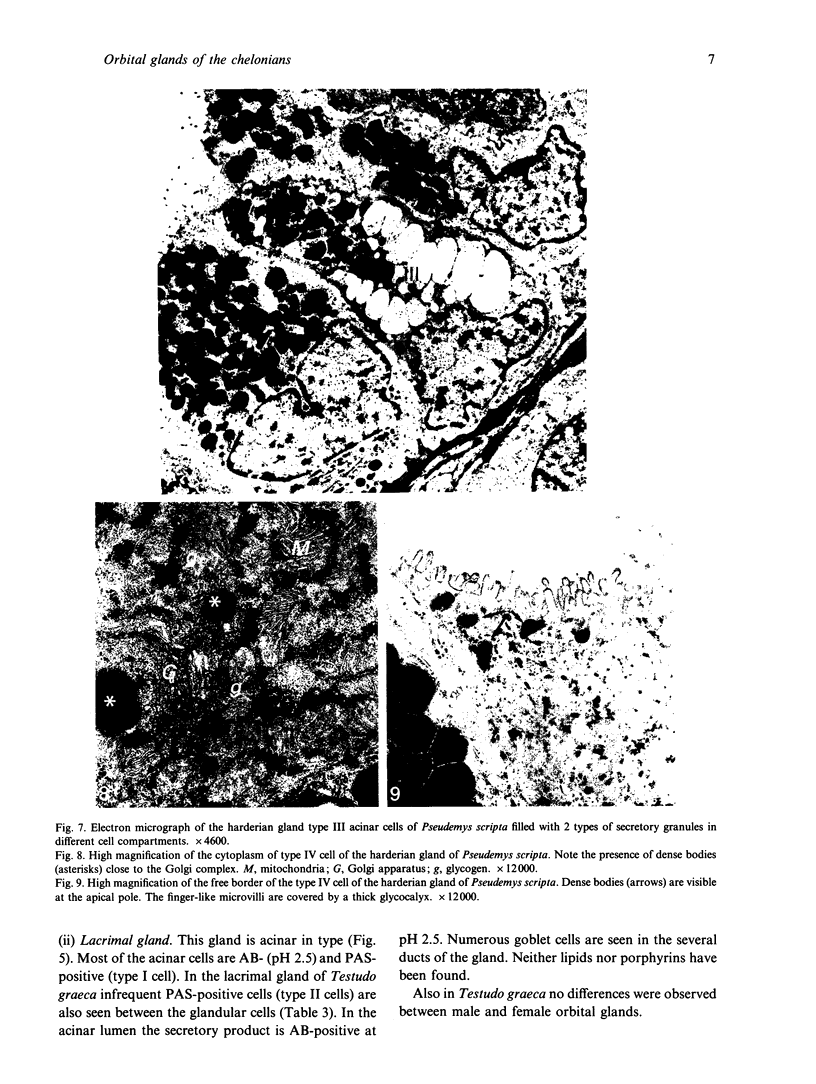
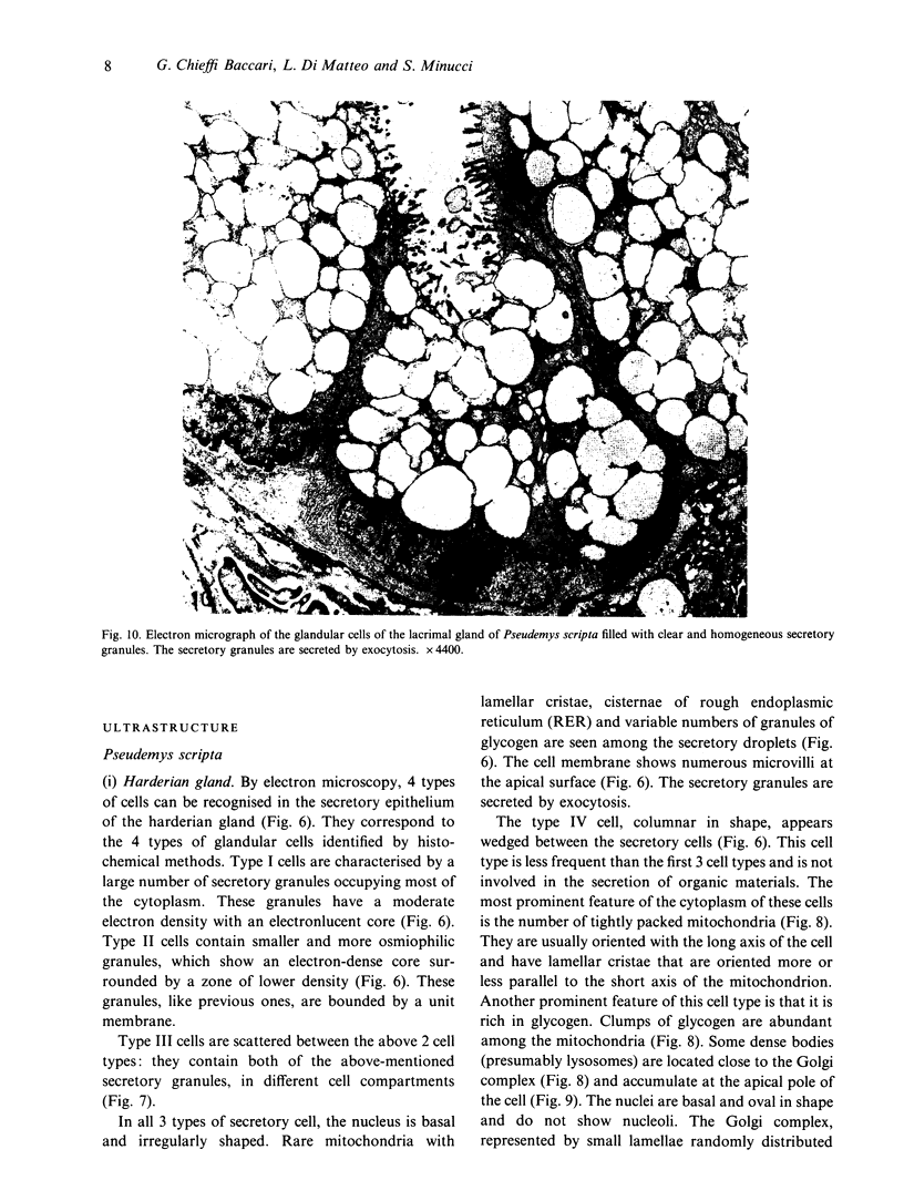
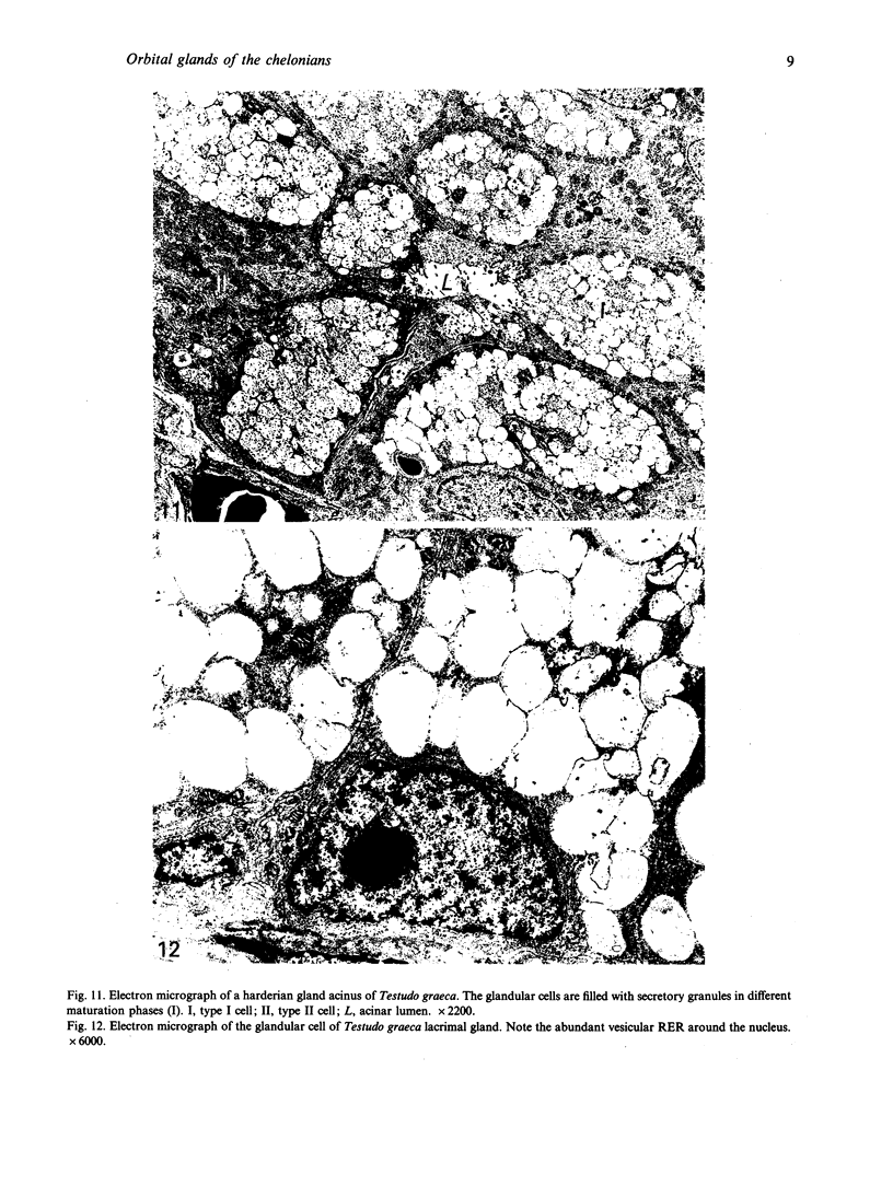
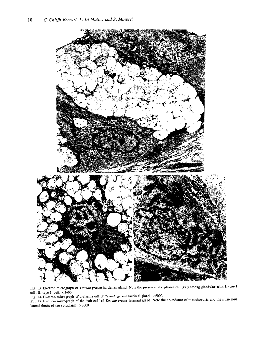
The orbital glands of the chelonians Pseudemys scripta and Testudo graeca were investigated at the histological, histochemical and ultrastructural levels. Four acinar cell types were seen in the harderian gland of P. scripta on the basis of histochemical reactions and ultrastructure. Secretory granules were of 2 types, one showing moderate electron density with an electronlucent core, the other being smaller and more osmiophilic with an electron-dense core. In the harderian gland of T. graeca only 2 glandular cell types were found; one type contained secretory granules with a dense core surrounded by a wide zone of lower density. Acinar cells of the anterior lacrimal gland in both species were of 2 types, one being of mucous type. In the harderian gland and in the lacrimal gland of both species, one cell type appeared not to be involved in the secretion of organic material. These cells contained numerous tightly packed mitochondria among which were abundant clumps of glycogen; the cell membrane was specialised at both edges. This cell type was similar ultrastructurally to the 'salt cells' described in the salt-secreting glands of various marine vertebrates, i.e. of the cells involved in transport processes. These combined histological, histochemical and ultrastructural studies have allowed us to distinguish orbital glands. In the past, the harderian and lacrimal glands in chelonians have often been mistaken for one another.
Full text
PDF

Images in this article


Selected References
These references are in PubMed. This may not be the complete list of references from this article.
- Abel J. H., Jr, Ellis R. A. Histochemical and electron microscopic observations on the salt secreting lacrymal glands of marine turtles. Am J Anat. 1966 Mar;118(2):337–357. doi: 10.1002/aja.1001180203. [DOI] [PubMed] [Google Scholar]
- BULGER R. E. FINE STRUCTURE OF THE RECTAL (SALT-SECRETING) GLAND OF THE SPINYDOGFISH, SQUALUS ACANTHIAS. Anat Rec. 1963 Sep;147:95–127. doi: 10.1002/ar.1091470108. [DOI] [PubMed] [Google Scholar]
- Baccari G. C., Minucci S., Di Matteo L., Chieffi G. Harderian gland and the lacrimal gland of the lizard Podarcis s. sicula: histology, histochemistry, and ultrastructure. Anat Rec. 1990 Mar;226(3):269–278. doi: 10.1002/ar.1092260302. [DOI] [PubMed] [Google Scholar]
- Burns R. B., Maxwell M. H. The structure of the Harderian and lacrimal gland ducts of the turkey, fowl and duck. A light microscope study. J Anat. 1979 Mar;128(Pt 2):285–292. [PMC free article] [PubMed] [Google Scholar]
- Cowan F. B. The homology of cranial glands in turtles: with special reference to the nomenclature of salt glands. J Morphol. 1973 Oct;141(2):157–169. doi: 10.1002/jmor.1051410204. [DOI] [PubMed] [Google Scholar]
- Cowan F. B. The ultrastructure of the lachrymal 'salt' gland and the Harderain gland in the euryhaline Malaclemys and some closely related stenohaline emydines. Can J Zool. 1971 May;49(5):691–697. doi: 10.1139/z71-108. [DOI] [PubMed] [Google Scholar]
- DOYLE W. L. Tubule cells of the rectal salt-gland of Urolophus. Am J Anat. 1962 Sep;111:223–237. doi: 10.1002/aja.1001110208. [DOI] [PubMed] [Google Scholar]
- ELLIS R. A., ABEL J. H., Jr INTERCELLULAR CHANNELS IN THE SALT-SECRETING GLANDS OF MARINE TURTLES. Science. 1964 Jun 12;144(3624):1340–1342. doi: 10.1126/science.144.3624.1340. [DOI] [PubMed] [Google Scholar]
- FARBER S. J. Mucopolysaccharides and sodium metabolism. Circulation. 1960 May;21:941–947. doi: 10.1161/01.cir.21.5.941. [DOI] [PubMed] [Google Scholar]
- Olcese J., Wesche A. The Harderian gland. Comp Biochem Physiol A Comp Physiol. 1989;93(4):655–665. doi: 10.1016/0300-9629(89)90480-5. [DOI] [PubMed] [Google Scholar]
- RHODIN J. Electron microscopy of the kidney. Am J Med. 1958 May;24(5):661–675. doi: 10.1016/0002-9343(58)90373-5. [DOI] [PubMed] [Google Scholar]