Abstract
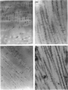
Isolated, purified small chondroitin (dermatan) sulphate proteoglycans from corneas of cow and rabbit and cow sclera were stained with Cupromeronic blue in 'model' experiments. The lengths and thicknesses of the images were compared with those of the same proteoglycans stained in the tissue, using the critical electrolyte concentration principle to give specificity for sulphated proteoglycans, and keratanase 1 or chondroitinase ABC digestion to distinguish between chondroitin and keratan sulphate. Corrections for orientation of the stained glycan filaments within the section plane were made to convert the observed lengths to true average lengths. Observed lengths of stained chondroitin (dermatan) sulphate were greater than those of keratan sulphate, both in models and tissues, in agreement with published data from biochemical and rotary-shadowing studies, in both species. Corrected (true) average lengths of stained isolated chondroitin (dermatan) sulphate proteoglycans were slightly, but not significantly, longer than expected from rotary shadowing or biochemical measurements. Keratan sulphate lengths were similarly somewhat longer. The data support the idea that Cupromeronic blue acts as a scaffold that helps maintain polyanion shape against distortion on staining. Stained filaments in tissues were sometimes over twice the length of isolated stained proteoglycans, suggesting that 2 glycan chains were aligned end-to-end. Thicknesses of proteoglycan filaments suggested that at least 2 glycan chains were aligned side-by-side, both in models and in tissues. A scheme for proteoglycan tertiary structure in cornea is proposed, in which glycan chains may bridge collagen fibrils in duplexed forms similar to those observed in rotary shadowed preparations.
Full text
PDF


Images in this article
Selected References
These references are in PubMed. This may not be the complete list of references from this article.
- Craig A. S., Parry D. A. Collagen fibrils of the vertebrate corneal stroma. J Ultrastruct Res. 1981 Feb;74(2):232–239. doi: 10.1016/s0022-5320(81)80081-0. [DOI] [PubMed] [Google Scholar]
- Fransson L. A. Interaction between dermatan sulphate chains. I. Affinity chromatography of copolymeric galactosaminioglycans on dermatan sulphate-substituted agarose. Biochim Biophys Acta. 1976 Jun 23;437(1):106–115. doi: 10.1016/0304-4165(76)90351-2. [DOI] [PubMed] [Google Scholar]
- Haigh M., Scott J. E. A method of processing tissue sections for staining with cu-promeronic blue and other dyes, using CEC techniques, for light and electron microscopy. Basic Appl Histochem. 1986;30(4):479–486. [PubMed] [Google Scholar]
- Jørgen H., Gundersen G. Estimation of tubule or cylinder LV, SV and VV on thick sections. J Microsc. 1979 Dec;117(3):333–345. doi: 10.1111/j.1365-2818.1979.tb04690.x. [DOI] [PubMed] [Google Scholar]
- Mörgelin M., Paulsson M., Malmström A., Heinegård D. Shared and distinct structural features of interstitial proteoglycans from different bovine tissues revealed by electron microscopy. J Biol Chem. 1989 Jul 15;264(20):12080–12090. [PubMed] [Google Scholar]
- Oeben M., Keller R., Stuhlsatz H. W., Greiling H. Constant and variable domains of different disaccharide structure in corneal keratan sulphate chains. Biochem J. 1987 Nov 15;248(1):85–93. doi: 10.1042/bj2480085. [DOI] [PMC free article] [PubMed] [Google Scholar]
- Scott J. E., Cummings C., Brass A., Chen Y. Secondary and tertiary structures of hyaluronan in aqueous solution, investigated by rotary shadowing-electron microscopy and computer simulation. Hyaluronan is a very efficient network-forming polymer. Biochem J. 1991 Mar 15;274(Pt 3):699–705. doi: 10.1042/bj2740699. [DOI] [PMC free article] [PubMed] [Google Scholar]
- Scott J. E., Cummings C., Greiling H., Stuhlsatz H. W., Gregory J. D., Damle S. P. Examination of corneal proteoglycans and glycosaminoglycans by rotary shadowing and electron microscopy. Int J Biol Macromol. 1990 Jun;12(3):180–184. doi: 10.1016/0141-8130(90)90029-a. [DOI] [PubMed] [Google Scholar]
- Scott J. E., Haigh M. 'Small'-proteoglycan:collagen interactions: keratan sulphate proteoglycan associates with rabbit corneal collagen fibrils at the 'a' and 'c' bands. Biosci Rep. 1985 Sep;5(9):765–774. doi: 10.1007/BF01119875. [DOI] [PubMed] [Google Scholar]
- Scott J. E., Haigh M. Keratan sulphate and the ultrastructure of cornea and cartilage: a 'stand-in' for chondroitin sulphate in conditions of oxygen lack? J Anat. 1988 Jun;158:95–108. [PMC free article] [PubMed] [Google Scholar]
- Scott J. E. Proteoglycan histochemistry--a valuable tool for connective tissue biochemists. Coll Relat Res. 1985 Dec;5(6):541–575. doi: 10.1016/s0174-173x(85)80008-x. [DOI] [PubMed] [Google Scholar]
- Scott J. E. Proteoglycan-fibrillar collagen interactions. Biochem J. 1988 Jun 1;252(2):313–323. doi: 10.1042/bj2520313. [DOI] [PMC free article] [PubMed] [Google Scholar]
- Ward N. P., Scott J. E., Cöster L. Dermatan sulphate proteoglycans from sclera examined by rotary shadowing and electron microscopy. Biochem J. 1987 Mar 15;242(3):761–766. doi: 10.1042/bj2420761. [DOI] [PMC free article] [PubMed] [Google Scholar]


