

Abstract
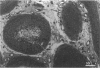
The morphology of the epididymal duct and, in particular, the epididymal microvasculature was examined at the light microscope level in young sexually-mature rats (3-5 months) and aged rats (18 months) to investigate the structural changes that may occur within the organ as a result of ageing, and which may predispose the organ to pathological changes. Quantitative data on the microvascular network of the epididymis (percentage of capillaries in the interstitial region, average area and surface density of the capillary lumen) were collected in 4 regions of the epididymis: the initial segment, caput, corpus and cauda. Epithelial cell height, epididymal lumen diameter, number of smooth muscle cells and percentage of smooth muscle surrounding the duct were also assessed within the same 4 regions. The data for both young and aged groups revealed a trend of decreasing capillary size from the initial segment of the epididymis to the cauda by 23%. Further, the percentage of capillaries within the interstitial region of the epididymis decreases dramatically (52%) in the same direction. The possible contribution of lymphatic capillaries to the data is discussed. The data revealed that none of the parameters assessed changed significantly up to 18 months of age. The quantitative data on the microvascular morphology of the epididymis presented in this study provide the basis for subsequent studies directed at the blood flow dynamics of the organ.
Full text
PDF


Images in this article

Selected References
These references are in PubMed. This may not be the complete list of references from this article.
- Abe K., Takano H., Ito T. Microvasculature of the mouse epididymis, with special reference to fenestrated capillaries localized in the initial segment. Anat Rec. 1984 Jun;209(2):209–218. doi: 10.1002/ar.1092090208. [DOI] [PubMed] [Google Scholar]
- Alsum D. J., Hunter A. G. Regional histology and histochemistry of the ductus epididymis in the rhesus monkey (Macaca mulatta). Biol Reprod. 1978 Dec;19(5):1063–1069. doi: 10.1095/biolreprod19.5.1063. [DOI] [PubMed] [Google Scholar]
- Ball R. Y., Mitchinson M. J. Obstructive lesions of the genital tract in men. J Reprod Fertil. 1984 Mar;70(2):667–673. doi: 10.1530/jrf.0.0700667. [DOI] [PubMed] [Google Scholar]
- Brehmer-Andersson E., Andersson L., Johansson J. E. Hemorrhagic infarctions of testis due to intimal fibroplasia of spermatic artery. Urology. 1985 Apr;25(4):379–382. doi: 10.1016/0090-4295(85)90493-5. [DOI] [PubMed] [Google Scholar]
- Brown P. D., Waites G. M. Regional blood flow in the epididymis of the rat and rabbit: effect of efferent duct ligation and orchidectomy. J Reprod Fertil. 1972 Feb;28(2):221–233. doi: 10.1530/jrf.0.0280221. [DOI] [PubMed] [Google Scholar]
- Chubb C., Desjardins C. Vasculature of the mouse, rat, and rabbit testis-epididymis. Am J Anat. 1982 Dec;165(4):357–372. doi: 10.1002/aja.1001650402. [DOI] [PubMed] [Google Scholar]
- Kormano M. Microvascular structure of the rat epididymis. Ann Med Exp Biol Fenn. 1968;46(2):113–118. [PubMed] [Google Scholar]
- Levine N., Marsh D. J. Micropuncture studies of the electrochemical aspects of fluid and electrolyte transport in individual seminiferous tubules, the epididymis and the vas deferens in rats. J Physiol. 1971 Mar;213(3):557–570. doi: 10.1113/jphysiol.1971.sp009400. [DOI] [PMC free article] [PubMed] [Google Scholar]
- Miller R. J., Killian G. J. Morphometric analyses of the epididymis from normal and vasectomized rats. J Androl. 1987 Sep-Oct;8(5):279–291. doi: 10.1002/j.1939-4640.1987.tb00962.x. [DOI] [PubMed] [Google Scholar]
- Mitchinson M. J., Sherman K. P., Stainer-Smith A. M. Brown patches in the epididymis. J Pathol. 1975 Jan;115(1):57–62. doi: 10.1002/path.1711150110. [DOI] [PubMed] [Google Scholar]
- Oshima S., Okayasu I., Uchima H., Hatakeyama S. Histopathological and morphometrical study of the human epididymis and testis. Acta Pathol Jpn. 1984 Nov;34(6):1327–1342. doi: 10.1111/j.1440-1827.1984.tb00558.x. [DOI] [PubMed] [Google Scholar]
- Pearson G. R., Slinger W. B. Arteriosclerosis of the spermatic arteries of a chimpanzee (Pan troglodytes). Vet Pathol. 1982 Nov;19(6):710–712. doi: 10.1177/030098588201900617. [DOI] [PubMed] [Google Scholar]
- Pérez-Clavier R., Harrison R. G., Macmillian E. W. The pattern of the lymphatic drainage of the rat epididymis. J Anat. 1982 Jun;134(Pt 4):667–675. [PMC free article] [PubMed] [Google Scholar]
- Regadera J., Nistal M., Paniagua R. Testis, epididymis, and spermatic cord in elderly men. Correlation of angiographic and histologic studies with systemic arteriosclerosis. Arch Pathol Lab Med. 1985 Jul;109(7):663–667. [PubMed] [Google Scholar]
- SETCHELL B. P., WAITES G. M., TILL A. R. VARIATIONS IN FLOW OF BLOOD WITHIN THE EPIDIDYMIS AND TESTIS OF THE SHEEP AND RAT. Nature. 1964 Jul 18;203:317–318. doi: 10.1038/203317b0. [DOI] [PubMed] [Google Scholar]
- Suoranta H. Changes in the small blood vessels of the adult human testis in relation to age and to some pathological conditions. Virchows Arch A Pathol Pathol Anat. 1971;352(2):165–181. doi: 10.1007/BF00548374. [DOI] [PubMed] [Google Scholar]
- Suzuki F. Microvasculature of the mouse testis and excurrent duct system. Am J Anat. 1982 Apr;163(4):309–325. doi: 10.1002/aja.1001630404. [DOI] [PubMed] [Google Scholar]
- Suzuki F., Nagano T. Microvasculature of the human testis and excurrent duct system. Resin-casting and scanning electron-microscopic studies. Cell Tissue Res. 1986;243(1):79–89. doi: 10.1007/BF00221855. [DOI] [PubMed] [Google Scholar]
- Turner T. T., Jones C. E., Howards S. S., Ewing L. L., Zegeye B., Gunsalus G. L. On the androgen microenvironment of maturing spermatozoa. Endocrinology. 1984 Nov;115(5):1925–1932. doi: 10.1210/endo-115-5-1925. [DOI] [PubMed] [Google Scholar]
- Turner T. T. Resorption versus secretion in the rat epididymis. J Reprod Fertil. 1984 Nov;72(2):509–514. doi: 10.1530/jrf.0.0720509. [DOI] [PubMed] [Google Scholar]
- Turner T. T. Transepithelial movement of 3H-androgen in seminiferous and epididymal tubules: a study using in vivo micropuncture and in vivo microperifusion. Biol Reprod. 1988 Sep;39(2):399–408. doi: 10.1095/biolreprod39.2.399. [DOI] [PubMed] [Google Scholar]
- WEIBEL E. R., GOMEZ D. M. A principle for counting tissue structures on random sections. J Appl Physiol. 1962 Mar;17:343–348. doi: 10.1152/jappl.1962.17.2.343. [DOI] [PubMed] [Google Scholar]