

Abstract
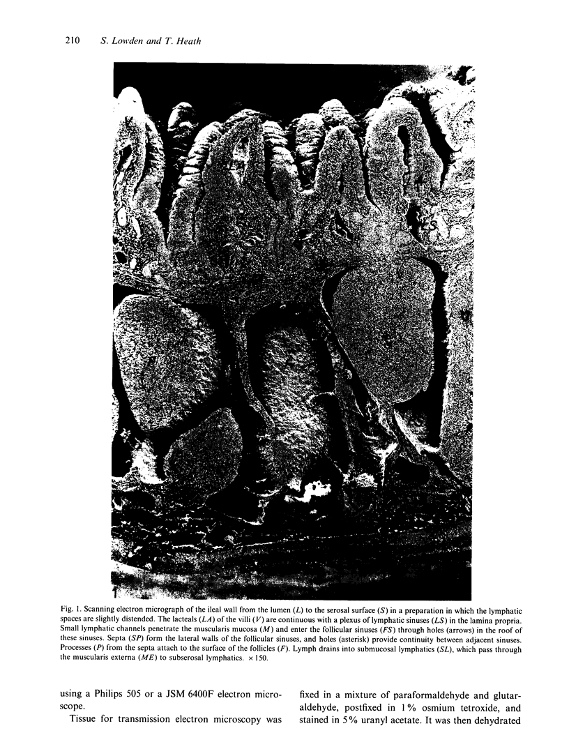
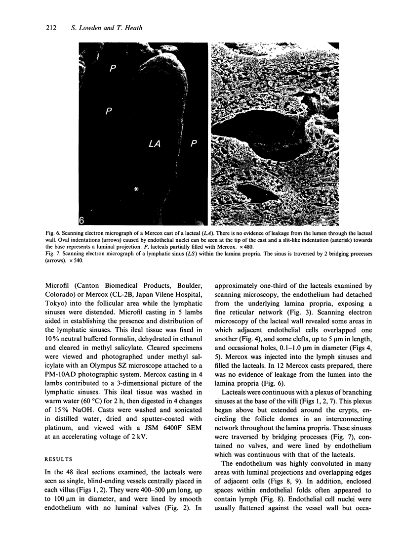
Lymphatic drainage of the ileum associated with Peyer's patches begins with lymph entering a single lacteal of the villus, probably through intercellular flaps, which prevent retrograde flow to the interstitium. These lacteals are continuous with an interconnecting plexus of branching sinuses which surrounds the crypts and follicle domes in the lamina propria. Small vessels emanating from this plexus penetrate the muscularis mucosa, where lymph can either flow within septal vessels to the deep submucosa, or enter the follicular sinuses to move freely around follicles, and through gaps in septal walls. All lymph enters a deep submucosal network where retrograde flow is prevented by valves. Lymph is then conveyed through vessels passing between the fibres of the muscularis externa to other lymphatics, which transport lymph from the ileal surface to the mesenteric nodes.
Full text
PDF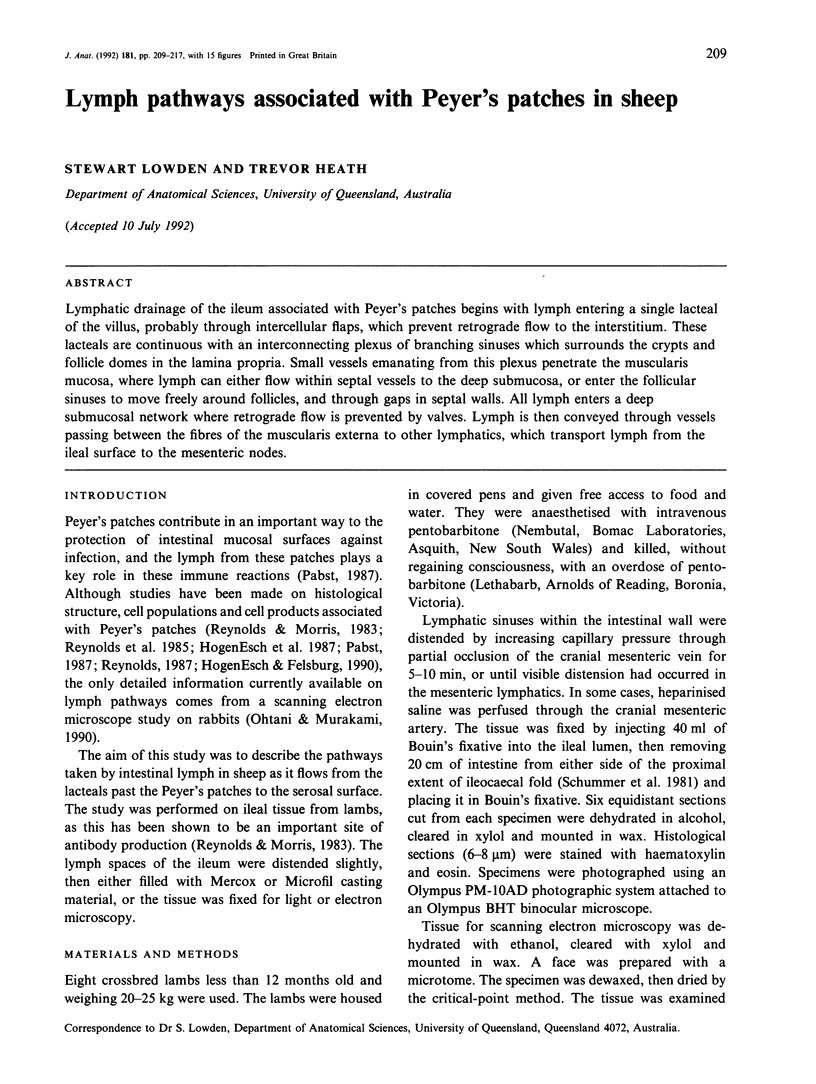


Images in this article

Selected References
These references are in PubMed. This may not be the complete list of references from this article.
- CASLEY-SMITH J. R., FLOREY H. W. The structure of normal small lymphatics. Q J Exp Physiol Cogn Med Sci. 1961 Jan;46:101–106. doi: 10.1113/expphysiol.1961.sp001502. [DOI] [PubMed] [Google Scholar]
- Collan Y., Kalima T. V. The lymphatic pump of the intestinal villus of the rat. Scand J Gastroenterol. 1970;5(3):187–196. [PubMed] [Google Scholar]
- HogenEsch H., Felsburg P. J. Ultrastructure and alkaline phosphatase activity of the dome epithelium of canine Peyer's patches. Vet Immunol Immunopathol. 1990 Feb;24(2):177–186. doi: 10.1016/0165-2427(90)90020-s. [DOI] [PubMed] [Google Scholar]
- HogenEsch H., Housman J. M., Felsburg P. J. Canine Peyer's patches: macroscopic, light microscopic, scanning electron microscopic and immunohistochemical investigations. Adv Exp Med Biol. 1987;216A:249–256. doi: 10.1007/978-1-4684-5344-7_29. [DOI] [PubMed] [Google Scholar]
- Leak L. V., Burke J. F. Fine structure of the lymphatic capillary and the adjoining connective tissue area. Am J Anat. 1966 May;118(3):785–809. doi: 10.1002/aja.1001180308. [DOI] [PubMed] [Google Scholar]
- Mohiuddin A. Blood and lymph vessels in the jejunal villi of the white rat. Anat Rec. 1966 Sep;156(1):83–89. doi: 10.1002/ar.1091560110. [DOI] [PubMed] [Google Scholar]
- Ohtani O., Murakami T. Organization of the lymphatic vessels and their relationships to blood vessels in rabbit Peyer's patches. Arch Histol Cytol. 1990;53 (Suppl):155–164. doi: 10.1679/aohc.53.suppl_155. [DOI] [PubMed] [Google Scholar]
- Ohtani O., Ohtsuka A., Owen R. L. Three-dimensional organization of the lymphatics in the rabbit appendix. A scanning electron and light microscopic study. Gastroenterology. 1986 Oct;91(4):947–955. doi: 10.1016/0016-5085(86)90699-2. [DOI] [PubMed] [Google Scholar]
- Ohtani O. Three-dimensional organization of lymphatics and its relationship to blood vessels in rat small intestine. Cell Tissue Res. 1987 May;248(2):365–374. doi: 10.1007/BF00218204. [DOI] [PubMed] [Google Scholar]
- Pabst R. The anatomical basis for the immune function of the gut. Anat Embryol (Berl) 1987;176(2):135–144. doi: 10.1007/BF00310046. [DOI] [PubMed] [Google Scholar]
- Reynolds J. D., Morris B. The evolution and involution of Peyer's patches in fetal and postnatal sheep. Eur J Immunol. 1983 Aug;13(8):627–635. doi: 10.1002/eji.1830130805. [DOI] [PubMed] [Google Scholar]
- Reynolds J. D. Peyer's patches and the early development of B lymphocytes. Curr Top Microbiol Immunol. 1987;135:43–56. doi: 10.1007/978-3-642-71851-9_3. [DOI] [PubMed] [Google Scholar]
- Reynolds J., Pabst R., Bordmann G. Evidence for the existence of two distinct types of Peyer's patches in sheep. Adv Exp Med Biol. 1985;186:101–109. doi: 10.1007/978-1-4613-2463-8_12. [DOI] [PubMed] [Google Scholar]