Abstract
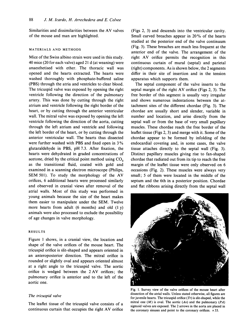
This paper reports a scanning electron microscope study of the morphology of the atrioventricular (AV) valves in the mouse. The leaflet tissue of the 2 AV valves consists of a continuous veil that shows no commissures or clefts. In all instances, the chordae that arise from the papillary system merge with the free border of the leaflet tissue. No distinct terminations of chordae were observed on the ventricular face of the valves. The leaflet tissue of the right AV valve can be divided into parietal and septal components on the basis of the insertion into the ventricular wall and of the papillary system. While the septal component is similar in shape, location and tension apparatus to the septal tricuspid leaflet in man, the parietal component appears to correspond to the anterior and posterior human leaflets. This segment of the valve is served by 3 papillary muscles that arise from the septal wall. The right AV valve is not a tricuspid structure from the morphological standpoint, but appears to function as such because of the particular attachment of the papillary muscles. The leaflet tissue of the mitral valve is served by 2 papillary muscles, anterior and posterior, which consist of muscular trabeculae extending from the heart apex to the base of the valve. These muscles remain associated with the ventricular wall. The leaflet tissue attaches directly to these papillary muscles, which give rise to a very small number of slender chordae. There are thus several important differences between the AV valves of the mouse and man.(ABSTRACT TRUNCATED AT 250 WORDS)
Full text
PDF


Images in this article
Selected References
These references are in PubMed. This may not be the complete list of references from this article.
- Anderson R. H., Wenink A. C. Thoughts on concepts of development of the heart in relation to the morphology of congenital malformations. Experientia. 1988 Dec 1;44(11-12):951–960. doi: 10.1007/BF01939889. [DOI] [PubMed] [Google Scholar]
- Gross L., Kugel M. A. Topographic Anatomy and Histology of the Valves in the Human Heart. Am J Pathol. 1931 Sep;7(5):445–474.7. [PMC free article] [PubMed] [Google Scholar]
- HARKEN D. E., ELLIS L. B., DEXTER L., FARRAND R. E., DICKSON J. F. The responsibility of the physician in the selection of patients with mitral stenosis for surgical treatment. Circulation. 1952 Mar;5(3):349–362. doi: 10.1161/01.cir.5.3.349. [DOI] [PubMed] [Google Scholar]
- Icardo J. M., Sanchez de Vega M. J. Spectrum of heart malformations in mice with situs solitus, situs inversus, and associated visceral heterotaxy. Circulation. 1991 Dec;84(6):2547–2558. doi: 10.1161/01.cir.84.6.2547. [DOI] [PubMed] [Google Scholar]
- Lam J. H., Ranganathan N., Wigle E. D., Silver M. D. Morphology of the human mitral valve. I. Chordae tendineae: a new classification. Circulation. 1970 Mar;41(3):449–458. doi: 10.1161/01.cir.41.3.449. [DOI] [PubMed] [Google Scholar]
- Layton W. M., Jr Heart malformations in mice homozygous for a gene causing situs inversus. Birth Defects Orig Artic Ser. 1978;14(7):277–293. [PubMed] [Google Scholar]
- Magovern J. H., Moore G. W., Hutchins G. M. Development of the atrioventricular valve region in the human embryo. Anat Rec. 1986 Jun;215(2):167–181. doi: 10.1002/ar.1092150210. [DOI] [PubMed] [Google Scholar]
- Roberts W. C. Morphologic features of the normal and abnormal mitral valve. Am J Cardiol. 1983 Mar 15;51(6):1005–1028. doi: 10.1016/s0002-9149(83)80181-7. [DOI] [PubMed] [Google Scholar]
- Silver M. D., Lam J. H., Ranganathan N., Wigle E. D. Morphology of the human tricuspid valve. Circulation. 1971 Mar;43(3):333–348. doi: 10.1161/01.cir.43.3.333. [DOI] [PubMed] [Google Scholar]
- Silverman M. E., Hurst J. W. The mitral complex. Interaction of the anatomy, physiology, and pathology of the mitral annulus, mitral valve leaflets, chordae tendineae, and papillary muscles. Am Heart J. 1968 Sep;76(3):399–418. doi: 10.1016/0002-8703(68)90237-8. [DOI] [PubMed] [Google Scholar]
- Wenink A. C., Gittenberger-de Groot A. C., Brom A. G. Developmental considerations of mitral valve anomalies. Int J Cardiol. 1986 Apr;11(1):85–101. doi: 10.1016/0167-5273(86)90202-0. [DOI] [PubMed] [Google Scholar]
- Wenink A. C., Gittenberger-de Groot A. C. Embryology of the mitral valve. Int J Cardiol. 1986 Apr;11(1):75–84. doi: 10.1016/0167-5273(86)90201-9. [DOI] [PubMed] [Google Scholar]
- Wenink A. C. Quantitative morphology of the embryonic heart: an approach to development of the atrioventricular valves. Anat Rec. 1992 Sep;234(1):129–135. doi: 10.1002/ar.1092340114. [DOI] [PubMed] [Google Scholar]
- Williams T. H., Folan J. C., Jew J. Y., Wang Y. F. Variations in atrioventricular valve innervation in four species of mammals. Am J Anat. 1990 Feb;187(2):193–200. doi: 10.1002/aja.1001870208. [DOI] [PubMed] [Google Scholar]


