

Abstract
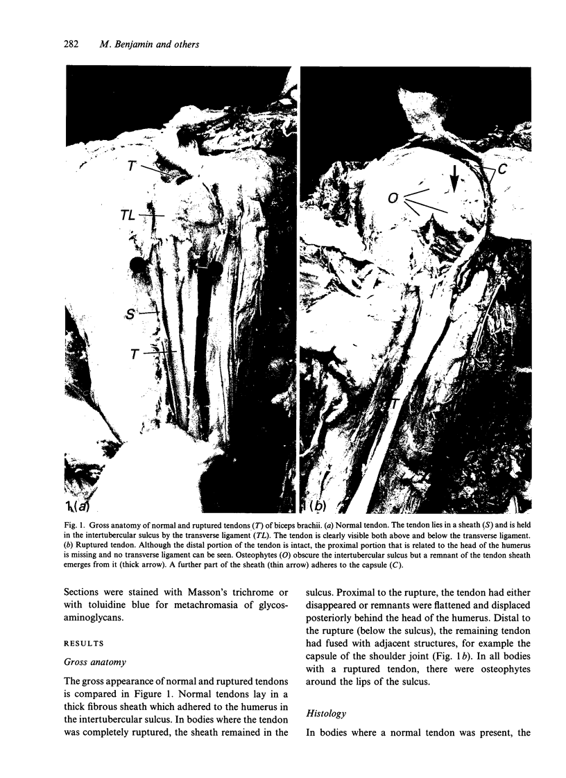
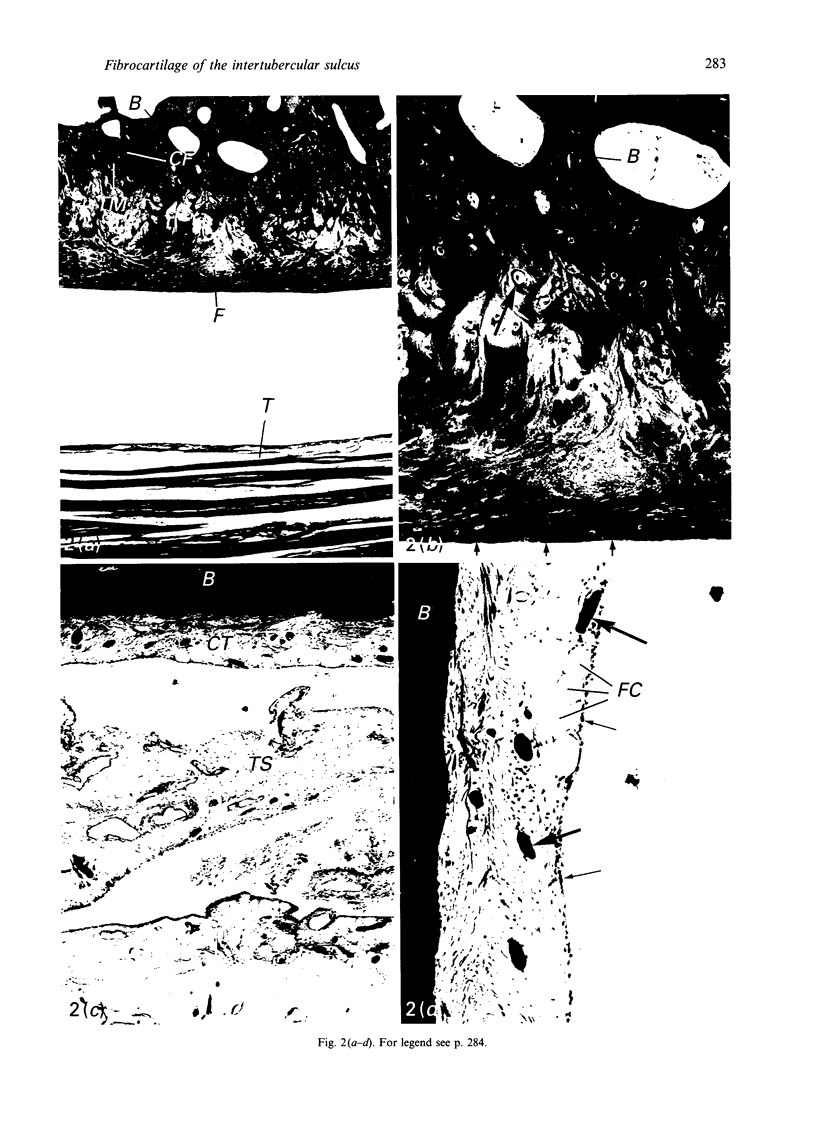
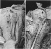
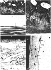
Fibrocartilage lines the intertubercular sulcus of the humerus and protects both the bone and the tendon of the long head of biceps brachii where the tendon passes through the sulcus. It provides a smooth, resilient, lubricated gliding surface on the bone. The fibrocartilage is highly metachromatic and organised into distinct superficial and deep zones. In the superficial zone, the cells are small and the fibres run parallel to the articular surface. In the deep zone, the cells are large and rounded and the coarse bundles of fibres are interwoven. In 6 of the 26 dissecting room cadavers examined the tendons were completely ruptured. In these, the fibrocartilage was replaced by loose connective tissue that resembled the synovium of the tendon sheath. The results suggest that bone fibrocartilage exhibits dynamic behaviour in response to changes in its environment, in the same manner as tendon fibrocartilage.
Full text
PDF

Images in this article
Selected References
These references are in PubMed. This may not be the complete list of references from this article.
- Benjamin M., Evans E. J., Copp L. The histology of tendon attachments to bone in man. J Anat. 1986 Dec;149:89–100. [PMC free article] [PubMed] [Google Scholar]
- Benjamin M., Tyers R. N., Ralphs J. R. Age-related changes in tendon fibrocartilage. J Anat. 1991 Dec;179:127–136. [PMC free article] [PubMed] [Google Scholar]
- Evanko S. P., Vogel K. G. Ultrastructure and proteoglycan composition in the developing fibrocartilaginous region of bovine tendon. Matrix. 1990 Dec;10(6):420–436. doi: 10.1016/s0934-8832(11)80150-2. [DOI] [PubMed] [Google Scholar]
- Evans E. J., Benjamin M., Pemberton D. J. Fibrocartilage in the attachment zones of the quadriceps tendon and patellar ligament of man. J Anat. 1990 Aug;171:155–162. [PMC free article] [PubMed] [Google Scholar]
- Ralphs J. R., Benjamin M., Thornett A. Cell and matrix biology of the suprapatella in the rat: a structural and immunocytochemical study of fibrocartilage in a tendon subject to compression. Anat Rec. 1991 Oct;231(2):167–177. doi: 10.1002/ar.1092310204. [DOI] [PubMed] [Google Scholar]
- Ralphs J. R., Tyers R. N., Benjamin M. Development of functionally distinct fibrocartilages at two sites in the quadriceps tendon of the rat: the suprapatella and the attachment to the patella. Anat Embryol (Berl) 1992;185(2):181–187. doi: 10.1007/BF00185920. [DOI] [PubMed] [Google Scholar]
- Vogel K. G., Koob T. J. Structural specialization in tendons under compression. Int Rev Cytol. 1989;115:267–293. doi: 10.1016/s0074-7696(08)60632-4. [DOI] [PubMed] [Google Scholar]